

Биоинженерия / ТИ_кость / Relative_influence_of_surface_topography_and_surfa
.pdfSee discussions, stats, and author profiles for this publication at: https://www.researchgate.net/publication/49804526
Relative influence of surface topography and surface chemistry on cell response to bone implant materials. Part 2: Biological aspects
Article in Proceedings of the Institution of Mechanical Engineers Part H Journal of Engineering in Medicine · December 2010
DOI: 10.1243/09544119JEIM901 · Source: PubMed
CITATIONS |
READS |
111 |
454 |
3 authors, including: |
|
Arnaud Ponche |
Maxence Bigerelle |
Université de Haute-Alsace |
Polytechnic University of Hauts-de-France |
70 PUBLICATIONS 1,386 CITATIONS |
186 PUBLICATIONS 3,470 CITATIONS |
SEE PROFILE |
SEE PROFILE |
Some of the authors of this publication are also working on these related projects:
Roughness View project
Steel coils annealing View project
All content following this page was uploaded by Arnaud Ponche on 29 July 2014.
The user has requested enhancement of the downloaded file.

Proceedings of the Institution of Mechanical Engineers, Part H: Journal of Engineering in
Medicine
http://pih.sagepub.com/
Relative influence of surface topography and surface chemistry on cell response to bone implant materials. Part 2: Biological aspects
K Anselme, A Ponche and M Bigerelle
Proceedings of the Institution of Mechanical Engineers, Part H: Journal of Engineering in Medicine 2010 224: 1487
DOI: 10.1243/09544119JEIM901
The online version of this article can be found at:
http://pih.sagepub.com/content/224/12/1487
Published by:
http://www.sagepublications.com
On behalf of:
Institution of Mechanical Engineers
Additional services and information for Proceedings of the Institution of Mechanical Engineers, Part H: Journal of Engineering in Medicine can be found at:
Email Alerts: http://pih.sagepub.com/cgi/alerts
Subscriptions: http://pih.sagepub.com/subscriptions
Reprints: http://www.sagepub.com/journalsReprints.nav
Permissions: http://www.sagepub.com/journalsPermissions.nav
Citations: http://pih.sagepub.com/content/224/12/1487.refs.html
>> Version of Record - Dec 1, 2010
What is This?
Downloaded from pih.sagepub.com by guest on March 16, 2013

SPECIAL ISSUE PAPER 1487
Relative influence of surface topography and surface chemistry on cell response to bone implant materials. Part 2: biological aspects
K Anselme1*, A Ponche1, and M Bigerelle2
1Institut de Sciences des Mate´riaux de Mulhouse (IS2M), CNRS LRC7228, Universite´ de Haute-Alsace, Mulhouse, France 2Laboratoire Roberval, CNRS UMR6253, Centre de Recherche de Royallieu, Universite´ de Technologie de Compie`gne, Compie`gne, France
The manuscript was received on 19 July 2010 and was accepted after revision for publication on 12 August 2010.
DOI: 10.1243/09544119JEIM901
Abstract: A current medical challenge is the replacement of tissue which can be thought of in terms of bone tissue engineering approaches. The key problem in bone tissue engineering lies in associating bone stem cells with material supports or scaffolds that can be implanted in a patient. Beside bone tissue engineering approaches, these types of materials are used daily in orthopaedics and dental practice as permanent or transitory implants such as ceramic bone filling materials or metallic prostheses. Consequently, it is essential to better understand how bone cells interact with materials. For several years, the current authors and others have developed in vitro studies in order to elucidate the mechanisms underlying the response of human bone cells to implant surfaces. This paper reviews the current state of knowledge and proposes future directions for research in this domain.
Keywords: surface topography, surface chemistry, cell adhesion, cell proliferation, cell differentiation, protein adsorption, bone, implant, roughness parameters
1INTRODUCTION
Understanding the way in which cells interact with materials is a crucial topic in the biomaterial and tissue engineering areas. Cells respond to nanostructures and microstructures, for example, with the extracellular matrix (ECM) where the nanometresized collagen fibrils are organized in three-dimen- sional (3D) meshes of micrometre-sized fibres. Moreover, because the ECM is composed of a variety of specific proteins, and in the case of bone, also of mineral components, the cellular environment exhibits a chemistry and mechanical behaviour with which the bone cells are used to interacting. A current medical challenge is the replacement of tissue which can be thought of in terms of bone
*Corresponding author: UHA, Institut de Science des Materiaux de Mulhouse, 15 rue jean starcky, BP2488, Mulhouse 68057, France.
email: karine.anselme@uha.fr
tissue engineering approaches. The key problem in bone tissue engineering lies in associating bone stem cells with material supports or scaffolds that can be implanted in a patient. This approach to promoting the growth of new bone tissue has the potential to replace bone grafts. The implantation of the cellcontaining scaffolds can occur immediately after their synthesis or it can be delayed until after a period of in vitro culture. This period allows the acceleration of the interactions of cells with the scaffold thus improving the survival chances of the hybrid material after implantation. Beside bone tissue engineering approaches, these types of materials are used daily in orthopaedics and dental practice as permanent or transitory implants such as ceramic bone filling materials or metallic prostheses. Consequently, it is essential to better understand how bone cells interact with materials. For several years, the current authors and others have developed in vitro studies in order to elucidate the mechanisms underlying the response of human bone cells to
JEIM901 |
Proc. IMechE Vol. 224 Part H: J. Engineering in Medicine |
Downloaded from pih.sagepub.com by guest on March 16, 2013
1488 |
A Ponche, K Anselme, and M Bigerelle |
|
|
implant surfaces [1–5]. This paper reviews the current state of knowledge and proposes future directions for research in this domain.
2DEFINITION OF MECHANISMS INVOLVED IN CELL–SURFACE INTERACTIONS
The majority of cells in the body (with the major exception of blood cells) adhere to an ECM. In bone tissue, the cells that form bone, the osteoblasts, and the cells that degrade bone, the osteoclasts, both adhere and grow on ECMs composed of proteins (90 per cent collagen and 10 per cent other proteins) and proteoglycans filled with calcium phosphate minerals. In vitro, these cells can survive only if they can adhere to a surface and thus only if this adhesion is achieved can the cells continue to grow and differentiate. The first interactions between the cell and the surface, both in vitro and in vivo, will define the quality of the cell–implant and tissue–implant interfaces. After only a few seconds of contact between the surface and the fluid (the culture medium in vitro or interstitial fluids or blood in vivo), the surface becomes coated by water and proteins. Thus, the remaining cells will sense the surface features of the ECM through this adsorbed layer. Cells first adhere through physicochemical interactions such as ionic forces and van der Waals forces and later through
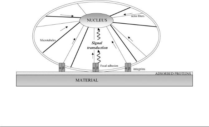
various biomolecules, both those adsorbed on the surface and those involved in cell response to the environment. The key molecules in this process are the cell’s surface receptors, called integrins, that are transmembrane molecules that in vivo mediate the interactions with ECM molecules. Integrins are heterodimers that consist of one alpha and one beta subunit that can be combined to form receptors specific for amino-acid sequences such as the ar- ginine–glycine–aspartic acid (RGD) recognition motif present in many ECM proteins. There are eight different alpha sub-units and 11 different beta subunits, that can be combined to form 24 different integrin receptors. These integrin receptors act as an interface between the intracellular and extracellular compartments. On the extracellular side they interact with the ECM and on the intracellular side they interact with the molecules of the cytoskeleton and with signalling molecules at the adhesion sites, called focal adhesions (Fig. 1). Thus, they can transmit a signal from the ECM to the nucleus through a biochemical signal transduction pathway. This signal transduction involves the accumulation and phosphorylation of several proteins such as FAK, Src, Rho GTPases, ERK, JNK etc. [6]. Alternatively, the signal can be transmitted mechanically since there exists a physical link between the focal adhesion sites and nuclear membrane created by the cytoskeleton [7]. Actually, integrins are physically ‘hardwired’ to the
Fig. 1 Schematic diagram of the proteins involved in adhesion of eukaryotic cells on materials. Integrins are transmembrane proteins that form with other intracellular proteins the focal adhesions. On these focal adhesions are connected actin fibres (thin lines) that physically join the cell membrane and nuclear membrane such as microtubules (bold lines). Signal transduction from cell membrane to nucleus can pass through a direct mechanotransduction way (straight arrows) or indirectly by biochemical signal transduction (twisted arrows)
Proc. IMechE Vol. 224 Part H: J. Engineering in Medicine |
JEIM901 |
Downloaded from pih.sagepub.com by guest on March 16, 2013

Relative influence of surface topography and surface chemistry. Part 2 |
1489 |
|
|
nucleus through the cytoskeleton. This involves the LINC complex (for linker of nucleoskeleton and cytoskeleton) and the coupling between KASH and SUN proteins such as nesprins with intermediate filaments or actin fibres [8]. This 3D configuration may act so as to order the nucleus and then coordinate transcription [9]. The potential use of nanoimprinting to directly imprint the topography onto the cell has been proposed [10].
When the integrin receptors interact with the ECM-adsorbed biomolecules, they cluster to form focal adhesions. At this point the cytoskeleton is reorganized to allow the spreading of cells to the substrate. The cytoskeleton is composed of three different molecules: actin fibers, tubulin microtubules, and vimentin, desmin, or keratin microfilaments. In the focal adhesions, these cytoskeletal molecules interact with signalling molecules such as talin, paxillin, vinculin, tensin, and protein kinases [11]. Focal adhesions are closed junctions in which the distance between the substrate surface and the cell membrane is between 10 and 15 nm. Some authors call them focal complexes when they are shorter than 1 mm, focal adhesions when they measure between 1 and 5 mm, and supermature adhesions when they are over 5 mm [12]. There is kinetic activity in these systems in that nascent close contacts transform into focal contacts before they become focal adhesions. The transition between these different phases is force dependent [5]. Moreover, in tissues or inside the 3D ECM, fibrillar adhesions have also been described [13].
Another group of essential proteins that are involved in a cell’s capacity to adhere, spread, divide, and migrate are the myosins [14]. They are actin-based motor proteins that translocate along actin fibres and allow their contraction. After adhesion and spreading, the cells secure their shape stability through a prestress state maintained by myosin motors transmitted by actin fibres throughout the cell body and balanced by microtubules [15, 16]. If needed, cells are able to migrate in order to find a more suitable place for their further development. Also, if the point on the substrate at which they are cultured has an anisotropic topography such as a groove, the cells migrate and orient themselves as a function of the axis of the groove. This phenomenon is called ‘contact guidance’ and has been only observed in vitro.
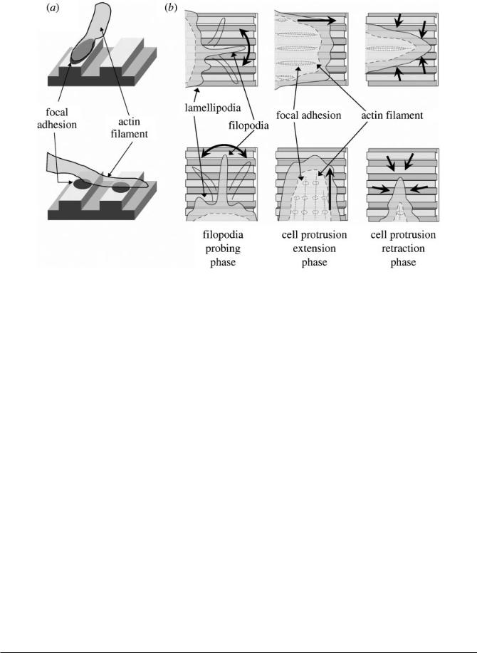
It has been demonstrated by several authors that filopodia are the cell’s tools for exploring its surroundings [17]. Recently, Fujita et al. [18] performed a time lapse analysis of cell alignment on nano-grooved patterns and demonstrated that the
filopodia probed the area surrounding the cell protrusion before movement into this area. However, it was also shown that when the cell protrusions extend perpendicularly to a groove axis they retract more rapidly than those parallel to the groove axis. Also, in protrusions parallel to the groove axis, actin filaments form wider focal adhesions at filament termination whereas when the protrusions are perpendicular to the groove axis, the focal adhesions are formed less easily and appear fragmented (Fig. 2). Thus, it appears that the major mechanism in cell alignment on grooves is not the filopodial sensing but rather the retracting phase of the cell protrusion that is related to the quality of the focal adhesions [18].
All these biochemical and mechanical interactions allow signal transduction to the nucleus that either induces cell death through apoptosis if the adhesion is not possible or cell growth and cell differentiation if cell adhesion and spreading can be achieved.
After adhesion and migration, cells divide and proliferate across the surface. They further differentiate which means that they synthesize the molecules they normally synthesize in their tissue of origin. Osteoblasts will synthesize collagen and the other proteins normally found in bone tissue. Also, they are able to induce their own in vitro mineralization in certain culture medium conditions and to produce a mineralized bone-like tissue [19].
Generally, the investigation of cell–surface interactions tends to be focussed on the initial cell adhesion phase and little attention is paid to cell– surface interactions after this point. It is the current authors’ contention that it is also necessary to consider the influences of surface topography and chemistry at the complex interfaces between the cell and the ECM [20]. Various methods have been developed to quantify the cell–surface adhesion strength. When the ECM is not involved, aspiration, centrifugation, and fluid flow techniques have been used to detach the cell populations. Micromanipulation methods based on micropipettes, microcantilevers, and optical or magnetic tweezers, have been used to detach isolated cells [3]. The cell–surface interactions can also be evaluated using an enzymatic detachment method. First, the adhesion of human osteoblasts after 24 h, called short-term adhesion, is quantified. At the end of this first phase, all the cells are spread on the surface and their cytoskeleton is well organized. However, at this point they have neither expressed b1 and a3 integrin molecules nor synthesized matrix proteins [21]. In addition, the long-term adhesion, which represents
JEIM901 |
Proc. IMechE Vol. 224 Part H: J. Engineering in Medicine |
Downloaded from pih.sagepub.com by guest on March 16, 2013
1490 |
A Ponche, K Anselme, and M Bigerelle |
|
|
Fig. 2 Model for cell alignment on the nanogrooved substrate. (a) Actin filaments parallel to the grooves form wide focal adhesions at filament terminations. However, termination of perpendicular filaments is fragmented because a focal adhesion is formed only on the ridge. (b) Filopodia movements are isotropic, i.e. no specific direction is observed for their extension and retraction against the nanogrooved structure. This finding suggests that filopodia probing does not play a major role in cell alignment. Cell protrusions extend isotropically, and those that are perpendicular to the nanogrooved pattern retract more rapidly than those parallel to the nanogrooved pattern. These cell protrusion dynamics force a cell to elongate and align along the nanogrooved pattern (Reproduced with permission from [18])
the strength of the cell–material interface formed during 3 weeks of culture involving the ECM proteins synthesized by the cells themselves and the cell–cell contacts, can also be quantified [20]. This long-term adhesion has been shown to be better correlated with topography, especially hybrid roughness parameters that quantify roughness organization, whereas short-term adhesion is more influenced by surface chemistry [22–28].
Another very important point to consider in the study of cell–surface interactions is the phenotype of the cells which is related to their original tissue type. Cell lines produced from normal healthy tissue are called primary cell lines. Their phenotype is very close to the one of cells inside the tissue but they are in general more difficult to culture in vitro. Moreover, they cannot be cultured for more than a few weeks and for only a limited number of passages (change from one culture dish to a new one). Other cell lines, called permanent cell lines, are produced
by the immortalization of primary cell lines or are derived from bone tumours. The major advantages of these permanent cell lines are their high proliferation capacity and the ability to culture them for almost indefinite periods. However, their phenotype can be slightly different from their initial or normal phenotype. It has been frequently observed that the cell phenotype has a strong influence on cell response to a material surface [29]. For example, it is clear that the adhesion mechanism of blood cells is different to that of cells originating from connective tissues such as fibroblasts, keratinocytes, or vascular cells since the extracellular environment in their tissues of origin are very different. Primary and permanent cell lines are also very different in term of their adhesiveness. Tumour cells tend to lose part of their ability to adhere to an ECM as the number of proliferations increases. It has been shown that both tumour cells and immortalized cells have a more diffuse cytoskeleton and are more deformable than
Proc. IMechE Vol. 224 Part H: J. Engineering in Medicine |
JEIM901 |
Downloaded from pih.sagepub.com by guest on March 16, 2013

Relative influence of surface topography and surface chemistry. Part 2 |
1491 |
|
|
normal cells [30]. The current authors recently obtained important results using two osteosarcomaderived cell lines that are frequently used for analysing the influence of surface topography on bone cells: SaOs-2 and MG63 [31]. Those cells cultured on poly-L-lactic acid (PLLA) micro-pillared substrates displayed a previously unreported extensive deformation of their nucleus. This deformation was not seen using bone-marrow-derived human mesenchymal stem cells (hMSCs). The shape of the nuclei reflected the shape of the micropillars; as if the cells had pushed their nuclei in-between the pillars. More surprising, whilst this deformation should impair future growth and differentiation, the two osteosarcoma cell lines demonstrated a proliferation and differentiation capacity comparable to the one they display on flat PLLA substrates. These results have been reproduced using immortalized cell lines originating from bone and other tissues thus demonstrating that the response of both cancerous and immortalized cell lines to topography can be abnormal [32]. Remarkably, this deformation is not due to any external pressure and is reminiscent of the type of deformation that a cancerous cell can encounter when it undergoes metastatic migration. The mechanical properties of cancerous cells can be related to their metastatic potential; cells that are more likely to form tumors in vivo are more deformable [33, 34]. Additionally, this change in shape of the nucleus is surprising given that the nucleus is thought to require a particular architecture for correct functioning. Indeed, it has been found that locating genes close to the nuclear lamina induces a switching off of the genes in question [35, 36]. Deformation of the nucleus increases the surface-to-volume ratio and, hence, the proportion of switched off genes should increase. Thus, it would be of interest to continue to study these deformed cells in order to test if they can act as a surrogate system for the biological behaviour of malignant cells during their metastatic dissemination. From this section of the review it is clear that noncancerous and non-immortalized cell lines must be preferred for use in the study of cell–surface interactions and in particular for the investigation of the influence of topography on bone cells.
Concerning the mechanisms of the cell response to surface topography, it has been proposed that cells react to the presence of discontinuities on the surface. Discontinuities have been defined as structures
with a radius of curvature less than the average length of a pseudopodium or of the distance part of the sensing elements that control cell movements [37].
Thus, the reaction to discontinuities involves focal adhesions and the actomyosin cytoskeleton and is related to the mechanisms of the mechanical sensitivity of cells. When a cell or a pseudopodium meets a concave or convex structure, the cell receptors and actomyosin cytoskeleton undergo varying degrees of deformation or compression [38] and this induces a reorganization of the attachment and cytoskeletal structures [39]. Dunn and Heath [40] have shown that fibroblasts were affected by a convex ridge in a prism substrate if its angle was greater than 4u. The cells displayed discontinuities in their cytoskeletal microfilament bundle system which coincided with a discontinuity in the substrate [40]. It has also been shown that the cytoskeleton is involved in cell interaction with grooved surfaces thus demonstrating cell sensitivity to concave surfaces [41]. Recently, a very interesting study has demonstrated that the response of cells to equivalent concave and convex structures made in polymethyldisiloxane was different. The cells preferentially adhered and migrated on the convex structures rather than on the concave structures [42]. This definitively confirms that the cells are able to identify and discriminate between concave and convex surfaces.
Another proposed mechanism for the cell response to surface topography is that the strains introduced into the cytoskeleton could in turn influence ion channels as observed in the stretch receptors of a variety of organisms [43].
Different hypothesis have been proposed for the mechanisms underlying the identification of discontinuities. One is based on the thermodynamics of the ECM adsorption. The discontinuities may differ in reactivity from nearby planar surfaces due to the existence of unsaturated bonds. This would alter the adsorption of ECM proteins and then modify the response of cells that would reflect the differences in adhesiveness related to surface topography [44]. Another hypothesis has proposed that the discontinuities represent energy barriers, the surface geometry and chemistry influencing the size and position of these energy barriers. Then cells modify their orientation, adhesion, or spreading in order to minimize their contact with high-energy discontinuities [45].
3RESPONSE OF CELLS TO SURFACE TOPOGRAPHY
Historically, the first in vitro studies of cell response to surface topography were performed on surfaces whose topography was controlled and measured at
JEIM901 |
Proc. IMechE Vol. 224 Part H: J. Engineering in Medicine |
Downloaded from pih.sagepub.com by guest on March 16, 2013

1492 |
A Ponche, K Anselme, and M Bigerelle |
|
|
the micron scale. When the objective was to compare surfaces with different roughness amplitudes, very frequently, the authors changed the process without considering the consequences of this change on the surface morphology or the surface chemistry [46–48]. Another cause for misunderstandings in this field is the practice of defining a surface by its manufacturing process instead of concisely defining it by its topographical parameters [19]. This practice has led to erroneous comparisons of surfaces since it is known that a process can induce different topographies when it is applied to different materials. Indeed, it is extremely rare in the literature that the surface topography is correctly characterized: by associating amplitude, frequency, and hybrid parameters [49]. In general, the amplitude parameters (Ra, Rt, Rz) are commonly considered but the frequency and hybrid parameters are more rarely quantified [50]. It should be noted that the current authors compute more than 100 roughness parameters and then statistically correlate these parameters with biological parameters in order to determine the surface parameter(s) to which the cells respond. Using this approach it has been shown that the parameters that describe the morphology of the topography and particularly its degree of organization are the parameters that better correlate with the adhesion of human primary osteoblasts [21, 51].
The proliferation and differentiation potential of MG-63-osteosarcoma-derived cells has been reported to be positively influenced by surface roughness although the attachment level is reduced [47, 52, 53]. These findings have been independently confirmed in several papers in the literature [27, 54– 59], however, there are papers that contradict these findings [60–62]. This disparity highlights the influence of the used cell model and the necessity for detailed characterization of the roughness of substrates to be performed in order to allow valid comparison of results. The topography of substrates is in general either poorly defined or the use of different surface treatments (polishing, turning, blasting, acid etching, plasma spraying etc.) renders comparisons invalid.
There have been extensive attempts to elucidate the mechanisms underlying the MG63 cells response to topography. It has been demonstrated that the cells exhibit a more differentiated phenotype on rough surfaces and that their response to systemic factors such as the vitamin D metabolite 1a, 25(OH)2D3 or 17b-estradiol is also stimulated by roughness [63–66]. In addition to how cells respond to exogenous factors, surface roughness has been shown to affect regula-
tory factors synthesized by cells such as prostaglandins (PGE1, PGE2), transforming growth factor beta1 (TGFb1) [52, 53, 63], and osteoprotegerin [67, 68]. It has been shown that this effect can be mediated by protein kinase A and phospholipase A2 [4, 65, 69, 70]. These effects have been reproduced using bone cell lines immortalized from mice or derived from rat calvaria that present a more differentiated maturation state than the MG-63 cells. The sensitivity to roughness appears to be higher for immature cells compared to more mature ones [64].
It has been demonstrated that a5b1 and a2b1 integrins are involved in the MG-63 cells response to titanium substrates with different surface roughness levels. In particular it was highlighted that a5b1 mainly influenced cell proliferation whereas a2b1 signalling was required for MG-63 cell differentiation [71–73]. However, contradictory results that show no expression of a5, a3, and a6 integrin subunits on sandblasted titanium substrates and a5 integrin subunits on polished substrates but with a2, a4, av, b1, and b3 integrin subunits being expressed on both substrates have been published in the literature [74]. Human primary osteoblasts express a3b1 integrins but not a2b1 integrins whatever the roughness of the titanium substrates [21]. However, expression of the b1 and b3 integrin subunits by human primary osteoblasts and MG-63 osteoblastic cells was observed in fibrillar adhesions visualized by immunofluorescence on smooth-structured titanium surfaces but not on coarser surfaces [75, 76]. Flow cytometric analysis was used to confirm a slight increase in the expression of b1 and b3 integrins on cells detached from coarse surfaces compared to smooth ones [76, 77]. These observations illustrate the difficulty in comparing results obtained by different experimental methods or using different cell lines. The role of integrins in the response of osteoblasts to bone materials has been comprehensively reviewed in [6].
In addition to substrates treated by the classical methods used for bone implant surface preparation, some experiments have been performed on grooved substrates in order to analyse the influence of roughness on anisotropic grooved surfaces such as can be found on screws or dental implants. A systematic orientation of cells in the direction of grooves has been observed [44, 78–82] and it has been elucidated that the cell orientation is not a function of the chemical nature of the substrate although the morphology of cells was influenced both by topography and chemistry [79]. By comparing isotropic rough surfaces obtained by sandblasting and acid etching to grooved surfaces, a higher osteoblastic cell adhesion
Proc. IMechE Vol. 224 Part H: J. Engineering in Medicine |
JEIM901 |
Downloaded from pih.sagepub.com by guest on March 16, 2013

Relative influence of surface topography and surface chemistry. Part 2 |
1493 |
|
|
and proliferation was isolated on the latter surfaces [78]. The dimension of the grooves is also critical and grooves measuring about 10 mm in width have been shown to significantly increase viability [81] and adhesion of osteoblastic cells [44]. Finally, the presence of a micro-topography inside the grooves has been shown to further increase the orientation of cells [82].
3.1Scale effect
As seen in part 1 a very important parameter to consider is the scale used to calculate the roughness parameter relative to the cell scale. Surfaces prepared by electro-erosion are particularly favourable for long-term adhesion of human osteoblastic cells. The topography of electro-eroded titanium surfaces has been analysed at two different scales: above the human osteoblastic cell scale (.50 mm) and below the cell scale (,50 mm). Above the cell scale, the surfaces were considered to be rough with a melted relief and smooth edges whereas below the cell scale, these surfaces were relatively smooth and presented a flat area permitting the adhesion of cells in a favourable environment that resembles a basin between reliefs [83]. Thus, it appears that rough surfaces are preferred by human bone cells; particularly when they can provide relatively flat areas at their own scale that help the anchoring of the cells. In order to control the surface chemistry, the electroeroded samples were coated with a thin (nanometre range) gold layer. The previous results were confirmed demonstrating that the cell response was solely a topography-related effect [83].
An interesting way of systematically exploring the dependence of surface chemistry on surface roughness is to use a single process to produce a gradient of roughness on a single sample. Kunzler et al. [84] used sandblasting followed by a chemical smoothing process to create a surface on an aluminium sheet whose Ra values changed between 1 and 6 mm. To ensure a constant surface chemistry, the samples were further replicated in an epoxy resin and coated with titanium. A linear increase of proliferation of rat calvaria osteoblasts was observed although human gingival fibroblasts showed a decreased proliferation. Thus, it appears that for this range of Ra and for the same surface organization, the cell response is linearly dependent on the roughness amplitude but the nature of this response is cell dependent. Again this study illustrates the importance of the cell phenotype. Here, the cell phenotypes influenced the cell response either by their different tissue
origin or by their different size and morphology since rat calvaria osteoblasts are smaller and present a more cubical morphology than human fibroblasts.
Recently, driven by the development of nanopatterning methods, the influence of nanotopography on cell response has started to be studied. This is particularly pertinent since the cells reaction to topography is mediated by proteins that have nanometre scales. Using nanofeatures prepared by colloidal lithography, Dalby et al. [85] studied the response of fibroblasts to nano-columns 160 nm high and 100 nm in diameter. They demonstrated a decreased cell adhesion and spreading on the nanocolumns characterized by smaller, fainter focal adhesions and a disorganization of the cytoskeleton. Andersson et al. [86] observed that on columns of 58, 91, 111, and 166 nm width, the larger the feature, the more spread the cells became. On surfaces with nano-pits with diameters of 35, 75, and 120 nm, fibroblasts were able to sense topography down to 35 nm using their filopodia [87]. On nano-grooved polystyrene substrates with groove depths from 5 to 350 nm and widths from 20 to 1000 nm, Loesberg et al. [88]concluded that depth was the most important parameter influencing cell alignment and that 35 nm was the threshold feature size for fibroblasts since below this value contact guidance was no longer apparent. This dimension can be related to the optimum distance for cell adhesion (58 nm) that was determined in elegant studies on surfaces presenting nanoparticles decorated with the RGD peptides involved in integrin recognition and separated by a controlled distance [89–91].
One area of considerable interest is the determination of the size threshold to produce an effect in cells. Dalby et al. [92] demonstrated that cells are able to respond to 13 nm tall islands and Dalby et al. [93] reported increased filopodia production and smaller focal contacts on 10 nm tall islands compared to the values obtained for flat surfaces. These observations were confirmed by Lim et al. [94] who showed that worm-like structures 14 nm tall increased human fetal osteoblastic cell attachment, spreading, integrin subunit expression, and focal adhesion protein synthesis compared to taller structures (29, 45 nm) and flat control surfaces. In conclusion, these results suggest that the size threshold for cell detection could be less than 10 nm; however, the control of the topography under this threshold remains an open problem.
These results obtained on model surfaces are useful for elucidating mechanisms for specific nanoscale cell features; however, they are not really re-
JEIM901 |
Proc. IMechE Vol. 224 Part H: J. Engineering in Medicine |
Downloaded from pih.sagepub.com by guest on March 16, 2013

1494 |
A Ponche, K Anselme, and M Bigerelle |
|
|
presentative of the implants’ actual nanotopography. In general, the materials used for implants have a random nanotopography, meaning that the surface does not present any organization or direction. An anodization process can be used to produce nanotubes or nanopores [95–98] and it has been shown that small nanotubes (,30 nm in diameter) promote adhesion of osteoblasts [99] or hMSCs [100] without noticeable differentiation whereas larger nanotubes (70–100 nm in diameter) elicite a dramatic cell elongation which induces cytoskeletal stress and selective differentiation. The same anodization process can be used to create random nano-sized patterns on oxide layers such as titanium dioxide and alumina [101, 102]. An experiment has been performed on titanium substrates micropatterned using electrochemical micromachining and further treated by acid etching and anodization in order to change the nanotopography of the microfeatures. Hemispherical cavities measuring 10, 30, and 100 mm in diameter were produced [101]. After anodization or acid etching of the 30 mm cavities, it was shown that MG63 cells adhered and proliferated better on the treated cavities than on the polished ones, especially after the anodization treatment. These studies confirm that implant surfaces should be controlled at every scale since the cells are able to identify and react to both nano-sized features and micron-sized features.
The concept of nanophase materials or nanomaterials has been proposed to describe materials with structural units that are between 1–100 nm [103, 104]. Several studies have reported improved in vitro osteoblast adhesion on the surfaces of nanophase materials created from a wide range of materials including ceramics [105, 106], metals [105, 107, 108], polymers [109, 110], and composites [111, 112]. This effect has been shown to be due to improved protein adsorption characteristics [113, 114] that are a consequence of their surface energy properties [103]. However, to date only a few in vivo studies have been performed to test the effect of nanophase materials on integration in bone tissue. Nanostructured Al2O3 topographic features applied to machined implants promoted a greater bone-specific gene expression in tissues adjacent to implants after implantation in rabbit tibias and an associated increase in bone-to-implant contact and torque removal [115]. Similarly, a greater amount of new bone formation was observed in contact with a tantalum-based scaffold coated with nanohydroxyapatite (nano-HA) than on scaffolds coated with conventional HA [103]. The results obtained on
nano-HA were inconsistent and appear to be dependent on the implantation method. Nano-HA increased bone formation when no gap existed between bone and implant but failed to enhance bone formation in a gap-healing model [116, 117].
3.2Organization of topography
As previously discussed, when cells are cultured on an anisotropic topography such as a groove, cells orientate themselves along the axis of the groove because of the contact guidance phenomenon. Also, when the surface consists of an organized array of pillars the cells are able to identify this organization and orient themselves in the orthogonal direction [41]. Normally, however, it is difficult to define the topographic organization for implant surfaces prepared by classical process (plasma spraying, sandblasting…) that display rather isotropic surfaces. As discussed in part 1 of this paper, numerous approaches have been proposed to solve the problem of how to define the degree of organization on a surface. It is the current authors experience that the parameters that best describe the organization are those that are correlated with long-term adhesion on the implant surface, for example, (see Fig. 3) the Order parameter that describes with the highest relevance the adhesion of human osteoblast cells on electro-eroded surfaces presented in [51] and [118]. The study of cell organization at the nanometre scale has been recently reported [119]. hMSCs were shown to differentiate better on surfaces presenting slight disorder of 120 nm diameter nanopits (displaced square placements) compared to a hexagonal, square, or even random placement. Their differentiation was comparable to the one obtained with an osteogenic medium containing stimulative factors such as dexamethasone and L-ascorbic acid. This confirms that cells are sensitive to surface organization at both the nanoscale and microscale and that they prefer relatively disordered surfaces. However, these experiments were done with bonederived cells that have in vivo a relatively disorganized environment. Other cells such as keratocytes, fibroblasts, or endothelial cells that have rather organized environment in their tissue of origin have shown a different behaviour in vitro [120].
The validity of the results obtained in vitro need to be confirmed in vivo since the key factor for using in vitro studies is an improved implant surface. In vivo, it has been shown that the roughness of implants permits the interlocking of bone tissue by increasing the bone-to-implant contact length as well as
Proc. IMechE Vol. 224 Part H: J. Engineering in Medicine |
JEIM901 |
Downloaded from pih.sagepub.com by guest on March 16, 2013