

Cardiology / English / Аритмии блокады
.pdfIII degree of AV block (complete heart block)
There is no electrical communication between the atria and the ventricles, and sinus node becomes the only pacemaker for the atria. The ventricles contract by their own automaticity in the centres of the second or third order (about 30-40 per min).
ECG signs of complete heart block:
1)atrial P waves and ventricular complexes QRS are recorded independently of each other;
2)the number of QRS is usually much smaller than the number of atrial P waves;
3)the shape of the ventricular complex does not change if the pacemaker arises from the AV node or His bundle;
4)with lower location of the pacemaker in the conduction system, the QRST complexes are altered.
The heart rate in persistent complete heart block may be sufficiently high (4050 beats/min) but the patient may be unaware of the disease for a long time. Examination of such patients reveals slow, rhythmic, and full pulse. The heart sounds are dulled but a loud first sound ("pistol-shot" sound according to Strazhesko) may be heard periodically. It occurs due to coincidence of the atrial and ventricular contractions. If the ventricular rhythm slows down significantly (to 20 beats/min and less), or the heart misses a beat when incomplete heart block converts into a complete one, i.e. when the impulses from the atria are not conducted to the ventricles, while their automaticity has not yet developed, attacks (the Morgagni-Adams-Stokes syndrome) may occur due to disordered blood supply, mainly to the central nervous system. During an attack the patient loses consciousness, falls, general epileptiform convulsions develop, the respiration becomes deep, the skin pallid, the pulse very slow or even impalpable. When the ventricular automaticity restores, the patient regains his consciousness and all other signs of the syndrome disappear. If automaticity is not restored for a time, fatal outcome is possible.
His bundle branch blocks
Intraventricular block usually develops as the right or left bundle-branch block. The left limb of the His bundle ramifies almost immediately to give left anterior and left posterior branches. Only one branch can therefore be blocked. Block of the right limb may be combined with block of the branches of the left limb. In complete block of either of the limbs, the impulse from the sino-atrial node is normally conducted through the AV node and the main part of the His bundle to meet an obstacle to its conduction in that ventricle whose branch is affected. The ventricle with the intact branch is therefore first excited and excitation is transmitted to the
ventricle with the affected branch. The ventricles are thus excited slowly and in an unusual way.
ECG signs of His bundle branch blocks:
(1)P wave does not change;
(2)ventricles contract rhythmically by the impulse from the sinus node;
(3)QRS complexes are markedly altered and widened ≥ 0.12-0.18 s and resemble complexes in ventricular extrasystole;
(4)The shape of the ventricular complexes depends on the particular bundle branch which is blocked.
ECG in left bundle branch block is characterized (Fig. Suppl. 32):
-wide and deformed QRS has the form of qR in I, II, V5-6; rS in III, aVF, V1-2 (the shape of the ventricular complexes resembles that of right ventricular extrasystoles);
-disconcordance of ST, T and the main wave of QRS; - levogram.
ECG in right bundle branch block is characterized (Fig. Suppl. 33):
-wide QRS in III, V1-2 has the form of rsR, rSR, RsR´(similar to “M”) (the shape of the ventricular complexes resembles that of left-ventricular extrasystoles);
-wide S in I, aVL,V5-6;
-negative ST and T in V1-2;
-dextrogram.
Normal sinus rhythm.
HR=60-80 in min, P(+) I, II, aVF, (-) aVR, PQ ≥0,12s
Sinus tachycardia.
HR>90 in min, regular rhythm.
Sinus bradycardia.
HR<60 in min, regular rhythm.
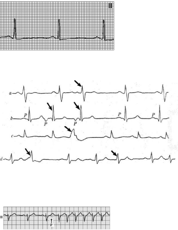
Extrasystolic arrhythmia:
a – atrial extrasystole, b - AV-junction extrasystole, c – ventricular extrasystole ,d - polytopic extrasystole.
Initiation of the atrioventricular nodal paroxysmal tachycardia (reentry tachycardia - abnormal P wave and the atrioventricular nodal delay (long PR)
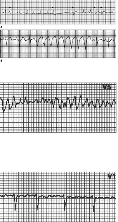
Ventricular paroxysmal tachycardia: A- Frequent ventricular extrasystoles precede PT; B- Episode of ventricular PT (at the same patient).
Ventricular fibrillation:
A rapid irregular ventricular rhythm due to multiple reentrant activity associated with essentially zero cardiac output.
Atrial fibrillation:
(1) P wave disappears; (2) multiple small irregular f waves; (3) QRS ventricular complexes follow are irregular, their shape does not change.
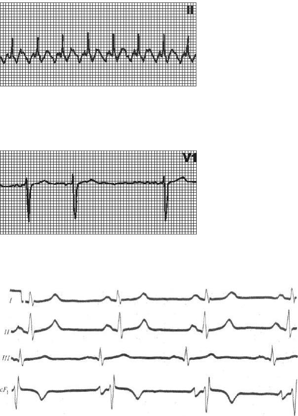
Atrial flutter:
(1) high F waves instead of the normal atrial P waves; (2) the number of F waves preceding each ventricular complex depends on the AV conduction; (3) QRS complexes follow at regular intervals.
Sino-atrial block:
(1) periodic missing of the heart complex (PQRST) in the presence of a regular sinus rhythm; (2) the length of diastole doubles.
Intra-atrial block:
(1)P waves are broadened ≥0.11 s and splitted;
(2)P wave in the V1 lead has two phases.
I degree of atrioventricular block:
(1) increased P-Q interval (> 0.21 s to 0.3-0.4 s and more) without missing QRS; (2) regular heart rhythm.
II degree of atrioventricular block with Samoilov-Wenckebach periods (Mobitz-1 type):
(2)gradual elongation PQ (Samoilov-Wenckebach periods);
(3)periodically missing ventricular contractions
II degree of atrioventricular block (Mobitz-2 type):
(1)periodic missing QRS without gradual elongation PQ (2:1);
(2)PQ can be normal or a little bit prolonged

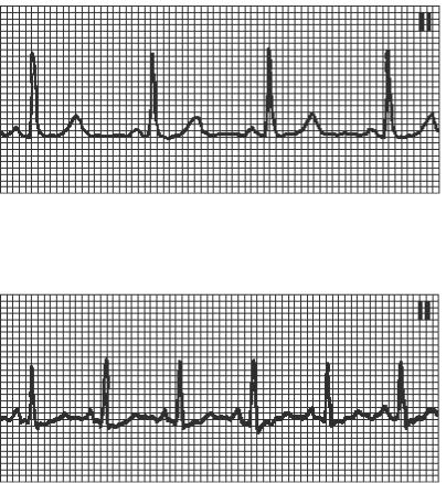
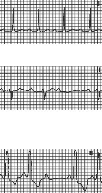
III degree of atrioventricular block (complete heart block):
(1)atrial P waves and ventricular complexes QRS are recorded independently of each other;
(2)the number of QRS is usually much smaller than the number of atrial P waves;
(3)the shape of the ventricular complex does not change if the pacemaker arises from the AV node or His bundle;
(4)with lower location of the pacemaker in the conduction system, the QRST complexes are altered.
Left bundle branch block:
(1) wide and deformed QRS has the form of qR in I,II, V5-6; rS in III, aVF, V1-2 ; (2) disconcordance of ST,T and the main wave of QRS; (3) levogram

Right bundle branch block:
(1)wide QRS III, V1-2 has the form of rsR, rSR, RsR´(similar to “M”);
(2)wide S I, aVL,V5-6; (3) negative ST and T in V1-2; (4) dextrogram