

New to the Fourth Edition
The Fourth Edition of Antibiotics Simplified expands on the drug classes covered in the previous editions while retaining the “key point” focus of the text that has made it successful. A new chapter—Chapter 6—has been added to Part I to (hopefully) simplify the complex nature of antibiotic resistance. The Fourth Edition includes expanded coverage of new agents, including the rapidly changing world of hepatitis C pharmacotherapy. New antibiotics have been added since the Third Edition, and the chapters for each class were updated with new clinical and scientific findings.
PART 1: Considerations with
Antibiotic Therapy

1: The Wonderful World of
Microbiology
Despite the promises of the household-products industry, almost every surface is covered in microorganisms almost all the time. Swab a countertop, your skin, or your dinner and you will find a little world—and that covers only the estimated 1% of bacteria that can be cultured! Obviously, trying to sterilize our patients (and our countertops) is futile; we have to try to target the bad organisms and let the rest happily crawl all over us—they greatly outnumber our own cells in our own bodies anyway.
In the microbial world, bacteria lie toward the “less like us” end of the spectrum (Figure 1–1). They are prokaryotes, not eukaryotes like fungi, protozoa, and humans. Viruses are even more different from us—they are basically just a package of genetic instructions in a cell-attacking protein coat (Figure 1–2). Differences between cells of microorganisms and humans in anatomy, biochemistry, and affinity of antibiotics for their targets are what allow for the safe and efficacious use of antibiotics. In this section we will concentrate on the microbiology of bacteria. Discussion of the unique characteristics of fungi, viruses, mycobacteria, and parasites will be covered in the sections where agents active against those organisms are introduced.
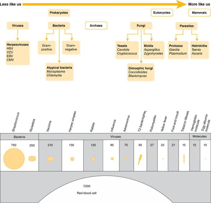
Figure 1–1 The Microbial World
Figure 1–2 Relative Sizes of Microorganisms
Differentiating bacteria that are responsible for infection from those just along for the ride can be difficult. Many bacteria that can cause human disease are also normal commensal flora, including Escherichia coli, Streptococcus pneumoniae, and Staphylococcus aureus. Thus, growth of one of these organisms from a culture is not necessarily synonymous with infection. Suspicion of infection is increased greatly if the organism grows from a normally sterile site, such as the bloodstream or cerebrospinal fluid (CSF). Indicators of infection in nonsterile sites (such as sputum and wound cultures)
are a high number of organisms, presence of inflammatory cells, and symptoms referable to the culture site (e.g., cough or dyspnea in a patient with a sputum culture growing S. pneumoniae, redness and pain in a patient with a skin culture growing S. aureus).
Definitive identification and susceptibility testing may take anywhere from hours to months, depending on the organism and the methods used. Microscopic examination and staining may allow for rapid preliminary identification. For bacteria, the most important of these techniques is the Gram stain. Being able to interpret preliminary results of microbiology testing will allow you to provide the most appropriate therapy for your patients as early as possible.
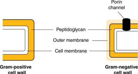
One of the most fundamental differences among types of bacteria is how they react to a Gram stain. Gram stain (crystal violet) is a substance that selectively stains the cell walls of Gram-positive bacteria but is easily washed away from Gram-negative bacteria. Why? In Gram-positive bacteria, the outermost membrane is a thick layer of peptidoglycan, a cellular substance that gives bacterial cells rigidity. In contrast, Gram-negative bacteria have an outer membrane of lipopolysaccharide that blocks the stain from adhering to the peptidoglycan within the cell (Figure 1–3).
Figure 1–3 Cell Walls of Gram-Positive and Gram-Negative Organisms
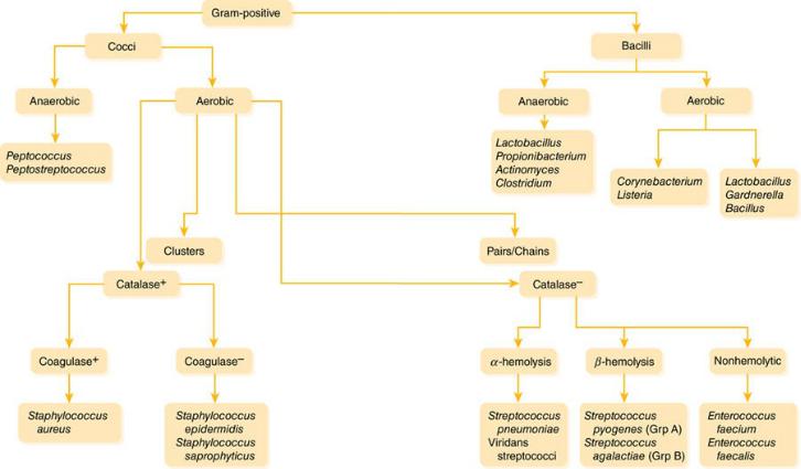
Rapid identification of Gram-positive bacteria based on morphology and preliminary biochemical tests can help to direct therapy.
1.Morphology: Most medically important Gram-positive pathogens are cocci (spheres) rather than bacilli (rods). The finding of Gram-positive bacilli should be interpreted within the clinical context: in blood cultures, Gram-positive bacilli often represent common skin contaminants (such as Propionibacterium, Corynebacterium, and Bacillus species), since the skin barrier must be bypassed to collect the culture. Detection of Gram-positive bacilli from necrotizing wound infections suggests clostridial infection, whereas the finding of Gram-positive bacilli in CSF cultures raises the concern for Listeria.
2.Colony clustering: Within the Gram-positive cocci, the staphylococci tend to form clusters, whereas the streptococci (including enterococci) typically appear in pairs or chains. Again, the clinical context aids in interpretation: The finding of streptococci in a respiratory culture suggests S. pneumoniae, while a report of “streptococci” from an intra-abdominal culture suggests Enterococcus (which may be identified preliminarily as a Streptococcus).
3.Biochemistry and appearance on agar: The rapid catalase test helps to differentiate staphylococci from streptococci. The coagulase test is useful for differentiating the more virulent (coagulase-positive)
S. aureus from its cousin the coagulase-negative Staphylococcus epidermidis. S. epidermidis is a frequent contaminant of blood cultures; if only one of a pair of blood samples is positive for coagulase-negative staphylococci, treatment may not be required. The pattern of hemolysis (clearing around colonies on agar plates) helps to differentiate among the streptococci: the oral flora (α-hemolytic S. pneumoniae and the viridans streptococci); pathogens of the skin, pharynx, and genitourinary tract (β-hemolytic Group A and β strep);
and the bugs of gastrointestinal origin (nonhemolytic enterococci: the more common Enterococcus faecalis and the more resistant
Enterococcus faecium).
Gram-negatives also contain peptidoglycan, but in smaller amounts, and it is not the outermost layer of the cell. Both Gram-positive and Gram-negative organisms contain an inner cell membrane that separates the cell wall from the cytoplasm of the organism.
Figures 1–4 and 1–5 show how you can identify different bacteria by differences in morphology, oxygen tolerance, and biochemical identification.
Figure 1–4 Gram-Positive Bacteria
Figure 1–5 Gram-Negative Bacteria
Preliminary identification is somewhat less useful with the Gram-negative bacteria because more extensive biochemical tests are usually needed to differentiate among them.
1.Morphology: Among Gram-negative pathogens, the bacilli predominate. The situation in which identification of Gram-negative cocci is most useful is in the setting of meningitis, where this finding would strongly suggest Neisseria meningitidis. Note also that some organisms have an intermediate or “coccobacillary” appearance, which may suggest organisms of the genera Haemophilus, Moraxella, or
Acinetobacter.
2.Glucose/lactose fermentation: The pathogens within the family Enterobacteriaceae (including E. coli, Klebsiella, Serratia, and
Enterobacter) generally ferment glucose/lactose; at this point the lab may identify them as “enteric Gram-negative rods” or “lactosefermenting Gram-negative rods.” In contrast, Pseudomonas,
Acinetobacter, Stenotrophomonas, and Burkholderia are “nonfermenters”; a report of “nonfermenting Gram-negative rods” should lead you to reassess and if necessary broaden your antibiotic coverage, because many of these organisms have a high level of antibiotic resistance.
3.Fastidious organisms: These organisms are picky eaters—they grow slowly and often require specially supplemented media. Thus, it may take a few days to a few weeks for them to grow from culture.

2: General Approach to Infectious
Diseases
The pharmacotherapy of infectious diseases is unique. In the pharmacotherapy of most diseases, we give drugs that have some desired pharmacologic action at some receptor or protein in the patient. To treat infections, we give antibiotics to exert a desired pharmacologic effect on the organism that is causing infection in the patient. With few exceptions, direct effects on the patients from antibiotics are not desired and are adverse effects. It is the third point in the triangle of infectious diseases pharmacotherapy, the pathogen, which makes each infection in each patient unique (Figure 2–1). The fact that the pharmacotherapy of infectious diseases involves organisms that change and “fight back” confuses many clinicians, but the approach to the patient with an infection is relatively simple and consistent. Understanding this approach is the first step in developing a useful expertise in infectious diseases and antibiotic use.
A note: technically the term antibiotic refers only to a subset of antibacterial drugs that are natural products. The terms anti-infective and antimicrobial encompass antibacterial, antifungal, antiviral, and antiparasitic drugs. However, because antibiotic is the more commonly used term, we will use it to refer to antimicrobials in general or antibacterials specifically.
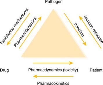
Figure 2–1 Relationships in the Infected Patient
Prophylactic Therapy
The use of antimicrobial chemotherapy—that is, the treatment of microorganisms with chemical agents—falls into one of three general categories: prophylaxis, empiric use, and definitive therapy. Prophylaxis is treatment given to prevent an infection that has not yet developed. Use of prophylactic therapy should be limited to patients at high risk of developing an infection, such as those on immunosuppressive therapy, those with cancer, or patients who are having surgery. These patients have weakened natural defenses that render them susceptible to infection. Because the likelihood of infection by some types of organisms in these patients is high and the consequences of infection are dire, we administer antimicrobial drugs to prevent infections from occurring. However, the world is not sterile and breakthrough infections do occur. The key to understanding antimicrobial prophylaxis is to remember that patients who receive it do not have an infection, but they are at risk for one.
Empiric Therapy
Unlike prophylactic therapy, empiric therapy is given to patients who have a proven or suspected infection, but the responsible organism(s) has or have not yet been identified. It is the type of therapy most often initiated in both outpatient and inpatient settings. After the clinician assesses the likelihood of an infection based on physical exam, laboratory findings, and other signs and symptoms, he or she should generally collect samples for culture and Gram staining. For most types of cultures, the Gram stain is performed relatively quickly. In the Gram stain, details about the site of presumed infection are revealed, such as the presence of organisms and white blood cells (WBCs), morphology of the organisms present (e.g., Gram-positive cocci in clusters), and the nature of the sample itself, which in some cases indicates if the sample is adequate. The process of culturing the sample begins around the time that the clinician performs the Gram stain. After a day or so, biochemical testing will reveal the identification of the organism, and eventually the organism will be tested for its susceptibility to various antibiotics.
However, this process takes several days, so empiric therapy is generally initiated before the clinician knows the exact identification and susceptibilities of the causative organism. Empiric therapy is our best guess of which antimicrobial agent or agents will be most active against the likely cause of infection. Sometimes we are right, and sometimes we are wrong. Keep in mind that empiric therapy should not be directed against every known organism in nature—just those most likely to cause the infection in question. In other words, broad-spectrum antibiotics are not a substitute for rational thought!
Definitive Therapy
After culture and sensitivity results are known, the definitive therapy phase of treatment can begin. Unlike empiric therapy, with definitive therapy we know on what organisms to base our treatment and which drugs should work against them. At this phase, it is prudent to choose antimicrobial agents that are safe, effective, narrow in spectrum, and cost effective. This helps us avoid unneeded toxicity, treatment failures, and the possible emergence of antimicrobial resistance, and it also helps manage costs. In general, moving
from empiric to definitive therapy involves decreasing coverage (assuming the empiric choice ended up being correct for the organism that grew), because we do not need to target organisms that are not causing infection in our patient. In fact, giving overly broad-spectrum antibiotics can lead to the development of superinfections, infections caused by organisms resistant to the antibiotics in use that occur during therapy.
The clinician who is treating an infected patient should always strive to make the transition to definitive therapy. Although it seems obvious, this does not always occur. If the patient improves on the first antibiotic, clinicians may be reluctant to transition to more narrow-spectrum therapy. Also, some infections may resolve with empiric therapy before culture results would even be available, as frequently happens with uncomplicated urinary tract infections (UTIs). In other cases, cultures may not be obtained or may be negative in spite of strong signs that the patient has an infection (e.g., clinical symptoms, fever, increased WBC count). Outpatient clinicians frequently skip the culture collection step, begin empiric therapy, and wait to see what happens. This may be because of time pressures or the perceived cost and inconvenience of obtaining cultures in patients with low-acuity infections. In most situations it is important that clinicians continuously consider the need to transition to definitive therapy. Overly broad-spectrum therapy has consequences, and the next infection is likely to be harder to treat. Excessive empiric antibiotic use is a big part of the reason there is an antimicrobial resistance crisis. Keep in mind the general pathway for the treatment of infectious diseases shown in Figure 2–2.
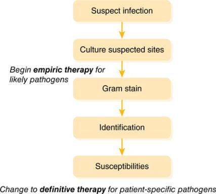
Figure 2–2 General Approach to Infectious Diseases
Examples of Therapy
Here are a few examples of each type of therapy:
Prophylaxis Therapy
 Trimethoprim/sulfamethoxazole (TMP/SMX) to prevent Pneumocystis jirovecii (formerly carinii) pneumonia in a patient on cyclosporine and prednisone after a liver transplant
 Azithromycin to prevent Mycobacterium avium intracellularae (MAI or MAC) in an HIV patient with a low CD4 count
 Cefazolin given before surgery to prevent a staphylococcal skin infection of the surgical site
Empiric Therapy
 Levofloxacin initiated for a patient with presumed community-acquired pneumonia
 Ceftriaxone given for the treatment of suspected pyelonephritis
 Voriconazole initiated for a neutropenic bone marrow transplant patient with shortness of breath and a radiograph suggestive of pulmonary aspergillosis
 Vancomycin, tobramycin, and meropenem for a patient with probable hospital-acquired pneumonia in the intensive care unit
Definitive Therapy
 Transitioning from piperacillin/tazobactam to ampicillin in a patient with a wound infection caused by Enterococcus faecalis, which is susceptible to both drugs
 Discontinuing ceftriaxone and initiating ciprofloxacin for a patient with a UTI caused by Klebsiella pneumoniae that is resistant to ceftriaxone but susceptible to ciprofloxacin
 Stopping caspofungin and initiating fluconazole for an improving patient with Candida in a blood isolate when the species is identified as Candida albicans (which is generally susceptible to fluconazole)
 Narrowing therapy from vancomycin, ciprofloxacin, and imipenem/cilastatin to linezolid alone for a patient with hospital-acquired pneumonia whose deep respiratory culture grew only methicillin-resistant
Staphylococcus aureus (MRSA) that is susceptible to linezolid
Case Study
Here is an example of treating a patient with an infection by the above pathway:
TR is a 63-year-old man with a history of diabetes, hypertension, and coronary artery disease who comes to the hospital complaining of pain, redness, and swelling around a wound on his foot. Close inspection reveals that he has an infected diabetic foot ulcer. He is admitted to the hospital (Day 1). A surgeon performs surgical debridement that evening and sends cultures from the wound during surgery as well as blood cultures. Another clinician initiates empiric therapy with vancomycin and ertapenem.
On Day 2, Gram stain results from the wound are available. There are many WBCs with many Gram-positive cocci in clusters but no Gram-negative rods (GNRs), so the clinician discontinues ertapenem. Blood cultures do not grow any organisms.
The following day (Day 3), culture results from the wound reveal many Staphylococcus aureus. Because vancomycin is usually effective against this organism, its use is continued.
On Day 4, susceptibility results from the wound culture return. The S. aureus is found to be susceptible to methicillin, oxacillin, cefazolin, clindamycin, TMP/SMX, and vancomycin. It is resistant to penicillin, ampicillin, tetracycline, and levofloxacin. Because the isolate from TR’s wound is methicillin-susceptible
S. aureus (MSSA), the clinician discontinues vancomycin and initiates definitive therapy with oxacillin.
Note how in TR’s case we began empiric therapy with a broad-spectrum regimen of vancomycin and ertapenem to cover the Gram-positive and Gram-negative aerobes and anaerobes that tend to cause diabetic foot infections but narrowed that therapy gradually as Gram stain and culture
data returned. Eventually we were able to choose a highly effective, narrowspectrum, inexpensive, and safe choice of definitive therapy that was driven by microbiology results. Both vancomycin and ertapenem were active against TR’s S. aureus as well, but both are broader in spectrum than oxacillin and represent less-ideal therapy choices. Note: ertapenem’s activity against this isolate is inferred from the susceptibility pattern even though it was not tested directly.
A Note on Rapid Diagnostics
Slowly, novel ways to determine the identification of microorganisms are making their way into clinical practice. Techniques that do not rely on culturing and the inherent delay that it represents are already commonly used to detect and quantify many viruses, such as polymerase chain reactions (PCR). These and other techniques are being used to identify other pathogens as well, such as strains of Candida (to determine likely fluconazole susceptibility), Clostridium difficile, and even MRSA. As they permeate clinical microbiology labs, hopefully the delays to effective therapy that current gold-standard culture and susceptibility testing cause will vanish.
3: Antibiotic Pharmacokinetics
The term antibiotic pharmacokinetics refers to how (and to what extent) antibiotics enter the body, where they go once they are “inside,” and how they get out. These three phases of pharmacokinetics are usually described as absorption, distribution, and metabolism/excretion (sometimes abbreviated “ADME”). The pharmacokinetics of antibiotics are key to the effectiveness of the drugs in clinical practice; there is no benefit for a patient to receive an antibiotic that is great at killing bugs if it never gets to the site of the infection at a high enough concentration to work. If you think about it, this is an issue for most types of human disease, but in infectious diseases it is very relevant clinically. You don’t have to wonder if phenytoin distributes to the central nervous system (CNS)—it does, or else it would be worthless for seizures. However, it is important to know that ceftriaxone distributes there well but cefazolin does not if you are treating meningitis. Figure 3–1 illustrates ADME phases on a concentration–time curve, with the concentration of the drug on the Y axis and the time since drug administration on the X axis. It also indicates the key pharmacokinetic parameters of peak concentration, trough concentration, and area under the concentration–time curve (“AUC”).
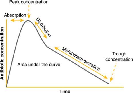
Figure 3–1 Pharmacokinetic Phases and Parameters
Absorption
Although the term “absorption” can be applied to any route of administration (e.g., absorption from intramuscular injection or inhalation), usually it is used to refer to the uptake of orally administered drugs into the bloodstream. The percentage of a nonintravenously administered drug that enters the bloodstream (such as an oral drug), relative to an intravenous formulation of the same drug, is termed the bioavailability. Antibiotics with oral formulations differ substantially in their bioavailability. For some antibiotics, bioavailability is at or near 100%; the same dose administered orally or intravenously will achieve similar levels. It is worth considering that several antibiotics have very good bioavailability, but their oral doses are substantially lower than the intravenous doses. This is usually because high oral doses lead to excessive gastrointestinal (GI) tract toxicity. Table 3–1 groups antibiotics into groups with high oral bioavailability and full or near intravenous–oral dose equivalence, those with good oral bioavailability but substantially lower oral than intravenous doses, and those with limited oral bioavailability. Some oral antibiotics have almost zero bioavailability and take advantage of this clinically to eradicate pathogens in the GI tract, where they achieve much higher concentrations than they would if given intravenously.
TABLE 3–1 Examples of Absorption for Different Oral Antibiotics
Drug |
Bioavailability |
Typical Intravenous |
|
Typical Oral Dose |
|
|
Dose |
|
|
|
|
|
|
|
Drugs with High Bioavailability and Similar Intravenous–Oral Doses |
|
|||
|
|
|
|
|
Metronidazole |
95–100% |
500 mg IV q8h |
|
500 mg PO q8h |
|
|
|
|
|
Levofloxacin |
95–100% |
500–750 mg IV q24h |
|
500–750 mg PO daily |
|
|
|
|
|
Linezolid |
95–100% |
600 mg IV q8h |
|
600 mg PO q12h |
|
|
|
|
|
Fluconazole |
95–100% |
200–400 mg IV daily |
|
200–400 mg PO daily |
|
|
|
|
|
Doxycycline |
95–100% |
100 mg IV q12h |
|
100 mg PO q12h |
|
|
|
|
|
Ciprofloxacin |
~80% |
400 mg IV q12h |
|
500 mg PO q12h |
|
|
|
|
|
Drugs with High Bioavailability but Different Intravenous–Oral Doses |
|
|||
|
|
|
|
|
Aminopenicillins |
~90% |
Ampicillin: 1–2 g IV |
|
Amoxicillin: 500 mg–1 g |
|
|
q4–6h |
|
PO TID |
|
|
|
|
|
First-generation |
~90% |
Cefazolin: 1–2 g IV |
|
Cephalexin: 500 mg PO |
cephalosporins |
|
q8h |
|
QID |
|
|
|
|
|
Drugs with Low Bioavailability and Different Intravenous–Oral Doses |
|
|||
|
|
|
|
|
Cefuroxime |
~40% |
750 mg IV q8h |
|
500 mg PO BID |
|
|
|
|
|
Acyclovir |
~25% |
5 mg/kg IV q8h |
|
400 mg PO TID |
|
|
|
|
|
It is important to consider factors that can affect the bioavailability of antibiotics. Three factors that can substantially influence absorption are food, gastric acidity, and chelating agents. Some antibiotics are better absorbed with food and some without, while for most antibiotics the presence or absence of food has minimal effect.
A small group of antibiotics are highly dependent on gastric acidity for adequate absorption; it is important to avoid concomitant use of drugs that
raise gastric pH (antacids, proton pump inhibitors, histamine-2 receptor antagonists) when patients are started on these drugs. Finally, two key classes of antibiotics—the tetracyclines and fluoroquinolones—can bind to coadministered minerals present in the gut such as calcium, iron, aluminum, and zinc. Administering these drugs along with mineral or some vitamin supplements can substantially reduce absorption. Table 3–2 provides examples of antibiotics where these factors need to be accounted for.
TABLE 3–2 Examples of Antibiotics Whose Absorption Is Significantly
Affected by Other Factors
Absorption |
Absorption |
Absorption Impaired by Drugs |
Absorption |
Improved with |
Impaired by |
That Increase Gastric pH |
Impaired by |
Food |
Food |
|
Minerals |
|
|
|
|
Posaconazole |
Voriconazole |
Itraconazole |
Fluoroquinolones |
suspension |
Itraconazole |
Posaconazole suspension |
Tetracyclines |
Itraconazole |
solution |
Atazanavir |
|
capsules |
Rifampin |
Rilpivirine |
|
Atazanavir |
Isoniazid |
|
|
Darunavir |
Pyrazinamide |
|
|
Rilpivirine |
|
|
|
|
|
|
|
Distribution
After a drug is absorbed or injected into the bloodstream, it moves into various tissues (e.g., bone, cerebrospinal fluid, lungs), a process known as distribution. The concentrations in these tissues can be similar to, lower than, or greater than the concentration of the antibiotic in the blood. A consequence is that a drug may be more or less effective in a particular tissue than would be expected based on its concentrations in the blood. For example, the concentrations of antibiotics in the cerebrospinal fluid are typically much lower than their bloodstream concentrations, limiting the effectiveness of many antibiotics in the treatment of meningitis. On the other hand, the macrolide antibiotics are more effective in lung infections than may be anticipated based on their blood levels, because they concentrate in pulmonary macrophages. With a few exceptions such as cerebrospinal fluid, it is difficult to obtain samples of human tissues to determine antibiotic
concentrations, and it is technically difficult to measure the concentrations in tissues like bone. Thus, data on drug distribution are often extrapolated from animal models, which may or may not be good surrogates for humans.
The extent to which antibiotics distribute into different tissues is largely determined by the physicochemical properties of the drug (lipophilicity, charge, molecular size, etc.). A key determinant of distribution is the degree to which an antibiotic binds to proteins in the bloodstream, most importantly albumin (you may hear this expressed as “fraction bound” or “fraction unbound”). Drug bound to proteins is not able to diffuse across membranes into different tissues; thus, antibiotics that are highly protein bound may be less likely to reach effective concentrations in certain tissues (such as the CNS). It is important to realize that the percentage degree of penetration of an antibiotic into a tissue is not the only determinant of effectiveness in that tissue. For example, ceftriaxone is a very highly protein-bound drug, and 5% or less enters into the CNS in patients with meningitis. However, large doses (2 g twice daily in adults) of ceftriaxone can be given safely to adults, resulting in high serum levels (peak levels of around 200 mg/L). In addition, the minimum inhibitory concentration (MIC) to ceftriaxone for organisms usually causing meningitis is typically very low (1 mg/L or less); thus, a concentration far in excess of the organism’s MIC can be obtained (200 mg/L × 5% = 10 mg/L). Also, the concentration–time curves in many tissues are different than they are in the bloodstream—more like rolling hills than peaks and valleys (think Appalachians versus the Rockies).
Patient characteristics may also significantly influence drug distribution. In order for a drug to distribute to a tissue, there must be adequate blood flow to that tissue. Conditions that reduce blood flow to tissues, either locally (e.g., peripheral vascular disease) or systemically (septic shock), can reduce antibiotic concentrations at the site of infection. Patients with severe infections can develop abscesses or areas of dead and devitalized tissue; distribution of antibiotics into these “protected” sites of infection can be significantly impaired. These patients are perfect setups for treatment failure and development of resistance and highlight the importance of appropriate surgical management of infections along with antibiotic treatment. Given the growing problem of obesity, another important consideration is the extent to
which drugs distribute into adipose tissue. Depending on the characteristics of the drug, it is possible to underdose patients who are morbidly obese (if the drug distributes extensively into adipose tissue and doses for standard weights are used) or overdose them (if a higher dose is used because of obesity, but the drug does not distribute well into excess adipose tissue). Thus, you may see recommendations for antibiotic dosing based on total or actual body weight, ideal body weight (an estimate of the patient’s body weight without their excess adipose tissue), or adjusted body weight (a value between the ideal and total body weight). This is an area that is relatively understudied.
Finally, it is important to note that with few exceptions, microbiological susceptibility testing does not account for distribution and is based on achievable bloodstream concentrations. For example, the microbiology lab may determine that an organism with an MIC of 4 mg/L is considered susceptible to a drug that achieves a concentration of 8 mg/L in the bloodstream, but it may only achieve a concentration of 1 mg/L in cerebrospinal fluid. Thus, that drug is likely to work for a bloodstream infection caused by the organism, but would fail for meningitis where cerebrospinal fluid concentrations are important. Thus, distribution is a key consideration when choosing antibiotics.
Metabolism/Excretion
Many antibiotics are excreted from the body, either in the urine or feces, in the same form as they were administered. In fact, shortly after penicillin was developed and supplies were scarce, doctors used to collect the urine of patients who received penicillin and recrystallize the drug for use in other patients! When a drug is excreted unchanged, it can reach very high concentrations in the area in which it is eliminated, making it potentially more effective for infections in those systems than would be anticipated based on blood concentrations. For example, the concentrations of nitrofurantoin achieved in the blood and tissues are generally inadequate to inhibit bacterial growth. However, it is removed from the bloodstream by the kidneys and accumulates in the bladder until its, ahem, final clearance. The concentrations achieved in the bladder are manyfold higher than those in the bloodstream, making nitrofurantoin an effective drug for treatment of bladder infections.
When the body does not inactivate the drug, an important consideration is to appropriately reduce the administered dose of the drug if there is damage to the organ responsible for excreting the drug. The most common example of this for antibiotics is the need to reduce the doses of most beta-lactams for patients with kidney dysfunction to avoid accumulation of toxic levels of the drug. Practitioners also need to be vigilant to increase doses if patients have improving renal function or treatment failure may occur.
Other drugs may be extensively transformed by the body prior to their excretion. These antibiotics that undergo extensive metabolism are considered to be substrates of drug-metabolizing enzymes. They have the potential to be subject to clinically important drug interactions, as other drugs may interfere with the enzymes that break the drugs down. Moreover, certain antibiotics have the potential to influence the metabolism of other drugs, either through inhibition of those enzymes (leading to a decrease in metabolism of the other drug) or induction (leading to an increase in metabolism of the other drug). A list of antibiotics with the greatest likelihood of clinically significant metabolic drug interactions is in Table 3–3, organized by whether the drug is a substrate, an inhibitor, or an inducer (and note that drugs may be in more than one category). Note that the several antibiotic classes are particularly prevalent: macrolides, azole antifungals, antituberculosis drugs, and antiretrovirals account for most of the antibiotics with significant drug interactions. Complex drug interactions can occur with these drugs: for example, the antiretroviral drug etravirine is simultaneously a substrate, an inhibitor, and an inducer of drug-metabolizing enzymes!
TABLE 3–3 Examples of Antibiotics with Significant Metabolic Drug
Interactions
Substrates |
Inhibitors |
Inducers |
|
|
|
Erythromycin |
TMP/SMX |
Rifampin |
Clarithromycin |
Metronidazole |
Rifabutin |
Telithromycin |
Fluconazole |
Efavirenz |
Atazanavir |
Voriconazole |
Nevirapine |
Darunavir |
Itraconazole |
Etravirine |
Efavirenz |
Posaconazole |
|
Elvitegravir |
Erythromycin |
|
Maraviroc |
Clarithromycin |
|
Rilpivirine |
Telithromycin |
|
|
Ritonavir |
|
|
Cobicistat |
|
|
Etravirine |
|
|
|
|

4: Antibiotic Pharmacodynamics
The term antibiotic pharmacodynamics refers to the manner in which antibiotics interact with their target organisms to exert their effects: Does the antibiotic kill the organism or just prevent its growth? Is it better to give a high dose of antibiotics all at once or to achieve lower concentrations for a longer time? Clinicians increasingly recognize such considerations as important in maximizing the success of therapy, especially for difficult-to-treat infections and in immunocompromised patients.
Susceptibility Testing
Typically, one judges the susceptibility of a particular organism to an antibiotic based on the minimum inhibitory concentration (MIC) for the organism–antibiotic combination. Classically, the microbiology laboratory determines the MIC by combining a standard concentration of the organism that the patient has grown with increasing concentrations of the antibiotic. Historically this was done in test tubes (Figure 4–1), but today it is done more commonly on microdilution plates or with automated systems. The mixture is incubated for about a day, and the laboratory technician examines the tubes or plates (with the naked eye or with a computer) for signs of cloudiness, indicating growth of the organism. The mixture with the lowest concentration of antibiotic where there is no visible growth is deemed to be the MIC. For each organism–antibiotic pair, there is a particular cutoff MIC that defines susceptibility. This particular MIC is called the breakpoint. Table 4–1 provides examples of how breakpoints differ for various organism/pathogen combinations and even based on the site of infection. Note that just because an antibiotic has the lowest MIC for a pathogen it does not mean it is the best choice—different antibiotics achieve different concentrations in the body in different places. Thus, antibiotic MICs for a single organism generally should not be compared across different drugs in selecting therapy.
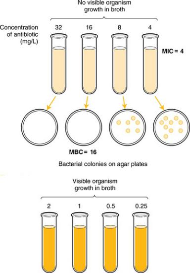
Figure 4–1 Susceptibility Testing of Antibiotics
TABLE 4–1 Examples of Antibiotic Susceptibility Breakpoints
Organism Antibiotics |
Susceptible |
Susceptible, |
Intermediate |
Resistant |
|
|
(Breakpoint) |
Dose- |
|
|
|
|
|
Dependent |
|
|
|
|
|
|
|
|
|
Escherichia Coli |
|
|
|
|
|
|
|
|
|
|
|
Ampicillin |
≤ 8 mg/L |
— |
16 mg/L |
≥ 32 mg/L |
|
|
|
|
|
|
|
Cefepime |
≤ 2 mg/L |
4–8 mg/L |
— |
≥16 mg/L |
|
|
|
|
|
|
|
Levofloxacin |
≤ 2 mg/L |
— |
4 mg/L |
≥ 8 mg/L |
|
|
|
|
|
|
|
Trimethoprim/sulfamethoxazole |
≤ 2/38 mg/L |
— |
— |
≥ 4/76 |
|
|
|
|
|
mg/L |
|
|
|
|
|
|
|
Streptococcus pneumoniae |
|
|
|
|
|
|
|
|
|
|
|
Ampicillin |
— |
— |
— |
— |
|
|
|
|
|
|
|
Cefepime (meningitis) |
≤ 0.5 mg/L |
— |
1 mg/L |
≥2 mg/L |
|
(Nonmeningeal) |
|
|
|
|
|
≤ 1 mg/L |
— |
2 mg/L |
≥ 4 mg/L |
||
|
|||||
|
|
|
|
|
|
Levofloxacin |
≤ 2 mg/L |
— |
4 mg/L |
≥ 8 mg/L |
|
|
|
|
|
|
|
Trimethoprim/sulfamethoxazole |
≤ 0.5/9.5 mg/L |
— |
1–2/19–38 |
≥ 4/76 |
|
|
|
|
mg/L |
mg/L |
|
|
|
|
|
|
MIC values are often misinterpreted by clinicians who are trying to choose the best therapy for their patients but may inadvertently ignore pharmacokinetic and pharmacodynamic differences between agents. For example, in Table 4–1, note that the breakpoint for levofloxacin against
Escherichia coli is 2 mg/L and for ampicillin 8 mg/L. So if an isolate of E. coli in the bloodstream of a patient has an MIC of 1 mg/L to levofloxacin but 2 mg/L to ampicillin, it does not mean that levofloxacin is a better choice for that patient. Levofloxacin is a concentration-dependent drug that is typically dosed in amounts of 500–750 mg daily. Ampicillin is a time-dependent drug that is typically dosed as 1–2 g every 4–6 hours. The much higher
concentrations of ampicillin achieved in the body (due to higher doses) mean that organisms with a higher MIC to ampicillin are still susceptible to it. In other words, the two numbers are not directly comparable. In fact, if the MIC was 8 mg/L to both drugs, the organism would be considered resistant to levofloxacin and susceptible to ampicillin.
Also note in Table 4–1 the categories of “susceptible,” “susceptible, dosedependent,” “intermediate,” and “resistant.” “Susceptible” and “resistant” are self-explanatory, but what do the other terms mean? “Intermediate” is a poorly defined range where successful therapy may be possible in some circumstances, such as when the drug is eliminated renally (for a urinary tract infection) or if higher dosing is given. It is more of a grey area made to separate the two more definitive terms than a scientifically derived definition. “Susceptible, dose-dependent” is just what it sounds like—the organism may be susceptible to high doses of the drug, but is likely to be resistant to low doses. This was created because many of these drugs are used in various doses, and a low dose of cefepime like 1 g IV q12h may be enough to treat an E. coli infection with an MIC of 2 mg/L, but a dose like 2 g IV q12h may be needed if the MIC is 4 mg/L. These may all sound like hard-and-fast rules, but they are really probabilities of how most patients will fare when faced with a certain bug–drug combination for their infections. Some will succeed even with high MICs, and some will fail even with low ones.
Finally, be aware that other methods of susceptibility testing exist, including disk diffusion and E-tests, but that broth dilution methods are generally considered the gold standard.
Static Versus Cidal
At the MIC the antibiotic is inhibiting growth, but it may or may not actually be killing the organism. Antibiotics that inhibit growth of the organism without killing it are termed bacteriostatic (or fungistatic in the case of fungi). If antibiotics are removed, the organisms can begin growing again. However, bacteriostatic antibiotics are usually successful in treating infections because they allow the patient’s immune system to “catch up” and kill off the organisms. Other antibiotics are considered bactericidal; their action kills the organisms without any help from the immune system.
For most infections, outcomes using appropriate bacteriostatic versus bactericidal drugs are similar; however, for certain infections bactericidal drugs are preferred. Such infections include endocarditis, meningitis, infections in neutropenic patients, and possibly osteomyelitis. The immune system may not be as effective in fighting these infections because of the anatomic location or the immunosuppression of the patient. Bactericidal activity is determined by taking a sample of the broth at various concentrations and below and spreading the broth on agar plates (Figure 4– 1). The bacterial colonies on the plates are counted, and the concentration corresponding to a 99.9% reduction (3-log) in the original bacterial inoculum is considered to be the minimum bactericidal concentration (MBC). When the MBC is four times or less than the MIC, the drug is considered to be bactericidal; if the MBC/MIC ratio is greater than four, it is considered bacteriostatic. MIC testing is standard in most laboratories; MBC testing is more difficult and is not commonly done in clinical practice. Table 4–2 lists drugs and indicates whether they are generally considered bacteriostatic or bactericidal; however, it should be noted that this activity can vary based on the pathogen being treated, the achievable dose, and the growth phase of the organism.
TABLE 4–2 Antibiotic Pharmacodynamic Parameters
Antibiotic Class |
Cidal or Static |
Predictive PK/PD Parameter |
|
|
|
Penicillins |
Bactericidal |
Time > MIC |
Cephalosporins |
|
|
Carbapenems |
|
|
Monobactams |
|
|
|
|
|
Vancomycin |
Bactericidal (slowly) |
AUC/MIC |
|
|
|
Fluoroquinolones |
Bactericidal |
Peak: MIC |
Aminoglycosides |
|
|
Metronidazole |
|
|
Daptomycin |
|
|
|
|
|
Macrolides |
Bacteriostatic |
AUC/MIC |
Tetracyclines |
|
|
Linezolid |
|
|
|
|
|
Pharmacokinetic/Pharmacodynamic Relationships
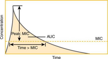
Besides differing in whether they kill microorganisms or merely inhibit their growth, antibiotics also differ in how they manifest their effects over time. Careful studies have revealed that for certain antibiotics, activity against microorganisms correlates with the duration of time that the concentration of the drug remains above the MIC (time-dependent activity). For other antibiotics, antibacterial activity correlates not with the time above the MIC but with the ratio of the peak concentration of the drug to the MIC (concentration-dependent or time-independent activity). For some antibiotics, the best predictor of activity is the ratio of the area under the concentration– time curve (AUC) to the MIC. Figure 4–2 illustrates these pharmacokinetic/pharmacodynamic (PK/PD) parameters schematically, and
Table 4–2 shows which parameter is most predictive of efficacy for antibiotic classes. The practical implications of these findings are in the design of antibiotic dosing schedules: aminoglycosides are now frequently given as a single large dose daily to leverage the concentration-dependent activity, while some clinicians are administering beta-lactam drugs such as ceftazidime as continuous or prolonged infusions because of their time-dependent activity. As target values for these parameters that predict efficacy are found, there may be an increase in the individualization of dosing of antibiotics to achieve these target values.
Figure 4–2 Pharmacokinetic/Pharmacodynamic Relationships

5: Adverse Consequences of
Antibiotic Use
Although antibiotics are undoubtedly one of the most beneficial discoveries of science, their use does carry risks. They can adversely affect patients by eliciting allergic reactions, causing direct toxicity, or altering the normal bacterial flora, leading to superinfections with other organisms. Antibiotic use is the primary driving force in the development of antibiotic resistance, which can affect not only the treated patients but other patients by transmission of resistant organisms. It is important to keep in mind all of these potential adverse consequences when using antibiotics.
Antibiotic Allergy
Through formation of complexes with human proteins, antibiotics can trigger immunologic reactions. These reactions may manifest immediately (such as anaphylaxis or hives) or be delayed (rashes, serum sickness, drug fever).
Because of their highly reactive chemical structure and frequent use, betalactam drugs are the most notorious group of drugs for causing allergic reactions. It is difficult to determine how likely it is that a patient with an allergy to a particular antibiotic agent will have a similar reaction to another agent within that class. While some (highly debated) estimates of the degree of cross-reactivity are available for beta-lactam drugs, estimates for crossreactivity within other classes (e.g., between fluoroquinolones) are essentially nonexistent. Because labeling a patient with an allergy to a particular antibiotic can limit future treatment options severely and possibly lead to the selection of inferior drugs, every effort should be made to clarify the exact nature of a reported allergy.
Antibiotic Toxicities
Despite being designed to affect the physiology of microorganisms rather than humans, antibiotics can have direct toxic effects on patients. In some cases, this is an extension of their mechanism of action when selectivity for microorganisms is not perfect. For example, the hematologic adverse effects of trimethoprim stem from its inhibition of folate metabolism in humans, which is also its mechanism of antibiotic effect. In other cases, antibiotics display toxicity through unintended physiologic interactions, such as when vancomycin stimulates histamine release, leading to its characteristic red man syndrome. Some of these toxicities may be dose related and toxicity often occurs when doses are not adjusted properly for renal dysfunction and thus accumulate to a toxic level. Proper dosage adjustment can reduce the risk of dose-related toxicities.
Superinfection
The human body is colonized by a variety of bacteria and fungi. These organisms are generally considered commensals, in that they benefit from living on or in the body but do not cause harm (within their ecologic niches). Colonization with commensal organisms can be beneficial, given that they compete with and crowd out more pathogenic organisms. They may even have a role in the prevention of other human diseases. When administration of antibiotics kills off the commensal flora, pathogenic drug-resistant organisms can flourish because of the absence of competition. This is considered a superinfection (i.e., an infection on top of another infection). For example, administration of antibiotics can lead to the overgrowth of the gastrointestinal (GI) pathogen Clostridium difficile, which is clinically resistant to most antibiotics. C. difficile can cause diarrhea and lifethreatening bowel inflammation. Similarly, administration of broad-spectrum antibacterial drugs can select for the overgrowth of fungi, most commonly yeasts of the genus Candida. Disseminated Candida infections carry a high risk of death. To reduce the risk of the impact of antibiotics on the commensal flora, and thus the likelihood of superinfection, antibiotics should be administered only to patients with proven or probable infections, using the most narrow-spectrum agents appropriate to the infection for the shortest effective duration.
Antibiotic Resistance
Thousands of studies have documented the relationship between antibiotic use and resistance, both at a patient level (if you receive an antibiotic, you are more likely to become infected with a drug-resistant organism) and a society level (the more antibiotics a hospital, region, or country uses, the greater the antibiotic resistance). The development of antibiotic resistance leads to a vicious spiral where resistance necessitates the development of broader-spectrum antibiotics, leading to evolution of bacteria resistant to those new antibiotics, requiring ever broader-spectrum drugs, and so on. This is particularly problematic because antibiotic development has slowed down greatly. Although we can see clearly the broad relationship between antibiotic use and resistance, many of the details of this relationship are not clear. Why do some bacteria develop resistance rapidly and others never develop resistance? What is the proper duration of treatment to maximize the chance of cure and minimize the risk of resistance?

6: Antibiotic Resistance
Though it may seem that the antibiotic era was introduced in the 1930s with the sulfonamides and penicillin, it had actually started millions of years earlier. Alexander Fleming only discovered one of the weapons of a war going on underfoot—literally, under our feet. In the soil and elsewhere, microbes are locked in life-and-death battles for dominance over each other for the limited resources they have access to. Among their weapons are antibiotics.
Causes of Antibiotic Resistance
The basic cause of antibiotic resistance is simple: antibiotic use. Some organisms are notorious for an intrinsic ability to express multiple types of resistance, such as Acinetobacter baumannii or Pseudomonas aeruginosa. Others have been generally treatable for many years and are only recently becoming highly drug-resistant through acquiring new resistance elements, such as Klebsiella pneumoniae. And some have remained highly susceptible to “old” antibiotics ever since their introduction, such as Streptococcus pyogenes and penicillin.
Where Does Antibiotic Resistance Come From?
In any species of bacteria, antibiotic resistance needs to have a point of origin. Resistance can emerge in the organism of interest through random mutations to the antibiotic’s target or other key elements. However, it is more common that a given species of bacteria acquired the genes, which enable a mechanism of resistance, from another species of bacteria that already had it through the transfer of mobile genetic elements. Bacteria are promiscuous little organisms and they are not picky—they often swap genes not only between their own species, but different species and even genera. There are several ways that genes are transmitted between bacteria, but the most important is by the transmission of plasmids via conjugation. Plasmids are loops of DNA that may contain multiple genes in them that encode for various processes (including antibiotic resistance), and they are highly portable. Since plasmids can contain multiple genes, they can encode for multiple types of resistance that are not related, such as resistance to cephalosporins via the production of a beta-lactamase and resistance to fluoroquinolone due to an efflux pump. With one act of gene swapping, a multiply-resistant bacterial strain is born.
Mechanisms of Antibiotic Resistance
The multiple mechanisms by which resistance occurs can be confusing, but here we are going to—wait for it—simplify it into four basic mechanisms, which are outlined in Figure 6–1.
Figure 6–1 Mechanisms of Antibiotic Resistance
Decreased permeability prevents the antibiotic from penetrating the bacterial cell, decreasing the intracellular concentration of the antibiotic. Enzymatic modification due to an enzyme produced by the bacteria destroys the antibiotic before it has a chance to reach its site of activity or even enter the cell. Target site changes can occur, leading to an elimination or modification of the antibiotic’s site of activity such that it cannot work. Active efflux occurs when efflux pumps in the bacteria pump out antibiotics, decreasing intracellular concentrations. Examples of how common mechanisms of resistance fall into these four broad categories are given in Table 6–1.
TABLE 6–1 Examples of Mechanisms of Antibiotic Resistance
Category |
Example |
|
|
Decreased permeability |
Cell wall changes |
|
Porin channel changes or loss |
|
Biofilm production |
|
|
Enzymatic modification |
Beta-lactamases (over 1400 different types!) |
|
Aminoglycoside-modifying enzymes |
|
Methylation |
|
|
Target site changes |
PBP 2a expression in Staphylococcus aureus |
|
PBP 2x expression in Streptococcus pneumoniae |
|
Ribosomal modification |
|
|
Active efflux |
Tetracycline efflux |
|
Fluoroquinolone efflux |
|
|
Knowing the mechanism of resistance for a particular bug–drug combination is not essential in most clinical situations. Multiple mechanisms of resistance are often present and being expressed simultaneously, which make it impossible to infer the mechanism of resistance just from the reported sensitivities. We have highlighted situations where it is more important throughout the antibacterial chapters in this text.
Antibiotic Pressure and Collateral Damage
Antibiotic use has consequences. Antibiotics are the only class of drug for which their use in one person affects their utility in another person, since microorganisms are transmissible. Antibiotic pressure is a term that reflects the fact that antibiotic-resistant strains may emerge when antibioticsusceptible strains are killed. If a patient is infected or colonized with resistant and susceptible strains of a bacteria, the use of an antibiotic will kill the susceptible strain and allow the resistant one to grow. This pressure is worth the trade-off when the patient has an infection and requires treatment, but excessive pressure placed on the microorganisms in the patient can be limited by how we use antibiotics. Collateral damage is a term used to describe the impact of antibiotic use on microorganisms in the body beyond the targeted organism. Every dose of antibiotics affects not just the pathogen causing an infection, but billions of other bacteria in and on the body, bacteria that are just minding their business and often helping us out by preventing colonization by more aggressive bacteria. Collateral damage will occur with any antibiotic, but can be minimized by using less-broad-spectrum antibiotics and, perhaps most importantly, minimizing the duration of use of all antibiotics when possible. One of the greatest concerns and most contentious issues in antibiotic resistance is the use of antibiotics in agriculture, which is where most antibiotics in the United States are used. The impact of this and the collateral damage that it has on the human microbiome is just beginning to be determined.
The ESKAPE Pathogens
Antibiotic resistance has been described in most bacteria that cause human infection, but some pathogens are particularly problematic. There have been several ways to describe the most serious antibiotic-resistance threats that plague mankind, but probably the most commonly utilized one is the acronym “ESKAPE” (Table 6–2).
TABLE 6–2 “ESKAPE” Pathogens and Other Resistance Threats
“ESKAPE” pathogens |
Enterococcus faecium |
|
Staphylococcus aureus |
|
Klebsiella pneumoniae |
|
Acinetobacter baumannii |
|
Pseudomonas aeruginosa |
|
Enterobacter species |
|
|
Organisms classified by CDC as an urgent health |
Clostridium difficile |
threat |
Carbapenem-resistant |
|
Enterobacteriaceae |
|
Drug-resistant Neisseria gonorrhoeae |
|
|
These diverse organisms have in common both, a high propensity for antibiotic resistance and limited options for treating their resistant phenotypes. The Centers for Disease Control and Prevention (CDC) published a report in 2013 titled “Antibiotic Resistance Threats in the United States, 2013” that details the estimated threat of resistance in the United States, available at http://www.cdc.gov. It estimated that 23,000 people in the United States die each year due to resistant bacterial infections, but that figure did not include the estimated 14,000 deaths per year from C. difficile infection.
Preventing Antibiotic Resistance—A Problem of Perspective
With all of this good news, what can we do to prevent antibiotic resistance? Convincing data show that limiting antibiotic use limits antibiotic resistance. This has been shown on many levels, from the patientto the hospitalto the country-level. Unfortunately, each of these levels has a different perspective. Patients want to get better and may not know that the antibiotic they are requesting is unlikely to work for a viral respiratory infection. Hospitals are concerned with both patient outcomes and costs, and diagnostic uncertainty drives much empiric antibiotic use. Many clinicians are afraid to miss a potential infection and end up using antibiotics for patients who actually are not infected. Only the societal viewpoint is high-level enough to really look at overall antibiotic use and how it impacts antibiotic resistance.
Guidelines
Decisions to use antibiotics are not made by society; they are made on a patient basis. It falls upon all of us to make therapy decisions with antibiotic resistance in mind. Some general guidance for utilizing antibiotics follows below.
Avoid Using Antibiotics to Treat Colonization or Contamination
A substantial percentage of all antibiotic use is directed toward patients who are not truly infected, but in whom organisms are recovered from culture. Isolation of Staphylococcus epidermidis from a single blood culture or
Candida species from a urinary culture in a catheterized patient are common situations in which patients should be scrutinized to determine whether an infection is truly present. Diagnostic improvements need to be developed and utilized when they are available. A simple test that could distinguish a cold from a bacterial infection could save society billions in avoided antibiotic use, resistance, and adverse effects.
Use the Most Narrow-Spectrum Agent Appropriate for the Patient’s Infection
Broader-spectrum agents multiply the number of bacteria affected by the drug, increasing the chances both for development of resistance and superinfection. “Broader” and “newer” are not synonymous with “better”: for example, good old penicillin kills susceptible organisms more rapidly than almost any drug on the market. The treating clinician’s goal always should be definitive, narrow-spectrum therapy.
Use the Proper Dose
Bacteria that are exposed to low concentrations of antibiotics are more likely to become resistant than those exposed to effective doses. After all, dead bugs don’t mutate! Further research in pharmacodynamics should make it easier to determine the proper dose for each patient and thus to reduce the likelihood that resistance will develop.
Use the Shortest Effective Duration of Therapy
Unfortunately, duration of therapy is one of the least-studied areas of infectious diseases. Examination of standard treatment durations says much more about how humans think than about how antibiotics and bacteria truly interact—durations are typically 5, 7, 10, or 14 days, more in line with our decimal system and the days in a week than with anything studied precisely. New studies are showing that shorter durations of therapy are often just as effective as prolonged courses and possibly less likely to select for resistance. As studies progress and determine additional factors that indicate when infections are sufficiently treated, it should be possible to define more accurately the length of therapy on a patient-by-patient basis. Many clinicians find that “old habits die hard,” however, they should remember that learning new evidence about duration of therapy is important as it emerges.