

Color Atlas of Gross Placental Pathology
.pdf
Section I
Diagnostic transvaginal endoscopy


1
Rationale of transvaginal laparoscopy in infertility
Stephan Gordts and Ivo Brosens
Introduction
Transvaginal laparoscopy (TVL) has been introduced as an alternative to standard laparoscopy (SL) for the exploration of patients with infertility. It is primarily a diagnostic procedure to be used in an office setting as a minimally invasive technique for the investigation of infertile patients with no obvious pelvic pathology. While SL is too invasive and probably not cost-effective to be performed in women without obvious pelvic pathology, the endoscopic exploration of the tubo-ovarian structures in infertility is in current practice frequently postponed or even omitted. As a consequence, diseases such as endometriosis, small endometriomas, or tubal disease are not diagnosed. However, even with the advent of in vitro fertilization (IVF) and the liberal referral to IVF we are not at the stage that we can ignore various uterine and pelvic pathologies associated with infertility, which may have an important impact on the reproductive outcome and can be corrected.
History
While attempts to use the transvaginal access for endoscopic exploration of the female pelvis go back to the beginning of the 20th century, the first successful technique of culdoscopy was introduced in the early 1940s by Decker.1 The endoscopic visualization of the pelvic organs was performed with the patient in the knee-chest or genupectoral position. It was originally conceived as a complex hospital procedure with the patient in an uncomfortable and unstable position requiring elaborate mechanical and manual bracing and with the physician in an unusual physical orientation to the target organs. Small wonder that, when laparoscopy was introduced in the 1960s, transabdominal access was seen as the answer to some of the problems. Laparoscopy soon became the standard method for gynecologic endoscopy, particularly when it was shown that the technique was superior to culdoscopy for surgical procedures such as tubal sterilization.2
However, pioneers of pelvic endoscopy like Raoul Palmer3 in Europe and Edward Diamond4 in the USA continued to promote culdoscopy as the method of choice in at least one special application – the diagnosis of infertility, because it provides a closer, clearer, and more detailed view of the fallopian tubes, ovaries, and surrounding pelvic structures than laparoscopy. In 1978 Diamond4 published a personal series of 4000 outpatient procedures of diagnostic culdoscopy in infertility. The results were impressive, the complications mild, and the failure rate was very low. Fimbrial phimosis and perifimbrial adhesions were more readily detected. Endometriosis could be seen on all the surfaces of the ovary, the distal end of the tube, the lateral pelvic wall, the utero-ovarian and uterosacral ligaments, and even in locations revealed with difficulty or not at all by laparoscopy. In particular, culdoscopy revealed the fine, filmy adhesions that are only rarely picked up by laparoscopy but which may be responsible for a significant amount of ovarian and tubal malfunction. Diamond concluded that the use of diagnostic culdoscopy as an outpatient procedure provides a better access for the diagnosis and treatment of infertility, especially when the pathology is not extreme enough to warrant laparoscopy. His advice was that the technique should be returned to gynecologic training programs, and he concluded:
True, culdoscopy requires laboriously won special skills, but its advantage to patient and physician are well worth the trouble. Once mastered, culdoscopy equips the gynaecologic endoscopist with a rapid and minimally traumatic outpatient option that supplies rich information not only in the initial diagnosis of infertility but also in circumstances where laparoscopy might be inappropriate.
While the technology of laparoscopy has continuously improved, the technology of culdoscopy has not advanced since the 1960s. Improvements such as dorsal decubitus,5 hydroflotation,6 and miniculdoscopy7 were suggested to revive culdoscopy, but received no further interest.
4 Atlas of Transvaginal Endoscopy
In 1998 Gordts et al8 described a new culdoscopic technique, called transvaginal hydrolaparoscopy (THL), for the exploration of infertile patients without obvious pelvic pathology. A somewhat similar technique using a disposable cannula was developed by Watrelot and called fertiloscopy.9 Both techniques combine the use of small-diameter instruments, dorsal decubitus, and hydroflotation.
Safety
SL requires general anesthesia and full operating room facilities. It is not an innocuous procedure, and the majority of laparoscopic complications occur during the transabdominal access.10,11 One-third of the major complications are caused by the instillation of pneumoperitoneum and insertion of trocar.12 Even in experienced hands, the bowel injury occurs as frequently during the access as during the surgical procedure and remains a major cause of morbidity and mortality.13 In particular, the delayed diagnosis of bowel injury at SL is a major cause of sepsis and mortality.
The use of the small-diameter cannula and the combined needle trocar system in TVL add to the safety of the transvaginal technique. A large survey of transvaginal pelvic endoscopy reported that all bowel injuries were diagnosed during the procedure and supported the view that the small, non-leaking injury in healthy tissue can be managed expectantly without consequences.14
Underwater view
SL is not an ideal technique for the exploration of the tuboovarian structures in infertility. The panoramic view of the pelvis is obtained by distending the abdomen with CO2 and by moving the bowels out of the pelvis using the Trendelenburg position and instrumental manipulation. While this panoramic view is essential in patients with major pelvic pathology or in acute conditions of pelvic pain or bleeding, it is very debatable whether the transabdominal approach and panoramic view of the pelvis are altogether needed for the exploration of infertility. The high intraabdominal pressure during SL causes collapse of structures like the fimbriae and superficial lesions such as polypoidal endometriosis, filmy adhesions, and neoangiogenesis. In addition, the CO2 pneumoperitoneum provokes pain with postoperative distress and induces acidosis, which is potentially harmful to the patient.
In transvaginal pelvic endoscopy the tubo-ovarian structures are directly accessible and the exploration is performed without additional manipulation. The aqueous distention medium keeps the organs afloat and enables the
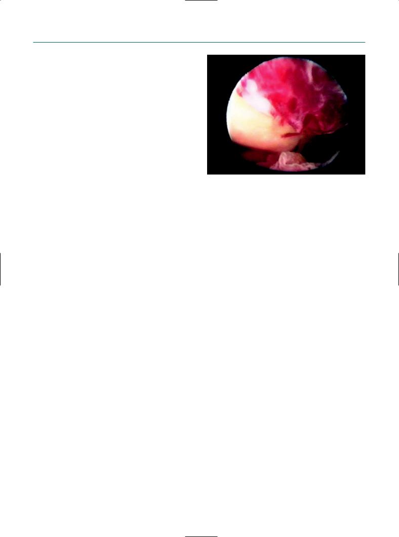
Figure 1.1
Cumulus mass capture by the fimbriae at the time of ovulation.
visualization of subtle tubo-ovarian structures in their natural position. The superiority of TVL over SL for exploring the tubo-ovarian architecture and physiologic processes, such as ovulation and ovum capture, was demonstrated when the first direct observation of the cumulus mass capture by the fimbriae at the time of ovulation was documented in the human15 (Figure 1.1).
Hydroflotation and direct access to the fimbriae allows for accurate inspection of the fimbrial mucosal folds and the infundibulum by fimbrioscopy. Full salpingoscopy or ampulloscopy is more difficult, but can be achieved without additional instrumentation in approximately 50% of the patients, being most successful in the periovulatory period.16 Salpingoscopy is not performed during SL in most centers because it requires training, is not an easy manipulation, and there is a need for an additional optical instrument and surgical assistance.
Finally, the use of an aqueous distention medium instead of CO2 pneumoperitoneum avoids postoperative irritation and allows the patient to leave the office in comfort after the diagnostic procedure.
Accurate diagnosis
Mild and moderate pelvic lesions affecting fertility include peritoneal endometriosis, ovarian endometrioma, tuboovarian adhesions, fimbrial agglutination and phimosis, and tubal mucosal adhesions.
Endometriotic disease is today defined by the presence of any laparoscopically visible endometrial implant >5 mm, any visible ectopic endometrial implant with evidence of inflammation or tissue damage, such as neoangio-
Transvaginal laparoscopy in infertility |
5 |
(a) |
(b) |
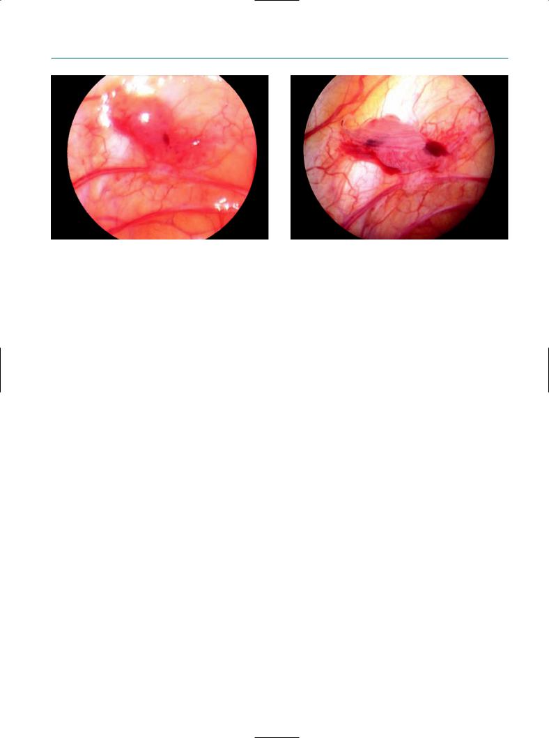
Figure 1.2
Effect of distention medium on appearance of peritoneal endometriotic lesion. (a) At standard laparoscopy using a CO2 pneumoperitoneun, the lesion appears as a polypoidal implant. (b) The same lesion seen during transvaginal hydrolaparoscopy using an aqueous distention medium shows a free-floating, vascularized adhesion on a red hemorrhagic implant surrounded by intense neoangiogenesis.
genesis or adhesions without another explanation, or an endometrioma of any size.17 For the accurate diagnosis we need a close and clear inspection of the lesion. Here, TVL offers major advantages over SL (Figure 1.2). First, endometriosis is frequently located at the caudal pole of the ovary and in the fossa ovarica. These locations are directly accessible at TVL. Without the need of supplementary manipulation, the whole ovarian surface and lateral pelvic wall can be inspected. Secondly, in mild and minimal endometriosis, 50% more ovarian adhesions are detected at TVL than at SL.18 Small ovarian endometriomas, which are missed at transvaginal ultrasound, are detectable at TVL by the presence of superficial lesions, neoangiogenesis, and free-floating adhesions at the site of invagination of the ovarian cortex or agglutination of the ovarian gyri. Finally, endometriotic adhesions between the ovary and lateral pelvic wall, which may rupture and collapse during the manipulation of the ovary and appear as a diffuse bleeding at SL, remain intact. In conclusion, as women should not be considered to have endometriotic disease unless the ectopic endometrial implants show evidence of activity or tissue damage, we need accurate inspection of the lesion. Unfortunately, no recommendations exist under which conditions and by which criteria the tissue damage, inflammation, or neoangiogenesis should be evaluated and scored.
Fimbrial abnormalities and adhesions are also easily detectable at TVL. Studies of the fallopian tube in pelvic inflammatory disease have shown that the endoscopic inspection of tubal mucosa by salpingoscopy is the most accurate technique in predicting the probability of pregnancy and the risk of ectopic pregnancy.19 In the presence
of Chlamydia antibodies, exclusion of any tubal mucosal pathology is important for further fertility management.
Accurate diagnosis of uterine lesions, tubo-ovarian adhesions, and active peritoneal and ovarian endometriotic lesions at an early stage is important in the management of infertility. These lesions are assumed to affect 20–30% of subfertile patients. Capelo et al20 found that one-third of the patients failing to conceive after four ovulatory cycles of clomiphene citrate had significant intrapelvic pathology. A multicentric study found that the performance of transvaginal endoscopy can mean avoiding laparoscopy in up to 93% of infertile women without clinical or ultrasound evidence of pelvic disease, as the relevant information can be obtained by this less-invasive procedure.21
Effective treatment
Endometriosis has been a major argument to opt for an endoscopy-based rather than ultrasound-based fertility investigation. The main question is whether minimal or mild endometriosis is a potential cause of delay in conception and whether surgical treatment is effective. A recent study of infertile women undergoing laparoscopy found that the time to natural conception differs significantly between women with unexplained infertility and infertile women with minimal or mild endometriosis.22 In that study, a group of 192 fully investigated infertile couples were followed up for up to 3 years following laparoscopy. No surgical therapy was undertaken to treat the

6 Atlas of Transvaginal Endoscopy
endometriosis found at that time. The authors found that the likelihood of pregnancy was significantly reduced in infertile women with minimal or mild endometriosis compared with those infertile women with a normal pelvis. Another recent study of 315 infertile patients with earlystage endometriosis and a control group of 152 infertile patients who had no endometriosis found significantly more fimbrial pathology, including agglutination, phimosis, and blunting, in the endometriosis group.23 Clearly, in addition to the endometriotic lesions the presence of other mild pathology deserves endoscopic investigation.
The view that the diagnosis and treatment of minor endometriosis in an early stage of subfertility is beneficial is supported by the results of the Canadian Collaborative Group on Endometriosis.24 In a study of 341 infertile patients with minimal and mild endometriosis who were randomized to laparoscopic ablation or expectant management, the authors found that laparoscopic ablation of minimal or mild endometriosis doubled the cumulative fecundity rate after a follow-up period of 36 weeks: 30.7% in the treatment group vs 17.7% in the no treatment group. A second Italian study could neither reject nor confirm this observation. The study included 101 infertile patients, but demonstrated no difference in fecundity rates after a fol- low-up period of 1 year.25 A recent review combining the results of these two randomized controlled trials into a meta-analysis showed that surgical treatment is more favorable than expectant management (OR for pregnancy = 1.7; 95% CI 1.1–2.5).26
In a recent meta-analysis of IVF outcome for patients with endometriosis, Barnhart et al27 recommended that patients with endometriosis of any stage should be referred for early aggressive infertility treatment, including IVF, to increase chances of conception. It remains an unfortunate fact that the diagnosis of endometriosis is still unduly delayed in many patients with infertility and pain.28
Timing the endoscopy-based investigation
The optimal approach in the management of female infertility requires that the timing and the method of the investigation are beneficial for the couple by avoiding both underand overtreatment. Unfortunately, infertility is a disorder in which the diagnosis and, consequently, reliable treatments are frequently unduly and excessively delayed.
The duration of infertility, or the time to conception, has been used as a major parameter for timing routine exploration and starting treatment. It has been assumed that the longer the interval, the lower is the probability of conception, and therefore investigations should normally not start before 1 year of infertility.29 On the other hand, a pro-
longed duration of infertility without preliminary endoscopic pelvic investigation has also been proposed as an indication for the use of assisted reproductive technology (ART). Therefore, in current practice, a delayed diagnosis may paradoxically favor both underand overconsumption of ART.
Recent prospective studies on fecundity have shown that human beings may be more fertile than has previously been estimated.30–32 In a recent debate Brosens et al33 proposed that in view of the availability of less-invasive and moreaccurate diagnostic tools and effective treatments, our current approach in timing the exploration of female infertility needs to be revisited. The issue is no longer when an invasive and expensive procedure like a laparoscopy should be performed, but at which stage a comprehensive minimally invasive fertility investigation is performed in order to inform the couple who worries about the delay in pregnancy. More than ever, the timing needs to be individualized, depending on factors such as age, medical, menstrual and sexual history, previous experience with contraceptive methods, use of fertility awareness methods for conception, and other individual factors. With the progress in minimally invasive exploration, the decision of timing the fertility investigation depends similarly as for other medical disorders in the first instance not on an abstract duration in time, but on the rational demand of the woman who worries about the cause of the delay of conception.
In older couples, some have argued that laparoscopy can be omitted from the infertility work-up when the hysterosalpingography is normal and there is no abnormal contributing history, and, as a consequence, the cost of fertility treatment is reduced without compromising success rates. However, Balasch34 argued that in relatively older women an evaluation would find more diseases known to cause infertility, such as pelvic adhesions and endometriosis. Two studies aimed at determining infertility factors in women of advanced reproductive age concluded that there is no unique pattern of infertility diagnosis in such patients.35,36 This supports the view that the routine investigation of infertility should not differ based on the age of the patient. Postponing the investigation in these women can be regarded as undertreatment when the couple is affected by a disorder or a combination of disorders for which an effective treatment, such as surgery or ART, may exist.
Conclusion
Today, the exploration of the female reproductive system, which traditionally included as the first step hysterosalpingography and at a later stage transabdominal laparoscopy, can be achieved in one step by minimally invasive transvaginal endoscopy. The procedure includes mini-hys-

Transvaginal laparoscopy in infertility |
7 |
teroscopy, transvaginal hydrolaparoscopy, fimbrioscopy, chromopertubation test and, in selected cases, salpingoscopy, and provides in a single procedure the most accurate and complete information on the reproductive organs. Therefore, transvaginal endoscopy is intended to replace hysterosalpingography as a first-line investigation, which avoids diagnostic laparoscopy in infertile patients without obvious pelvic pathology. The approach has the benefit of restoring in the infertile patient the normal stratification of a surgical procedure, which proceeds from diagnosis to full and accurate information of the patient, and of performing the surgical procedure after informed consent.
The clinical implementation of a new diagnostic tool, however, requires assessment of various aspects of the technique, including feasibility, safety, diagnostic accuracy, patient acceptability, and cost–benefit analysis. Several of these issues are discussed in detail in other chapters of this book.
References
1.Decker A. Culdoscopy – A New Technique in Gynecology and Obstetric Diagnosis. Philadelphia: WB Saunders, 1952.
2.McCann MF, Cole LP. Risks and benefits of culdoscopic female sterilization. Int J Gynaecol Obstet 1978; 16: 242–7.
3.Palmer R. Les Explorations Fonctionelles Gynécologiques, 2nd edn. Paris: Masson, 1974: 226–8.
4.Diamond E. Diagnostic culdoscopy in infertility: a study of 4,000 outpatient procedures. J Reprod Med 1978; 21: 23–30.
5.Mintz M. Actualisation de la culdoscopie transvaginale en décubitus dorsal. Un nouvel endoscope à vision directe muni d’une aiguille à ponction incorporée dans l’axe. Contracept Fertil Sex 1987; 15: 401–4.
6.Odent M. Hydrocolpotomie et hydroculdoscopie. Nouv Presse Méd 1973; 2: 187.
7.van Lith DAF, van Schie KJ, Beekhuizen W. Diagnostic miniculdoscopy preceding laparoscopy when bowel adhesions are suspected. J Reprod Med 1997; 23: 87–90.
8.Gordts S, Campo R, Rombauts L et al. Transvaginal hydrolaparoscopy as an outpatient procedure for infertility investigation. Hum Reprod 1998; 13: 99–103.
9.Watrelot A, Dreyfus JM, Andine JP. Evaluation of the performance of fertiloscopy in 160 consecutive infertile patients with no obvious pathology. Hum Reprod 1999; 14, 707–11.
10.Jansen FJ, Kapiteyn K, Trimbos-Kemper T et al. Complications of laparoscopy: a prospective multicentric observational study. Br J Obstet Gynaecol 1997; 104: 595–600.
11.Querleu D, Chapron C, Chevalier L et al. Complications of gynaecologic laparoscopic surgery – a French multicentre collaborative study. Gynaecol Endosc 1993; 2: 3–6.
12.Oshinsky GS, Smith AD. Laparoscopic needles and trocars: an overview of designs and complications. J Laparoendosc Surg 1992; 2: 117–25.
13.Brosens I, Gordon A, Campo R et al. Bowel injury in gynecologic laparoscopy. J Am Assoc Gynecol Laparosc 2003; 10: 9–13.
14.Gordts S, Watrelot A, Campo R et al. Risk and outcome of bowel injury during transvaginal pelvic endoscopy. Fertil Steril 2001; 76: 1238–41.
15.Gordts S, Campo R, Rombauts L et al. Endoscopic visualisation of
the process of fimbrial ovum retrieval in the human. Hum Reprod 1998; 13: 1425–8.
16.Gordts S, Campo R, Rombauts L et al. Transvaginal salpingoscopy: an office procedure for infertility investigation. Fertil Steril 1998; 70: 523–6.
17.Zondervan KT, Cardon LR, Kennedy SH. What makes a good casecontrol study? Design issues for complex traits such as endometriosis. Hum Reprod 2002; 17: 1415–23.
18.Brosens I, Gordts S, Campo R. Transvaginal hydrolaparoscopy but not standard laparoscopy reveals subtle endometriotic adhesions of the ovary. Fertil Steril 2001; 75: 1009–12.
19.Marana R, Catalano GF, Muzii L. Salpingoscopy. Curr Opin Obstet Gynecol 2003; 15: 333–6.
20.Capelo FO, Kumar A, Steinkampf MP et al. Laparoscopic evaluation following failure to achieve pregnancy after ovulation induction with clomiphene citrate. Fertil Steril 2003; 80: 1450–3.
21.Watrelot A, Nisolle M, Chelli H et al. International Group for Fertiloscopy Evaluation. Is laparoscopy still the gold standard in infertility assessment? A comparison of fertiloscopy versus laparoscopy in infertility. Results of an international multicentre prospective trial: the ‘FLY’ (Fertiloscopy-LaparoscopY) study. Hum Reprod 2003; 18: 834–9.
22.Akande VA, Hunt LP, Cahill DJ et al. Differences in time to natural conception between women with unexplained infertility and infertile women with minor endometriosis. Hum Reprod 2004; 19: 96–103.
23.Abuzeid M, Mitwally MF, Schwark S et al. A possible mechanical factor in infertile patients with early stage endometriosis. Fertil Steril 2005; 84(Suppl 1): S197.
24.Marcoux S, Maheux R, Bérubé S. Laparoscopic surgery in infertile women with minimal or mild endometriosis. The Canadian Collaborative Group on Endometriosis. N Engl J Med 1997; 337: 217–22.
25.Parazzini F. Ablation of lesions or no treatment in minimal-mild endometriosis in infertile women: a randomized trial. Hum Reprod 1999; 14: 1332–4.
26.Olive DL, Pritts EA. The treatment of endometriosis: a review of the evidence. Ann NY Acad Sci 2002; 955: 360–72.
27.Barnhart K, Dunsmoor-Su R, Coutifaris C. Effect of endometriosis on in vitro fertilization. Fertil Steril 2002; 77: 1148–55.
28.Dmowski WP, Lesniewicz R, Rana N et al. Changing trends in the diagnosis of endometriosis: a comparative study of women with pelvic endometriosis presenting with chronic pelvic pain or infertility. Fertil Steril 1997; 67: 238–43.
29.van der Steeg JW, Steures P, Hompes PGA et al. Investigation of the infertile couple: a basic fertility work-up performed within 12 months of trying to conceive generates costs and complications for no particular benefit. Hum Reprod 2005; 20: 2672–4.
30.Wang X, Chen C, Wang L et al. Conception, early pregnancy loss, and time to clinical pregnancy: a population-based prospective study. Fertil Steril 2003; 79: 577–84. .
31.Gnoth C, Godehardt D, Godehardt E et al. Time to pregnancy: results of the German prospective study and impact on the management of infertility. Hum Reprod 2003; 18: 1959–66.
32.Gnoth C, Godehardt E, Frank-Herrmann P et al. Definition and prevalence of subfertility and infertility. Hum Reprod 2005; 20: 1144–7.
33.Brosens I, Gordts S, Valkenburg M et al. Investigation of the infertile couple: when is the appropriate time to explore female infertility? Hum Reprod 2004; 19: 1689–92.
34.Balasch J. Investigation of the infertile couple in the era of assisted reproductive technology: a time for reappraisal. Hum Reprod 2000; 15: 2251–7.
35.Balasch J, Fábregues F, Jové IC et al. Infertility factors and pregnancy outcome in women above age 35. Gynecol Endocrinol 1992; 6: 31–5.
36.Miller JH, Weinberg RK, Canino NL et al. The pattern of infertility diagnoses in women of advanced reproductive age. Am J Obstet Gynecol 1999; 181: 952–7.


2
Diagnostic hysteroscopy
Rudi Campo and Carlos Roger Molinas
Introduction
Although operative hysteroscopy has progressively been accepted for the treatment of intrauterine pathologies, diagnostic hysteroscopy is still not widely and routinely used. Whereas almost all urologists utilize office cystoscopy to evaluate bladder pathology, it is estimated that less than 20% of gynecologists utilize office hysteroscopy to evaluate uterine pathology.1 Conventional hysteroscopy, defined as a procedure performed with an instrument of 5.0 mm total diameter and with CO2 as a distention medium and in which the insertion of the hysteroscope is facilitated by the use of a speculum and a tenaculum, has not been proven to be a technique accessible for all gynecologists and applicable in a routine set-up. Recently, well-conducted scientific studies have highlighted some important elements that can explain this underutilization of hysteroscopy as a first-line diagnostic procedure both in the office and in the conventional inpatient clinic. Nagele et al2 have proved that CO2 induces significantly more pain than a watery solution when used as distention medium.2 Furthermore, a watery distention medium has the advantage of cleaning the environment, leading to a better and easier visualization of the uterine cavity than with the conventional CO2. In a prospective randomized trial (PRT) we have recently proved the importance of the instrument diameter for both patient compliance and visualization quality.3 In the same study we also demonstrated that both the experience of the surgeon and the anatomical difficulties determined by patient’s parity play a key role when a conventional hysteroscope is used. With the use of a mini-hysteroscope, however, neither surgeon’s experience nor patient’s parity influence the results, offering a significant improvement for patient compliance and visualization quality. Office hysteroscopy is wrongly associated to a large extent with the disadvantages of conventional hysteroscopy and unfortunately many physicians, including gynecologists who do not witness the recent technical developments, believe that office hysteroscopy is similar to conventional hysteroscopy but performed in an office setting. In addition, the benefits of incorporating hysteroscopy as a first-line diagnostic tool
for the investigation of abnormal uterine bleeding (AUB)4,5 and infertility6–9 are still not completely assumed by the medical community, whereas the lack of expertise to perform the procedure is evident.
The office approach for diagnostic hysteroscopy
In order to propose the systematic use of diagnostic hysteroscopy and to avoid the still well-established delay in indication, it is mandatory to perform the technique in the office, ideally at the same time as transvaginal sonography (TVS). The most important challenge for the office approach is to be able to perform the procedure with an acceptable patient compliance. This should not be underestimated, since many patients still prefer the inpatient approach, believing that it will be pain-free.10 Several alternatives have been proposed for pain reduction during conventional office diagnostic hysteroscopy, but the results are inconclusive.11–16
The scientific evidence gathered over the last years and the major technical improvements in the manufacturing of high-quality small-bored scopes (minihysteroscopes) have provided an answer to the question of how diagnostic hysteroscopy should be implemented successfully in an office environment2,17–21 (Table 2.1).
Instruments for office diagnostic hysteroscopy
Hysteroscope
While the diagnostic hysteroscopes used in the past had a total outer diameter of 5.0 mm, recent technical advances have allowed miniaturizing the instruments without compromising the quality of visualization. Today, two systems are suitable for performing hysteroscopy in the office: