

Color Atlas of Gross Placental Pathology
.pdf60 Atlas of Transvaginal Endoscopy
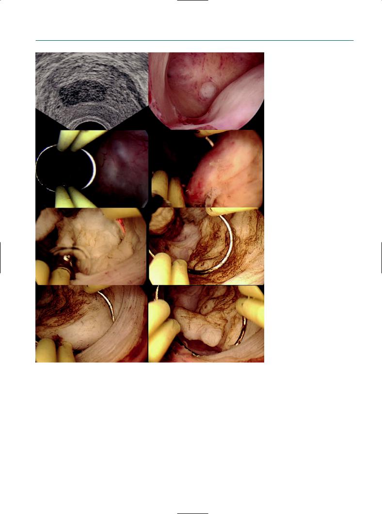
ance of a polyp during resection is its soft structure and the image of multiple small white spots when cutting through the tissue. They represent the glandular channels or cystic dilated endometrial glands within the polypous structure.
Before surgery, the base of the tumor has to be defined by turning around the lesion using the 30° angle of the optic.
The prominent vessels at the base are first coagulated in order to avoid excessive bleeding and blurring of the vision. Finding the demarcation of and cleavage plane at the base of the polyp can be achieved by gently pushing the resectoscope’s loop electrode against its base. This is very helpful, also and especially in the case of hysteroscopic myomectomy. The tumor is then resected progressively by cutting small slices. Although these chips float around or fall down on the posterior uterine wall and can obstruct the surgeon’s vision, it is incorrect to take them out after each cut, because moving the resectoscope in and out of the uterine cavity too often increases the risk of air embolism. It is better to remove all the fragments at the end of the procedure or when the view becomes impaired. At the end of the resection, the base of the tumor is coagulated to avoid excessive blood loss. This can be done with the resection loop or with the rollerball.
For very large sessile polyps it is advisable to remove the superficial part of the entire polyp before entering the myometrium. Indeed, as soon as the removal of the base of the polyp is started at one spot, the risk of fluid overload increases. So it is safer to reserve this part of the resection to the end of the procedure. But nowadays it is preferable to use bipolar loop electrodes together with a physiologic saline solution as a distention medium for the hysteroscopic resection of large polyps and submucous or intramural fibroids, in order to avoid the risk of (hence the need for monitoring) fluid overload and toxicity of anionic solutions such as dextran, glycine, or sorbitol–mannitol (Purisole) altogether.
Endometrial polyps usually lie completely within the uterine cavity and do not extend intramurally. Therefore, they are an ideal structure for the beginner in operative hysteroscopy to train in intrauterine resection as the risk of uterine perforation is usually very low. Resection may occasionally be difficult when a polyp is very mobile and moving away from the cutting loop or when it is located very deeply in the cornual region of the uterine cavity.
Myomas
If an intrauterine myoma is suspected, a diagnostic hysteroscopy should first be performed to evaluate the situation and the type of myoma (see also Table 6.2). With the use of a 30° optic, the exact localization and extension of
the myoma is defined by slow rotation. The tubal ostia are also located as helpful landmarks. Sometimes it is not possible to see both ostia, as the myoma may be shadowing one of them. In very large myomas it may even be impossible to gain access to one of the tubal ostia when the myoma is obscuring the view. In the case of profuse uterine bleeding, which is often found in these cases, it can become necessary to use a double-flow hysteroscope to clear the uterine cavity from bloody debris. After diagnostic hysteroscopy, the cervix is dilated to Hegar 8–9.5 (depending on the size of the instrument) for insertion of the resectoscope.
The use of an ‘active type’ of working element where, in the 0 position, the loop is out of the working channel, and is brought into the sheath by active pulling with fingers 2–4, seems more physiologic and safer than the use of the ‘passive handgrip’ where the cutting loop is brought into the sheath by retrograde movement of the operator’s thumb.
Type 0 myoma
After again clearly identifying the location of the myoma, the loop is brought behind the myoma, a part of the myoma is ‘hooked’ on it, and then the loop is pulled through the myoma while activating cutting current.
A sequential movement, consisting of (1) pulling the loop halfway to the sheath, followed by (2) the retraction of the entire resectoscope, is performed. When doing so, one must follow the outline and curve of the uterine cavity, but this technique allows longer slices to be cut over the whole surface in the case of large myomas (and the same principle can be applied to the endometrial ablation with the cutting loop electrode).
Having cut the first slice, one can usually identify whitish, fibrous tissue as typical for myoma tissue in contrast to the described polyp type of lesion. Systematically, one slice after the other is cut. The chips created are pushed into the cornual region opposite to the myoma. The more chips produced, the more one’s vision can become impaired.
The resected chips are best evacuated under direct vision by gently grasping them with the loop electrode and holding them against the optic while removing the whole instrument.
The decrease of distention pressure causes bleeding from the endometrium, the myometrial vessels, and the surface of the remaining part of the myoma. Therefore, after reintroducing the resectoscope, the uterine cavity should first be flushed to allow viewing again; then, resection is continued in a systematic manner until the complete base of the myoma is removed. The resection is followed by careful
Fertility-enhancing hysteroscopic surgery |
61 |
coagulation of the base in order to minimize fluid resorption and to avoid excessive bleeding.
Type I or II myoma
In the case of a type I or type II fibroid it is necessary to carry out a part of the resection inside the uterine wall (Figure 6.17). Usually one can distinguish very well between myoma tissue and normal surrounding myometrium: the myoma has a more dense structure, with whitish color; the myometrium is composed of more connective fibrous tissue with a slightly pinkish appearance.
Figure 6.17
Several stages of an operative hysteroscopy for the removal of a type 2 submucous myoma, in this case with the use of a bipolar loop electrode (Karl Storz GmbH, Tuttlingen, Germany) in Hartmann’s solution, i.e. without the risk of fluid overload like with macromolecular distention media.
The deeper the resection is performed into the wall, the greater becomes the risk of uterine perforation. In this situation, one can sometimes enucleate the base of the myoma from its capsule just by blunt dissection. This has the advantage of completing the enucleation without risk of uterine perforation and bowel laceration by the activated cutting loop. This is especially true when vaginal ultrasound indicates a thin safety margin of healthy myometrium and/or when one attempts to resect an intramural fibroid. Only experienced hysteroscopic surgeons can perform these difficult procedures.
In cases with deep intramural resection, it is advisable to interrupt the operative procedure in between to evacuate the

62 Atlas of Transvaginal Endoscopy
cavity. When reintroducing the resectoscope, one can often see that the image has changed completely: the former deep hole in which the base of the myoma is still present now seems to protrude into the uterine cavity and hence can be resected much more easily than before. This may be caused by uterine contractions, and sometimes one can try to further enhance this mechanism by giving drugs, such as oxytocin or ergometrine, that stimulate uterine contractility.
In difficult cases one should consider the performance of simultaneous transabdominal ultrasonography or laparoscopy as safety measures. The hysteroscopic appearance of whitish, bloodless area of tissue or the laparoscopic recognition of white decoloration of the uterine serosa indicate that perforation is likely to occur soon. The residual thickness of the myometrium can be measured exactly by ultrasound.
Although the endoscopic image of bleeding is sometimes very impressive, vaginal bleeding after hysteroscopic myoma resection is only minimal and usually stops within the first 3 postoperative days.
Hysteroscopic myoma resection usually gives excellent results. Nearly all patients regain eumenorrhea and relief of dysmenorrhea after a minimally invasive procedure that does not cause a great deal of discomfort. Therefore, hysteroscopic resection is the method of choice for the treatment of intrauterine submucous fibroids.
References
1.Campo R, Molinas CR, Rombauts L et al. Prospective multicentre randomized controlled trial to evaluate factors influencing the success rate of office diagnostic hysteroscopy. Hum Reprod 2005; 20: 258–63.
2.Fusi L, Cloke B, Brosens JJ. The uterine junctional zone. Best Pract Res Clin Obstet Gynaecol 2006; Epub.
3.Campo R, Van Belle Y, Rombauts L, Brosens I, Gordts S. Office mini-hysteroscopy. Hum Reprod Update 1999; 5: 73–81.
4.Acién P. Incidence of Müllerian defects in fertile and infertile women. Hum Reprod 1997; 12: 1372–6.
5.The American Fertility Society classifications of adnexal adhesions, distal tubal occlusion, tubal occlusion secondary to tubal ligation, tubal pregnancies, müllerian anomalies and intrauterine adhesions. Fertil Steril 1988; 49: 944–55.
6.Kaufman RH, Adam E, Binder GL, Gerthoffer E. Upper genital tract changes and pregnancy outcome in offspring exposed in utero to diethylstilbestrol. Am J Obstet Gynecol 1980; 137: 299–308.
7.Kaufman RH, Noller K, Adam E et al. Upper genital tract abnormalities and pregnancy outcome in diethylstilbestrol-exposed progeny. Am J Obstet Gynecol, 1984; 148: 973–84.
8.Kaufman RH, Adam E, Hatch EE et al. Continued follow-up of pregnancy outcomes in diethylstilbestrol-exposed offspring. Obstet Gynecol 2000; 96: 483–9.
9.Acién P. Reproductive performance of women with uterine malformations. Hum Reprod 1993; 8: 122–6.

7
Bipolar resectoscope: the future perspective of hysteroscopic surgery
Luca Mencaglia, Emmanuel Lugo, and Cristiana Barbosa
Introduction
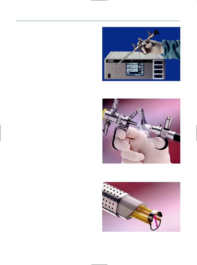
The gynecologic resectoscope, born from its urologic equivalent, is commonly used in gynecologic practice to resect or remove intracavitary pathology and also to perform endometrial ablation. The resectoscope consists of a telescope (2.9 and 4 mm, preferably with a 12° angle of view to always keep the loop within the viewing field), an electrical loop to perform passive cuts, and two sheaths for continuous-flow suction and irrigation of liquid distention medium. Besides the cutting loop, other instruments such as microknives or series of coagulation or vaporization electrodes of various shapes can be connected to the resectoscope.
There are essentially two types of resectoscopes which differ in outer diameter: 7.5 mm and 9.2 mm. The 7.5 mm resectoscope should be used in the case of a narrow cervical canal or difficult dilatation, whereas the 9.2 mm resectoscope facilitates the performance of major surgery.1
Electrosurgery in hysteroscopy
Biological tissue contains a more or less high concentration of electrolytes, making it sufficiently conductive to be treated by electrosurgery. The thermal effect of high-fre- quency current is used for separating (cutting) and coagulating tissue (desiccation of tissue). High-frequency (HF) currents must be used on humans, since low-frequency currents can stimulate nerve and muscle cells as a result of electrochemical processes (electrolysis). These effects are small enough to be disregarded with frequencies >100 kHz.2
A monopolar or bipolar current system can be adopted.
Monopolar resectoscope
Conventional hysteroscopic surgery uses a monopolar electrocautery system in which the current passes from the
active electrode through the patient’s body towards the return plate. The distention media used is glycine 1.5% or sorbitol–mannitol (non-electrolyte irrigation fluid). The monopolar resectoscope is connected to a monopolar electrosurgery generator of high frequency, automatically controlled by an acoustic alarm system. In a monopolar system, the electrons flow from an electrosurgery generator to an active electrode (electrode of the loop). From the electrode, the current flow is transmitted to the tissue, then to the plate (neutral electrode), and then it returns to the generator. This system is potentially dangerous since electrons flow through the body, outside the surgeon’s visual control, before returning to the generator. The new generators, however, decrease the incidence of electrical damage. In these generators, the cut current flow is automatically regulated by the tissue resistance. The unipolar loop can be used as coagulation, cut and combined (coag-cut) current. The coagulation current/flow is characterized by intermittent current flow periods that cause cellular dehydratation, resulting in tissue emostasis. The non-modulated cut flow is a continuous flow, with high intracellular temperature, causing cellular explosion. Non-modulated flow can be used also for coagulation and should be preferred because the voltage is lower and continuous.1
Bipolar resectoscope
In bipolar electrosurgery, the current flow through the tissue is restricted to the area between the two electrode loops that is under visual control of the surgeon. In this case, saline solution can be used as the distention media, because there is no risk of current dispersion. The generator produces a high initial voltage spike that establishes a voltage gradient in a gap between the bipolar electrodes. When the activated bipolar electrode is not in contact with the tissue, the electrolyte solution in the uterus dissipates it. When the loop is sufficiently close to the tissue, the high bipolar voltage spike
64 Atlas of Transvaginal Endoscopy
arc between the electrodes converts the conductive sodium chloride solution into a non-equilibrium vapor layer or plasma effect containing energy-charged sodium particles. Once formed, this plasma effect can be maintained at lower voltages (100–350 RMSV).3 With tissue contact, there is disintegration of tissue via molecular dissociation. Energetic species of the charged ions from the plasma effect result in disruption of carbon–carbon and carbon–nitrogen bonds. There is also electron impact dissociation of water molecules into exited fragments of H+ and OH– ions. The bottom line is rupture of cell membranes, which translates into visible cutting. Clinically, there is a precise tissue effect with minimal collateral damage, as the charged ions have an estimated penetration depth in tissue of only 50–100 µm (0.5–1 mm).4,5 The depth of coagulation is determined principally by the electrode configuration and by the system design, as well as by the technique used by the operator (time and pressure of contact).6
Figure 7.1
Autocon II 400.
Equipment
The new working element developed by Karl Storz System, the Autocon II 400 (Figure 7.1), is for use in laparoscopy, resectoscopy, and open surgery. It can be equipped with four different HF output sockets: monopolar, bipolar, multifunction, and neutral electrode sockets. The user can assign a socket with the following parameters: cutting mode, coagulation mode, or cutting and coagulation activation type. A mode is characterized by different generator settings such as frequency, peak voltage, and modulation. A total of 10 cutting and 9 coagulation modes are available. However, each specific socket is only available for a part of these modes. The user can also assign the following parameters to a mode: power limitation cutting, cutting effect, power limitation coagulation or coagulation effect.
Using a bipolar system, in the saline time-c-cut mode as the saline coagulation mode, the HF voltage form is unmodulated sinusoidal, the rated frequency is 350 kHz (at RL = 500 ohms) ± 10%, the crest factor is 1.4, the rated load resistance is 50 ohms, the maxim HF peak voltage is 190 Vp, the number of effects available are 8, the maxim power output 300 W ± 20%, and the parameters standard of use are 180 (in effect 4) for cut and 120 (in effect 4) for coagulation. It is important to point out that peak voltages are much lower than monopolar systems.
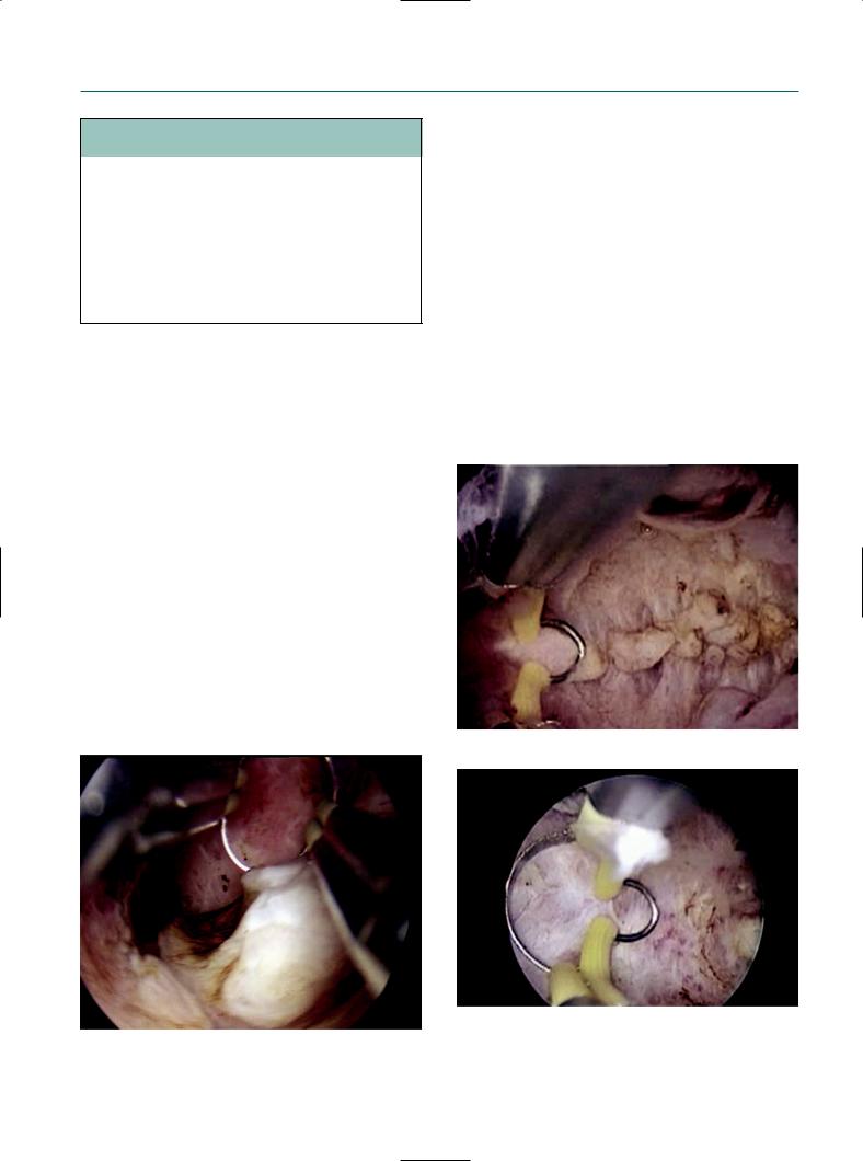
The bipolar components (Figures 7.2 and 7.3) are compatible with existing resectoscopes (optics, sheath). The sheath, designed by Karl Storz Endoskope, is completely isolated. The direct current returns via the electrode, preventing a current flow via the sheath, and guaranteeing a high level of safety. There are a variety of cutting loops (GP, GPV, GD, GDV) of 24 Fr on the market, but other bipolar loops for resection in gynecology and urology are still being developed.
Figure 7.2
The bipolar element of the resectoscope.
Figure 7.3
The direct current return via the electrode prevents a current flow via the sheath.
Bipolar resectoscope |
65 |
Table 7.1 Bipolar resection in 57 patients
Indication |
Number of |
Fibroids >4 cm |
|
patients |
|
Myomas G0 |
9 |
1 |
Myomas G1 |
15 |
7 |
Myomas G2 |
10 |
2 |
Endometrial ablation |
12 |
|
Polyps |
8 |
2 |
Uterine septum |
3 |
2 |
Total |
57 |
|
Clinical data
In 2004, Loffer7 reported a preliminary experience with the VersaPoint bipolar resectoscope in gynecology. A vaporizing electrode in saline solution distending medium was shown to be effective in removing submucous myomas in 15 patients. Golan et al8 in 2001 reviewed outcomes of operative hysteroscopy, using bipolar electrical energy (VersaPoint) in saline solution in 116 women with intrauterine pathology. They proposed this new technique as a potential replacement for monopolar resection. Furthermore, many studies have demonstrated the benefits in urology. Recently, Singh et al9 reported the use of a bipolar system in transurethral prostate resection (TURP) with less postoperative dysuria. In 2004, Wendt-Nordahl et al10 underlined the advantage of the bipolar system in TURP, with the Vista System, and stressed the theoretical benefit to avoid the risk of the TUR syndrome.
Our preliminary experience in gynecology consists of 57 patients (Table 7.1) treated with a Karl Storz 26 Fr bipolar
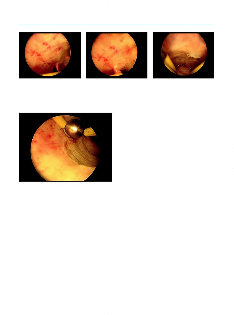
Figure 7.4
resectoscope and Autocon II 400 HF unit with parameters standard selected 180 (in effect 4) for cut and 120 (in effect 4) for coagulation. The resection loops are completely insulated from the sheath of the resectoscope and are reusable loops. Saline solution (NaCl 0.9%) was used as distention medium with no complications. Cutting power and coagulation appear better than with monopolar resection. In particular, the first cut in the case of fibroid tissue doesn’t give any problem thanks to the plasma effect. Furthermore, the vision during resection is not disturbed by the presence of air bubbles. We included complex cases in our series to evaluate the technical characteristics of the instruments, including an important number of uterine fibroids, that in some cases were >4 cm and G1 and G2 (partially or totally intramural localization). Results in terms of time of surgery, intraoperative bleeding, and complete removal of the pathology were better than with traditional monopolar resection (Figures 7.4–7.7).
a
b
Figure 7.5
Endometrial polyp resection with bipolar resectoscope.
Metroplasty of uterine septum with bipolar resectoscope.
66 Atlas of Transvaginal Endoscopy
a |
b |
Figure 7.6
Endometrial ablation with bipolar resectoscope.
Figure 7.7
Bipolar roller ball.
Conclusion
In our experience, and also in the analysis of the published data, it seems clear that the bipolar resectoscope presents some advantages in comparison with the monopolar resectoscope.
Current flow
The current flow through tissue is restricted to the area between the two electrodes’ loops that is under the direct vision of the surgeon. Current can be regulated at all times and set to the lowest possible value: optimal flow of current for minimally invasive treatment. The plasma effect of bipolar current allows better cut and coagulation. In the monopolar technique, the current passes through many
c
tissues outside the surgeon’s visual control before it can return to the generator.5,11 The risk of thermal injuries at distant organs or tissues, by direct contact of instruments, imperfection of insulation, or diffusion of the electric current, is reduced in the bipolar technique.12,13 It has minor risks of interference with other electronic equipment – electrocardiogram (ECG), pacemakers, etc. – simultaneously connected to the patient.5,14 Furthermore. there is reduced stimulation of peripheral nerves, including the obturator nerve, because of no flow of current through the body of the patient.
Distention media
According to Kolmer and Norlen15 and Koshiba et al,16 the incidence of overflow syndrome in gynecology and TUR syndrome in urology vary considerably in the literature, ranging from 0.18% to 10.9%. Mebust et al17 reported an incidence of 2% of TUR syndrome in 3885 patients. In 1996, Kudela et al18 reported the risk of fluid overload syndrome during the hysteroscopy monopolar procedure and underlined the necessity of adhering to safety measures that include selection of a suitable medium (hypotonic electrolyte-free solutions – glycine or sorbitol–mannitol solution), controlling duration of surgery, respecting the correct surgical indications and procedures and, in particular, performing continuous control of the balance of the distention medium. In 2003, Estes and Maye19 pointed out the danger of hypotonic, electrolyte-free distention media and their potential of being absorbed in volumes large enough to cause hyponatremia and hypervolemia. The main concerns in urologic and gynecologic conventional monopolar resection are fluid absorption with hyponatremia, hypervolemia, and glycine toxicity. This syndrome is very dangerous for the patient, leading to neurotoxic coma and death in the worst cases. Most of the morbidities

Bipolar resectoscope |
67 |
of the overflow syndrome are related to the use of hypotonic non-electrolyte irrigation fluid. For this reason, close and continuous perioperative monitoring of the balance of the distention medium by a nurse and frequent laboratory investigations are required. The bipolar resection system enables resection using saline solution. The use of saline solution for the distention media of the uterine cavity is the principal advantage of this technology, thus avoiding the use of a hypotonic non-electrolyte solution that can cause fluid overload during the surgical procedure. Saline solution is easily metabolized, is not toxic and can be used with higher quantity, and it is also less expensive than conventional hypotonic non-electrolyte solutions. Singh et al,9 in a bipolar vs monopolar TURP randomized controlled study, reported a significant difference in serum sodium concentration postoperatively. In bipolar TURP, the change in serum sodium was –1.2 mEq/L (not different from preoperative serum Na concentration), whereas in the monopolar group the mean decrease was 4.6 mEq/L. In three patients serum Na was >125 mEq/L at risk of TUR syndrome. However, the balance of the distention medium using saline solution should also be under control. Starkman and Santucci20 retrospectively reviewed 43 patients undergoing TURP: 18 consecutive patients treated with monopolar TURP and 25 with bipolar TURP. These investigators found an unexpected case of hyponatremia and pulmonary edema in a bipolar TURP patient. Patel et al6 also expressed concern for potential problems with hypervolemia and hyponatremia. They suggest warming the saline solution and emptying the bladder from time to time during surgery. In our experience, no overflow syndrome occurs.
Tissue alterations
In traditional monopolar resection, the tissue’s electrical resistance creates temperature as high as 400°C, which leads to desiccation with significant collateral and penetrative tissue damage.6 High-frequency current generated by a bipolar instrument tends to remain superficial; Luciano et al4,5 reported a 0.5–1 mm depth of penetration compared with the 3–5 mm seen in a monopolar system, allowing a better control of the cut and lower possibility of accidental injury. The technique allows maintenance of the current between the active electrode and the adjacent return electrode. The plasma effect of the loop prevents a sticking effect. In this case, tissue damage is minimized and the tissue temperature range is from 40°C to 70°C. Improved tissue analysis secondary to reduced carbonization of tissue has also been reported with better histologic interpretation. It has also been repeatedly reported that improved visibility aids the identification of surgical landmarks during the procedure.
Less bleeding during resection
Optimized resection current provided by the Autocon II 400 allows a better coagulation during resection, with reduced bleeding. Furthermore, the coagulation capacity, by itself, is much more powerful in the bipolar system than in the monopolar. This avoids time-consuming recoagulation after resection for coagulation and contributes to close the superficial capillary vascularization reducing also intravasation.
Better visibility
Minor air bubbles and less bleeding during resection allow a better vision during surgery, reducing the length of surgery and improving results.
Reduced costs
Compatible components of existing Karl Storz resectoscopes (optics, sheaths) can be used. Only the bipolar element of the resectoscope must be acquired. Resection loops are reusable and have a duration and a cost comparable with the traditional monopolar instruments.
The outcomes of studies in gynecology and urology with the bipolar system demonstrate its versatility and the possibility of rapid replacement of the old monopolar system. The bipolar system is technically superior, cost-effective, and safer compared with the monopolar system. If we take into consideration also the medicolegal aspects, it will be very dangerous to maintain the old system, especially in case of complications. Preliminary findings of decreased morbidity with the bipolar system, using saline solution, force us to consider other factors involved in possible complications, such as duration of the surgery as well as the experience of the surgeon. These variables must be included in our future investigations. However, the decrease of ‘theoretical risk’ of overflow syndrome that favors the bipolar system does not allow us to avoid close perioperative monitoring of distention medium balance and laboratory investigations.
Larger prospective randomized clinical trails examining cost-effectiveness and long-term outcome need to be performed, although it seems already clear that this technology will replace conventional monopolar electrosurgery in the near future.
References
1.Mencaglia L, Cavalcanti L. Histeroscopia Cirùrgica. Rio de Janeiro: UnionTask Press, 2004: 15–28.

68 Atlas of Transvaginal Endoscopy
2.Soderstrom RM. Principles of electrosurgery during endoscopy. In: Sammarco MJ, Stovall TG, Steege JF, eds. Gynecologic Endoscopy. Baltimore: Williams & Wilkins, 1996: 179–92.
3.Stadler KR, Woloszko J, Brown IG. Repetitive plasma discharges in saline solutions. Appl Phys Lett 2001; 79: 4503–5.
4.Luciano AA, Whitman G, Maier DB et al. A comparison of thermal injury, healing patterns, and postoperative adhesion formation following CO2 laser and electromicrosurgery. Fert Steril 1987; 48: 1025–9.
5.Luciano AA. Power sources. Obstet Gynecol Clin N Am 1995; 22: 423–43.
6.Patel A, Adshead J. First clinical experience with new transurethral bipolar prostate electrosurgery resection system: controlled tissue ablation (coblation technology). J Endourol 2004; 18: 959–64.
7.Loffer FD. Preliminary experience with the VersaPoint bipolar resectoscope using a vaporizing electrode in a saline distending medium. J Am Assoc Gynecol Laparosc 2004; 7: 498–502.
8.Golan A, Sagiv R, Berar M, Ginath S, Glezerman M. Bipolar electrical energy in physiologic solution – a revolution in operative hysteroscopy. J Am Assoc Gynecol Laparosc 2001; 8: 252–8.
9.Singh H, Desai M, Shrivastav P, Vani K. Bipolar versus monopolar transurethral resection of prostate: randomized controlled study. J Endourol 2005; 19: 333–8.
10.Wendt-Nordahl G, Hacker A, Reich O et al. The Vista System: a new bipolar resection device for endourological procedures: comparison with conventional resectoscope. Eur Urol 2004; 46: 86–90.
11.Riedel HH, Semm K. There is no place in gynecological endoscope for unipolar of bipolar high frequency current. Endoscopy 1982; 14: 51–4.
12.Levy BS, Soderstrom RM, Dail DH et al. Bowel injuries during laparoscopy. Gross anatomy and histology. J Reprod Med 1985; 30: 168–72.
13.Di Giovanni M, Vasilenko P, Belsky D et al. Laparoscopic tubal sterilization. The potential of thermal bowel injuries. J Reprod Med 1990. 35: 951–4.
14.Odell RC. Electrosurgery: principles and safety issues. Clin Osbtet Gynecol 1995; 38: 610–21.
15.Kolmer T, Norlen H. Transurethral resection of the prostate: a review of 1111 cases. Int Urol Nephrol 1989; 21: 47–55.
16.Koshiba K, Egawa S, Ohori M et al. Does transurethral resection of prostate pose a risk to life? 22-year outcome. J Urol 1995; 153: 1506–9.
17.Mebust WK, Holtgrewe HL, Cockett ATK, et al. Transurethral prostatectomy: immediate and postoperative complications. A cooperative study of 13 participating institutions evaluating 3,885 patients. J Urol 1989; 141: 243–7.
18.Kudela M, Lubusky D, Dzvincuk P. [Risk of fluid overload syndrome during hysteroscopy procedures]. Ceska Gynekol 1996; 61: 291–3 [in Czech].
19.Estes CM, Maye JP. Severe intraoperative hyponatremia in a patient scheduled for elective hysteroscopy: a case report. AANA J 2003; 71: 203–5.
20.Starkman JS, Santucci R. Comparision of bipolar transurethral resection of the prostate with standard transurethral prostatectomy: shorter stay, earlier catheter removal and fewer complications. BJU Int 2005; 95: 69–71.

8
Endometriosis: features at transvaginal laparoscopy
Patrick Puttemans
Introduction
As mentioned in previous chapters of this atlas, the workspace and environment offered by transvaginal hydrolaparoscopy are significantly different from the workspace and working conditions every gynecologist is used to when performing a conventional or standard laparoscopy. First of all, the workspace of transvaginal laparoscopy (TVL) is quite small, meaning that all the organs and tissues, hence all pathologies and lesions, are visualized from a physical distance (i.e. between lens and target) of some millimeters to a couple of centimeters at the most. The images that are obtained in this way can be defined as detailed close-ups that are quite different from the panoramic overview offered by standard laparoscopy. A second difference is the reversed anatomy and topography, not from top to bottom, but exactly the opposite so to speak. The combination of the limited workspace and the reversed approach makes a systematic evaluation of all anatomical regions of the female pelvis very important (Table 8.1). If this systematized way of exploring the pelvis is neglected, then significant lesions of endometriosis may be missed. On the other hand, and
Table 8.1 Systematized exploration of the female pelvis via transvaginal laparoscopy
1.Look up the posterior side of the uterus = anatomical landmark at the start of the procedure
2.Go from there to the ovarian ligament and the isthmic part of the Fallopian tube
3.Explore the tubo-ovarian relationship (to rule out the presence of tubo-ovarian adhesions or salpingitis isthmica nodosa)
4.Inspect the whole ovarian surface carefully, using the 30° angle of the optic
5.Explore the ovarian fossa, a common predilection site for endometriosis
6.Turn the optic 180° to explore the uterosacral ligament and the pouch of Douglas
7.Repeat this whole procedure at the other side
specifically for endometriosis, the angle at which the ovarian fossa is approached is ideal to explore one of the commonest predilection sites of the disease without any manipulation of the ovary, even in the presence of connecting adhesions between ovary and fossa. This contrasts with standard laparoscopy, where one needs to grasp and rotate the ovary at its ligament in order to visualize the ovarian fossa.
And a third difference is the salineor Ringer’s lactate-filled environment, i.e. a natural, pressure-less and transparent milieu, in contrast to the artificial, gas filled, and pressurized CO2 pneumoperitoneum at standard laparoscopy.
In view of the features of both peritoneal and ovarian endometriosis described in this chapter, transvaginal hydrolaparoscopy definitely offers a number of advantages over standard laparoscopy, including the recognition of a new and distinct clinical entity in the field of ovarian endometriosis. However, with regard to endometriosis, the technique also presents a number of disadvantages.
Features of endometriosis at transvaginal laparoscopy
The implants
Transvaginal hydrolaparoscopy is a highly accurate technique for the diagnoses of endometriosis and endometriotic adhesions in infertile patients without major pelvic pathology. Inspection under fluid has been reported to improve the visualization of red or ‘subtle’, non-fibrotic lesions of endometriosis by the three-dimensional optical effect of the fluid environment.1 These implants have a vesicular or papular form,2 are sessile or pedunculated yet always turgescent and erect in the aqueous environment (Figures 8.1a–8.1c), and demonstrate a marked increase of the vascularization of the surrounding peritoneum, often with small blood vessels in the stalk and on their surface. Inspection under water (‘hydroflotation’) facilitates the visualization of these capillary networks within and towards the