

Color Atlas of Gross Placental Pathology
.pdf80 Atlas of Transvaginal Endoscopy
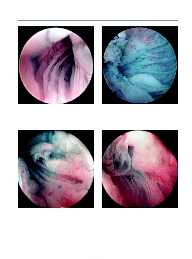
Figure 9.7 |
Figure 9.8 |
The endoscope was inserted into the ampulla by the guidance |
The distal part of the tubal mucosa was observed. |
of chromotubation. |
|
Figure 9.9 |
Figure 9.10 |
The normal-looking tubal mucosa under transvaginal |
The ampullary mucosa was clearly visible. |
salpingoscopy. |
|
Role of transvaginal salpingoscopy |
81 |
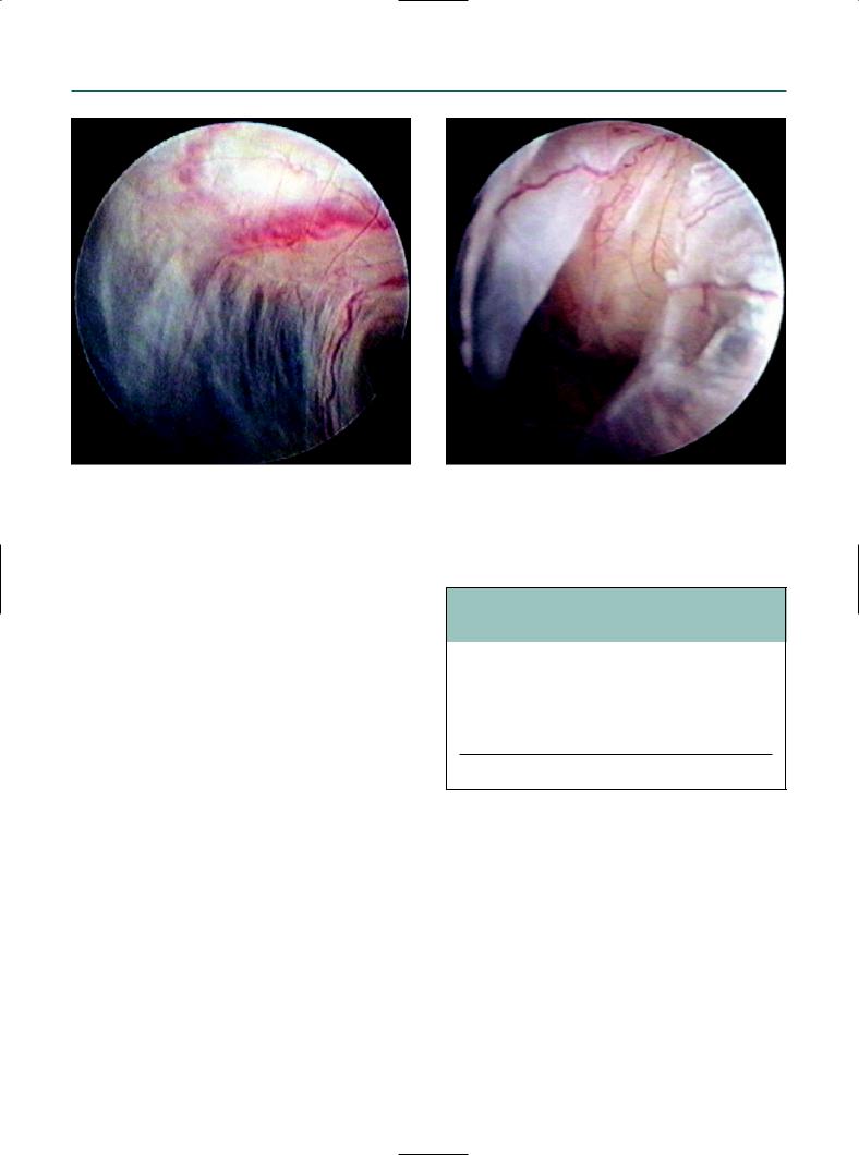
Figure 9.11
The salpingoscope was deeply inserted into the ampulla.
Figure 9.13
Figure 9.12
Chromotubation finding in the normal tubal mucosa.
Figure 9.14
The normal-looking tubal mucosa. |
The normal-looking tubal mucosa. |
82 Atlas of Transvaginal Endoscopy
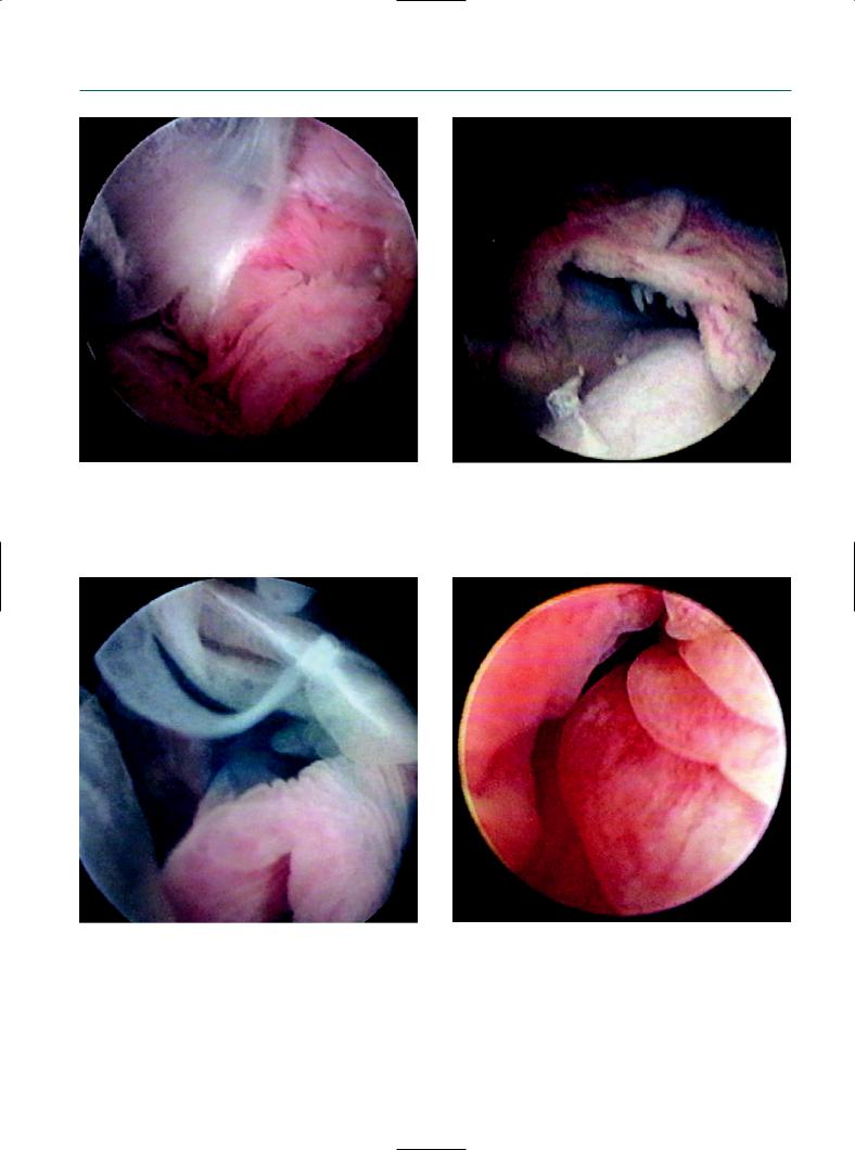
Figure 9.15
Unsuccessful salpingoscopy in a case of extensive peritubal adhesion by previous C. trachomatis infection.
performed in 55 (47.8%) of 115 regular tubes, which was significantly higher than that of 34 (33.3%) in 102 convoluted tubes or hydrosalpinges (p = 0.03). Deformed fimbriae or fimbrial adhesions prevented the cannulation of the distal tubal segment.
However, a history of C. trachomatis infection did not always influence the success of a salpingoscopy, because extensive peritubal adhesion by previous C. trachomatis infection may disturb visualizing the tubes themselves (Figures 9.15–9.17). Therefore, we speculate that the successful salpingoscopy rate was similar between women with and without previous C. trachomatis infection. Typical tubal damage by C. trachomatis infection includes verruca and atypical vessel formation, peritubal adhesion, and endosalpingial edema (Figures 9.18–9.20).
The correlation between laparoscopic and salpingoscopic findings remains controversial. In the present study, the 89 tubes that were observed by a salpingoscopy, were divided into three categories according to the laparoscopic findings as described above: regular 53 (59.6%); convoluted 34 (38.2%), and hydrosalpinx 2 (2.2%). They were also divided into five categories according to the salpingoscopic findings according to Puttemans et al,14 as shown in Table 9.2: grade I 66 (74.2%), grade II 19 (21.3%), grade III 3 (3.4%), grade IV 1 (1.1%), and grade V 0 (0%). Statistical analysis demonstrated a significant correlation between salpingoscopic and laparoscopic findings (r = 0.50; p <0.0001). These data were supported by the previous
Figure 9.16
Extensive peritubal adhesion by previous C. trachomatis infection.
Table 9.2 Classification of salpingoscopic findings
Grade I normal folds
Grade II distended fold pattern
Grade III focal lesions (adhesions, polyps, stenosis) Grade IV extensive lesions with or without destruction of
mucosa
Grade V complete loss of mucosal fold pattern
Reproduced from Puttemans et al,14 with permission.
reports demonstrating a positive correlation between salpingoscopic and laparoscopic findings.15,16 On the contrary, several groups arrived at the opposite conclusions.14,17,18 We consider that TVL and salpingoscopy should be simultaneously performed if possible in order to refer to the determination of the next treatment strategies.
Role of transvaginal salpingoscopy
We previously reported the usefulness and prognostic value of TVL in infertile women, and concluded that TVL
Role of transvaginal salpingoscopy |
83 |
Figure 9.17
Disturbance of visualization of the tubes due to previous C. trachomatis infection.
Figure 9.19
Peritubal adhesion by C. trachomatis infection with normal chromotubation.
Figure 9.18
Typical tubal damage by C. trachomatis infection. Ischemic appearance of fimbria.
Figure 9.20
Endosalpingial edema caused by C. trachomatis infection.

84 Atlas of Transvaginal Endoscopy
is useful in selecting a future treatment strategy.5,6 Women with severe tubal disease were recommended in vitro fertil- ization–embryo transfer (IVF-ET).
In this study, 16 patients who experienced a salpingoscopy bilaterally were carefully monitored for at least 6 months with appropriate infertility treatment, excluding IVF-ET. Pregnancies were established in 10 (62.5%) of 16 patients, with four pregnancies by timed intercourse and six by artificial insemination with the husband’s sperm (AIH). Tubal pregnancies were not included. The number of pregnancies did not differ between patients with regular and those with convoluted tubes. For example, four pregnant women (80%) with salpingoscopic grade I tubes had a convoluted tubal appearance at laparoscopy. This indicates that the salpingoscopy may be a better predictor of future fertility outcome than TVL alone. Previous reports also suggested that the simultaneous performance of salpingoscopy and laparoscopy could predict the reproductive outcome better than laparoscopy alone.2,15–19
In conclusion, these findings suggest that endoluminal assessment by transvaginal salpingoscopy can be simultaneously performed in some infertile women, especially with patent tubes or with regular tubes undergoing TVL. Further studies are being carried out to clarify the usefulness of intratubal exploration as a tubal infertility assessment.
References
1.Brosens I, Boeckx W, Delattin P et al. Salpingoscopy: a new preoperative diagnostic tool in tubal infertility. Br J Obstet Gynaecol 1987; 94: 768–73.
2.Marchino GL, Gigante V, Gennarelli G et al. Salpingoscopic and laparoscopic investigations in relation to fertility outcome. J Am Assoc Gynecol Laparosc 2001; 8; 218–21.
3.Gordts S, Campo R, Rombauts L, Brosens I. Transvaginal hydrolaparoscopy as an outpatient procedure for infertility investigation. Hum Reprod 1998; 13: 99–103.
4.Gordts S, Campo R, Rombauts L, Brosens I. Transvaginal salpingoscopy: an office procedure for infertility investigation. Fertil Steril 1998; 70: 523–6.
5.Shibahara H, Fujiwara H, Hirano Y et al. Usefulness of transvaginal hydrolaparoscopy in investigating infertile women with Chlamydia trachomatis infection. Hum Reprod 2001; 16: 1690–3.
6.Fujiwara H, Shibahara H, Hirano Y et al. Usefulness and prognostic value of transvaginal hydrolaparoscopy in infertile women. Fertil Steril 2003; 79: 186–9.
7.Shibahara H, Hirano Y, Ayustawati et al. Chemokine bioactivity of RANTES is elevated in the sera of infertile women with past Chlamydia trachomatis infection. Am J Reprod Immunol 2003; 49: 169–73.
8.Shibahara H, Takamizawa S, Hirano Y et al. Relationships between Chlamydia trachomatis antibody titers and tubal pathology assessed using transvaginal hydrolaparoscopy in infertile women. Am J Reprod Immunol 2003; 50: 7–12.
9.Hirano Y, Shibahara H, Takamizawa S et al. Application of transvaginal hydrolaparoscopy for ovarian drilling using Nd:YAG laser in infertile women with polycystic ovary syndrome. Reprod Med Biol 2003; 2: 37–40.
10.Shibahara H, Hirano Y, Kikuchi K et al. Postoperative endocrine alterations and clinical outcome of infertile women with polycystic ovary syndrome after transvaginal hydrolaparoscopic ovarian drilling. Fertil Steril 2006; 85: 244–6.
11.Watrelot A, Dreyfus JM, Andine JP. Evaluation of the performance of fertiloscopy in 160 consecutive infertile patients with no obvious pathology. Hum Reprod 1999; 14: 707–11.
12.Suzuki T, Shibahara H, Hirano Y et al. Feasibility and clinical significance of endoluminal assessment by transvaginal salpingoscopy under transvaginal hydrolaparoscopy in infertile women. J Minim Invasive Gynecol 2005; 12: 420–5.
13.Marchino GL, Gigante V, Gennarelli G et al. Salpingoscopic and laparoscopic investigations in relation to fertility outcome. J Am Assoc Gynecol Laparosc 2001; 8: 218–21.
14.Puttemans P, Brosens I, Delattin P et al. Salpingoscopy versus hysterosalpingography in hydrosalpinges. Hum Reprod 1987; 2: 535–40.
15.Marana R, Rizzi M, Muzii L et al. Correlation between the American Fertility Society classifications of adnexal adhesions and distal tubal occlusion, salpingoscopy, and reproductive outcome in tubal surgery. Fertil Steril 1995; 64: 924–9.
16.Surrey ES, Surrey MW. Correlation between salpingoscopic and laparoscopic staging in the assessment of the distal fallopian tube. Fertil Steril 1996; 65: 267–71.
17.De Bruyne F, Hucke J, Willers R. The prognostic value of salpingoscopy. Hum Reprod 1997; 12: 266–71.
18.Marana R, Catalano GF, Muzii L et al. The prognostic role of salpingoscopy in laparoscopic tubal surgery. Hum Reprod 1999; 14: 2991–5.
19.Heylen SM, Brosens IA, Puttemans PJ. Clinical value and cumulative pregnancy rates following rigid salpingoscopy during laparoscopy for infertility. Hum Reprod 1995; 10: 2913–16.
10
Operative transvaginal laparoscopy
Stephan Gordts
Introduction
The technique of diagnostic transvaginal laparoscopy (TVL) as an outpatient procedure was described by us for the first time in 1998.1,2 It was meant to offer the subfertile patient without obvious pelvic pathology the possibility of a minimally invasive endoscopic exploration of the pelvis.
As a distention medium, pre-warmed Ringer’s lactate is used; it contributes to an accurate and correct visualization and to floating of the organs. Consequently, tubo-ovarian structures can be inspected in their natural position without supplementary manipulation.
After gaining more experience with the technique, it became obvious that the easy access to the ovarian surfaces, the fossa ovarica, and the Fallopian tubes opened the
potential for some operative procedures. With the endoscopic instruments in the same axis, the transvaginal approach creates excellent possibilities for a minimally invasive dissection of the ovary under direct vision.
Technique and instruments
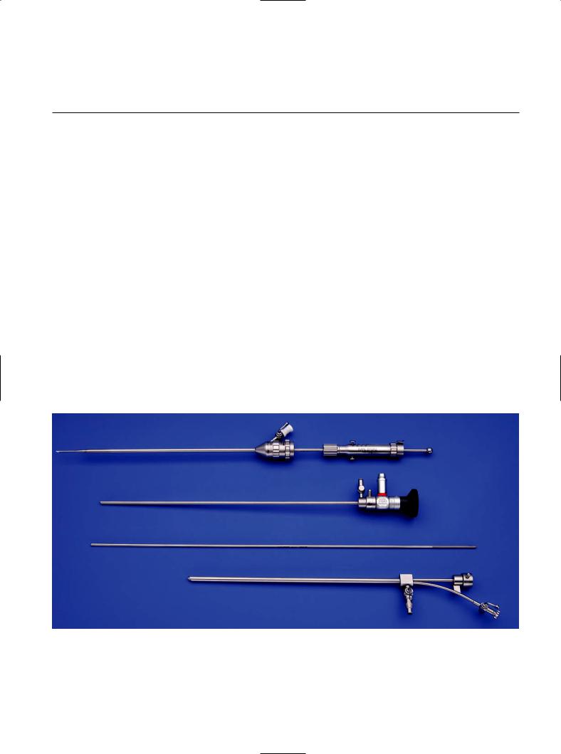
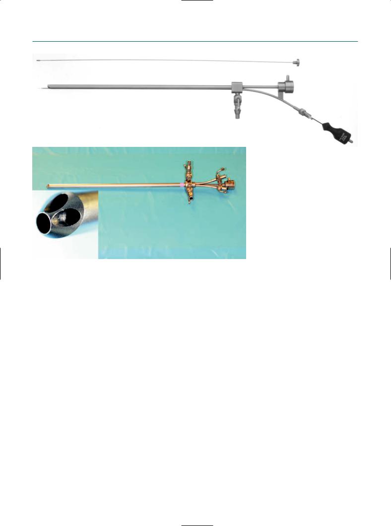
As for the diagnostic procedure, the patient lies in a dorsal decubitus position and access to the pelvis is gained through the same needle puncture technique. After verification of the correct localization of the diagnostic instruments, a specially developed obturator allows easy exchange between the diagnostic and the operative instruments (Figure 10.1). A 2.9 mm endoscope with a 30° optical angle is inserted in
Figure 10.1
Set for transvaginal endoscopy, showing:
•the assembled needle
•the 2.9 mm endoscope with the diagnostic trocar of 3.7 mm
•the obturator allowing a switch from the diagnostic sheet to the operative sheet
•the operative trocar with one channel.
86 Atlas of Transvaginal Endoscopy
a
Figure 10.2
The operative trocars: (a) one channel with the bipolar needle; (b) two channels allowing the introduction of 5 Fr instruments.
b
the operative trocar sheet with one or two operative channels (Figure 10.2), with outer diameters of 5 and 7 mm, respectively. The operative channels allow the insertion of 5 Fr instruments such as scissors, biopsy, and grasping forceps and bipolar coagulation probes. The two-channel operative trocar is mostly used in the case of ovarian endometrioma, as it allows the insertion of the bipolar probe through one channel and scissors or forceps through the other one.
Before starting the procedure, about 100 ml of warm Ringer’s lactate is instilled in the pouch of Douglas to give the necessary distention.
As we are working in an aqueous distention medium, preventive and meticulous hemostasis with bipolar coagulation is mandatory to avoid insufficient or disturbed view. During the entire procedure, a continuous flow of prewarmed Ringer’s lactate is used.
Although tubo-ovarian structures are easy accessible, the lack of panoramic view will restrict the technique to minor operative procedures. The technique is indicated in cases of superficial peritoneal and ovarian endometriosis, small endometriomas, tubo-ovarian adhesiolysis, and drilling of ovarian capsule in cases with clomiphene-resistant polycystic ovaries. Acute situations such as intra-abdominal bleeding or infection are an absolute contraindication.
Superficial endometriosis and ovarian endometrioma
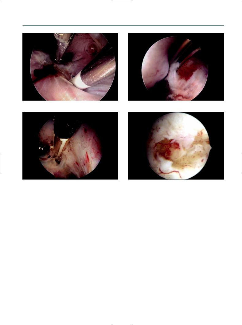
About 50–60% of the pathologic findings at TVL are due to the presence of superficial endometriosis.3 These lesions are mostly associated with free-floating ovarian or peritoneal adhesions or fixed adhesions with the fossa ovarica. The findings at TVL correspond very well with the regurgitation and implantation theory of Sampson4 followed by adhesions and invagination of the ovarian cortex, as demonstrated by Hughesdon and Brosens5,6 (Figure 10.3). Therefore the endometriosis in an ovarian endometrioma is superficial, as it is lining the invaginated cortex. The access to the ovary is achieved without manipulation, since there is a direct exposure of the fossa ovarica. At this site, endometrioma are frequently adherent to the lateral pelvic wall, the uterosacral ligament. or the posterior leaf of the broad ligament.
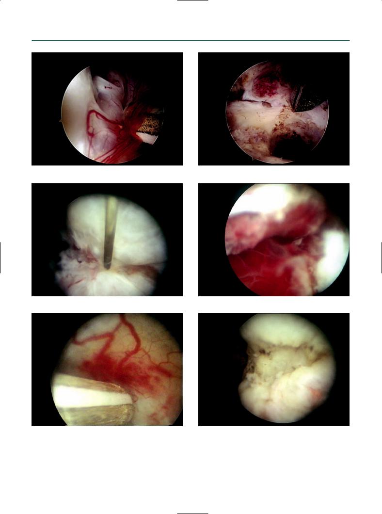
The technique for reconstructive ovarian surgery (Figure 10.4) at TVL is based upon the same principles as at standard laparoscopy (SL): the first step consists of an adhesiolysis with full mobilization of the ovary and identification of the site of inversion; at the second step, the cyst is opened at the site of inversion. Access into the endometriotic cyst at
Operative transvaginal laparoscopy |
87 |
a |
b |
c |
d |
Figure 10.3
(a) Dissection and opening of a superficial peritoneal lesion using the bipolar probe and scissors. (b) After removal of the superficial peritoneal adhesions, endometrial-like tissue can be visualized at the base of the lesion. (c) A 5 Fr bipolar probe used for coagulation and dissection. (d) Partial coagulated small endometrioma: note the invagination of the cortex, with, at the base, still the presence of some neovascularization, and the non-collapsing of the walls.
this side is essential to minimize ovarian trauma and to maintain patency of the pseudocyst. This technique differs from fenestration, as the opening of the pseudocyst is performed by adhesiolysis and resection of the fibrotic ring at the site of inversion. After aspiration and rinsing, the site of inversion is widely opened. At the third step, a superficial and abrasive coagulation is performed of the vascularized endometriotic lesions lining the wall.7 Because of the watery distention medium and in the absence of a high intraabdominal pressure, the neoangiogenesis remained visible, facilitating the identification of endometrial implants. We found no difference in recurrence rate of endometriomas between microsurgical resection of the wall and the present simplified technique of eversion or marsupialization and coagulation.8 In the case of an endometrioma >5 cm, the
treatment was performed in two steps. This is also our normal strategy in the case of a classical operative laparoscopy.9 During the first procedure, adhesiolysis with resection of the fibrotic area at the site of inversion is performed with coagulation of the visible vascularized endometriotic implants. During the second step, adhesiolysis is performed whenever necessary and the fulguration of the reduced pseudocyst is completed. Our preliminary results showed no recurrence of an endometrioma in operated patients with a follow-up of 12–36 months. After opening of the endometrioma, there is no collapse of the wall as this is formed by the rigid ovarian cortex; this is in contrast with true intraovarian cysts (Figure 10.4e).
In our series of operative TVL procedures, no conversion to laparoscopy was necessary. Compared with the
88 Atlas of Transvaginal Endoscopy
a |
b |
c |
d |
e |
f |
Figure 10.4
Technique for reconstructive ovarian surgery. Dissection of fossa ovarica using the (a) bipolar probe and (b) scissors. (c) A bipolar needle is used for incision of the endometrioma at the place of invagination. (d) After opening, endometrial-like tissue is clearly visible at the base of the pseudocyst. (e) A bipolar probe is used for coagulation. Invagination is clearly visible after coagulation with the bipolar probe. (f) After coagulation, no carbonization occurs.

Operative transvaginal laparoscopy |
89 |
morbidity after an SL, the morbidity after the transvaginal procedures was very low and most of the patients had no sensation of pain afterwards and at most complained of a light tenderness in the lower abdomen. All patients returned home the same day and resumed their full activity 1 or 2 days later. The 1-day hospitalization and the low morbidity of the procedure makes a second-step procedure also more acceptable for the patient.
The underwater inspection of small ovarian endometriotic lesions confirms the invagination theory of Hughesdon and Brosens.5,6 What initially appear as small brown superficial ovarian lesions reveal at closer inspection to be invaginated areas of the ovarian cortex covered by small adhesions. After opening and at the base of these invaginated areas, typical endometrial-like implants with their neovascularization can be identified (Figures 10.3 and 10.4).
The disadvantage that was noted during the pioneering years does not apply to the same extent today: with time, experience, and a constant effort on behalf of the manufacturing company (Karl Storz, Tüttlingen, Germany) to provide ever-better prototypes of the instruments, operative TVL now allows us to treat stage I, II, and even specific cases of stage III endometriosis of the adnexa. However, some limitations are still present:
1.Despite the availability of a very efficient bipolar coagulation probe, a problem with hemostasis, i.e. uncontrolled bleeding of blood vessels, might occur and blur the vision in such a way that it becomes impossible and dangerous to continue the procedure. The chocolate content of an endometrioma, however, is not a cause of a blurred vision, since it is washed out quite efficiently by the pressurized infusion of Ringer’s lactate.
2.The inability to expose cleavage planes between different types of tissue in the same way as with the help of an assistant or resident during operative laparoscopy via three or four suprapubic ports; here, the surgeon is alone and has to rely mostly on the visual information coming from the operating field. There is also, but to a much lesser degree, some form of tactile feedback when touching tissues. Prototypes of operating sheaths with two (instead of one) operating channels proved to be very promising since one channel can then be used to hold or fix tissue planes, while the other channel serves to operate (take a biopsy, incise, excise, coagulate, etc.)
3.The progress during operative TVL with the current instruments is still more time consuming than during a conventional operative laparoscopy; one can only proceed at the pace of millimeters rather than of centimeters.
This means that, at this moment, it is probably wiser and less time consuming, even with a vast surgical experience, to treat ovarian endometriomas of >40–50 mm in diame-
ter by conventional operative laparoscopy. Nevertheless, operative transvaginal hydrolaparoscopy is definitely the ideal tool to detect and treat the small ovarian endometrioma of ≤15 mm, meaning that in most of these instances the detection of such a small endometrioma will always be followed by its treatment during that same procedure.
Ovarian capsule drilling
The TVL is also very suitable for performing drilling of the ovarian capsule in patients with polycystic ovary syndrome (PCOS) resistant to medical therapy. One of the major concerns of laparoscopic drilling of the ovaries was the risk of postoperative adhesion formation. The CO2 pneumoperitoneum has been shown to induce acidosis and enhance adhesion formation by its deleterious effect on the oxygenation of the peritoneal mesothelial layers.10 Although further examinations are necessary to prove the diminished risk of adhesion formation if procedures are performed under water using a pre-warmed solution of Ringer’s lactate as a distention medium, data from experimental work in mice has shown that less adhesions were induced if the same surgical procedure was performed under water compared with the use of a pneumoperitoneum with CO2.11,12
After identification of the ovarian surface, drilling of the ovarian capsule is performed using a bipolar needle with a diameter of 1 mm and a length of 0.8 cm (Figure 10.5a). The needle is placed perpendicular to the ovarian surface and is gently pushed against the ovarian surface (Figure 10.5b and c). The capsule is perforated using a bipolar cutting current, allowing an easy insertion of the needle. A coagulating current is used for 5 seconds, followed by the removal of the needle. Although there are no data available with regard to the numbers of holes to be performed, our policy is to make about 10–15 small holes on each ovarian surface, preferentially on the anterolateral site (Figure 10.5d). When the current is switched on, the continuous flow of Ringer’s lactate is stopped, allowing a more accurate performance of the bipolar current and a maximal effect of energy delivery. The total procedure doesn’t last longer than 30 minutes. The small needle diameter minimizes the defect on the ovarian capsule and, in the absence of carbonization in a watery environment, risks for postoperative adhesion formation will be reduced.
Our results are in line with the results of others13,14 and are comparable with those obtained after drilling through SL.15 Restoration of monofollicular cycles, avoidance of multiple pregnancies, and the higher risks for complications are all factors in favor of a surgical treatment of PCOS. At the same time, a complete exploration of the female pelvis can be carried out. In the absence of the postoperative onset of ovulatory cycles, ovulation induction