

113
4
The Locomotor System (Musculoskeletal System)
Contents
Axes, Planes, and Orientation 114
Axes and Planes of the Body 114
Nomenclature of Positions and
Directions 114
General Anatomy of the Locomotor
System 116
The Bones 116
The Joints 117
− Immovable Joints (Synarthroses) 117
−Movable Joints (Synovial Joints, Diarthroses) 118
−Slightly Movable Joints (Amphiarthroses) 120
− |
Types of Joint |
120 |
|
|||
− |
Joint Mechanics |
122 |
|
|||
Function and Structural Principles |
||||||
of Skeletal Muscle |
123 |
|
||||
The Tendons |
127 |
|
|
|||
Auxiliary Structures of Muscles |
||||||
and Tendons |
127 |
|
|
|||
Special Anatomy of the Locomotor |
||||||
System 129 |
|
|
|
|
||
The Skeleton of the Trunk |
129 |
|||||
− |
The Spinal Column 130 |
|||||
− |
The Rib Cage (Thorax) |
138 |
||||
|
|
|
|
|
|
|
|
|
|
|
|
|
|
The Muscles of the Trunk |
140 |
||
− |
The Back 141 |
|
|
− |
The Chest Wall |
141 |
|
− |
The Abdominal Wall |
147 |
|
− |
The Diaphragm |
149 |
|
− |
The Pelvic Floor |
149 |
|
The Upper Extremity 153
−The Shoulder Girdle—Bones, Joints, Muscles 153
−The Free Upper Limb—Bones, Joints, Muscles 156
The Lower Extremity 166
−The Pelvic Girdle and the Pelvis—Bones, Joints, Muscles 166
−The Free Lower Limb—Bones, Joints, Muscles 171
Head and Neck 184 |
|
|
− |
The Neck (Collum) |
184 |
− |
The Head (Caput) |
185 |
− |
General Aspects of the Skull 186 |
|
− |
The Muscles of the Skull 193 |
|
−The Jaw Joint (Temporomandibular Joint) 194
Summary 197

114 4 The Locomotor System (Musculoskeletal System)
Axes, Planes, and Orientation
Axes and Planes of the Body
Any number of axes and planes may be drawn through the human body. It is customary, however, to define three main axes running perpendicular to each other in three spatial coordinates (Fig. 4.1):
A longitudinal axis (vertical axis, cephalocaudal axis) of the body, which in the upright posture runs perpendicular to the base
A horizontal axis (transverse axis) running from left to right and perpendicular to the longitudinal axis
A sagittal axis running from front to back and perpendicular to both the other axes
Hence it is possible to define three principal planes:
A sagittal plane, defined as any plane that is oriented along the sagittal axis (the vertical plane that divides the body into two equal halves is called the median plane)
A transverse plane, defined as any plane running transversely across the body
A frontal plane (coronal plane) that includes all planes oriented parallel to the forehead
Nomenclature of Positions and Directions
The following designations of positions and directions serve to describe accurately the positions of parts of the human body:
For the trunk
Cranial, cephalad, or superior: Toward the head
Caudad or inferior: |
Toward the coccyx (tailbone) |
Ventral or anterior: |
Toward the front (abdomen) |
Dorsal or posterior: |
Toward the rear (back) |
Medial: |
Toward the median plane |
Lateral: |
Away from the median plane |
Internal: |
Inside the body |
External: |
Outside the body |
Peripheral: |
Away from the trunk |
Axes, Planes, and Orientation 115
|
Fig. 4.1 Major axes and |
|
planes of the human body. |
Median |
Left anterior view |
(sagittal) |
|
plane |
|
Longi- |
|
tudinal |
|
axis |
|
Hori- |
|
zontal |
|
(transverse) |
|
plane |
|
Coronal |
|
plane |
|
Sagittal axis |
|
Transverse axis |
|
For the extremities |
|
Proximal: |
Toward the trunk |
Distal: |
Toward the extremities of the limbs |
Radial |
Toward the radius (thumb side) |
Ulnar: |
Toward the ulna (side of the little finger) |
Tibial: |
Toward the shin (side of the big toe) |
Fibular: |
Toward the fibula (side of the little toe) |
Palmar (volar): |
Toward the palm of the hand |
Plantar: |
Toward the sole of the foot |
Dorsal (of hand and foot): |
Toward the back of the hand or foot |
|
(upper side of foot) |

116 4 The Locomotor System (Musculoskeletal System)
General Anatomy of the Locomotor System
The skeleton, the supporting framework of the body, is formed by bony and cartilaginous elements, connected by connective tissue structures. Its parts are moved or held in defined positions or postures by the skeletal musculature. The overarching term locomotor system includes the skeleton and the musculature. The passive locomotor system consists of the skeleton and its joints (articulations), while the active motor system includes the striated muscles, the tendons, and their auxiliary structures (muscle fasciae, bursae, tendon sheaths, and sesamoid bones). Beside their supporting function, the skeletal elements and their joints serve to provide levers for the muscles during locomotion. The skeletal elements, joints, and skeletal musculature together form the organs of locomotion. In addition, the skeletal elements function to protect other organ systems (bones of the skull, vertebral canal, chest cage).
The Bones
The bony skeleton consists of bones of various structures and shapes. In the adult human, the skeleton is composed of about 200 individual bones, which are connected by cartilaginous, fibrous, and synovial joints. Each bone, with the exception of the cartilaginous joint surfaces and areas where flat tendons are attached, is enclosed in a connective tissue sheath, the periosteum, like a stocking.
The shape of each bone is determined genetically, but its structure depends largely on the type and extent of the mechanical demands placed on it. According to their external shape, bones are divided into long, short, flat, and irregular bones. Examples of long bones (pipe bones) are the bones of the free extremities, with the exception of the wrist and ankle bones. Long bones are composed of a shaft (diaphysis) and an epiphysis at each end. During growth, each diaphysis and the corresponding epiphysis are separated by the so-called epiphyseal cartilage (epiphyseal plate) (see Fig. 3.10, p. 85). The short bones include the cubeshaped bones of the wrist and ankle.
Among the flat bones are the ribs, the breast bone, the shoulder blade, and the bones of the skull. The irregular bones include the vertebrae and

General Anatomy of the Locomotor System 117
the bones at the base of the skull. Some of the bones in the skull (frontal, cribriform plate, upper jaw) contain air-filled cavities. Sesamoid bones are bones embedded in tendons (e. g., the kneecap). Finally, certain extra bones, occurring especially in the hand and foot, are called accessory bones. Their presence in a radiographic image can lead to diagnostic errors (as displaced fragment due to a fracture).
The Joints
Joints are connections between cartilaginous and/or bony parts of the skeleton. They enable movement between the individual segments of the trunk and the extremities, and transmit force. They are divided, according to the type of connection, into immovable and movable.
Immovable Joints (Synarthroses)
So-called immovable joints, or synarthroses, are those joints in which parts of the skeleton are separated by a different tissue such as cartilage or connective tissue. According to the intervening tissue these are divided into (Fig. 4.2):
Syndesmoses (fibrous joints)
Synchondroses (cartilaginous joints)
Synostoses (bony joints), which are not true joints but bony fusions
Syndesmoses. In syndesmoses, two bones are connected by connective tissue (Fig. 4.2). Examples are the interosseous membrane between the ulna and radius of the lower arm; the membranous fontanelles of the newborn skull, and the sutures between the bones of the skull. The connective tissue anchoring of the roots of teeth in the upper and lower jaw, known as a peg and socket joint (gomphosis), is also a syndesmosis.
Synchondroses. The connecting tissue in synchondroses is cartilage (Fig. 4.2). Examples are the fibrocartilaginous intervertebral disks between vertebrae or the symphysis pubis at the junction of the two pubic bones. The connection of the bony diaphysis of a juvenile long
118 4 The Locomotor System (Musculoskeletal System)
Bone
Syndesmosis (fibrous joint)
Connective tissue |
|
|
Synchondrosis |
|
(cartilaginous |
Cartilage |
joint) |
|
|
|
Synostosis |
|
(bony joint) |
Bone |
|
Fig. 4.2 Simplified representation of the different immovable joints (synarthroses)
bone to the epiphysis by its cartilaginous epiphyseal plate is also a synchondrosis.
Synostoses. In a synostosis individual bones are fused secondarily by bone tissue (Fig. 4.2). A typical example is the sacrum, which originally consists of five separate vertebrae that fuse to each other when growth is complete. Another example is the hip bone, which, until growth is completed, consists of three separate bones: the pubis, the ilium, and the ischium.
Movable Joints (Synovial Joints, Diarthroses)
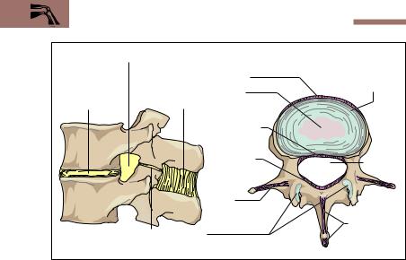
In synovial joints the bones are separated by a joint space (Fig. 4.3). They are also distinguished by hyaline cartilage covering the joint surfaces and by a joint capsule that encloses a joint cavity. Some joints feature interarticular disks (menisci), articular lips, or intra-articular ligaments. For instance, the menisci of the knee are made of fibrocartilage, are semilunar in shape, and incompletely partition the knee joint. Disks are also a feature of the mandibular joint and the sternoclavicular joint among others. The function of disks in joints is to increase the contact between two opposing surfaces.
General Anatomy of the Locomotor System 119
Cancellous bone |
Joint space |
Hyaline cartilage of joint |
Bone marrow |
|
|
|
cavity |
Compact bone |
|
|
|
|
|
Fibrous joint capsule |
|
Tendon sheath |
Joint cavity |
|
|
Long flexor tendon |
Inner joint membrane |
||
(synovial membrane) |
|||
Fig. 4.3 Structure of a movable joint as exemplified in the metatarsophalangeal joint of the big toe
Joint Cartilage. The smooth surface of joint cartilage consists mostly of hyaline cartilage (Fig. 4.3), the mechanical and “shock absorbing” properties of which are essentially due to its extracellular matrix. Important constituents of extracellular matrix are collagen fibers, macromolecules (protein saccharides), and water. The thickness of joint cartilage varies considerably. It averages 2−3 mm, but in some places (joint surface of the patella) joint cartilage can reach 8 mm. Since this cartilage does not contain blood vessels, it must receive nutrients by diffusion from the synovial fluid. Optimal nutrition requires regular movement (loading and unloading) of the cartilage, so that the synovia is pressed into the cartilage. Lack of movement and unphysiologically high tensions lead to degenerative changes (osteoarthritis) in joint cartilage, especially in older

120 4 The Locomotor System (Musculoskeletal System)
people. Because there is no perichondrium, the regenerative power of joint cartilage is insignificant (see p. 79).
Joint Capsule and Synovial Fluid. The joint capsule (Fig. 4.3) is a continuation of the periosteum. It is made up of an outer dense white fibrous layer (fibrous membrane) and an inner loosely structured membrane rich in vessels and nerves (synovial membrane), which may contain a varying amount of fat. The outer fibrous membrane is often reinforced with ligaments, which may reinforce the capsule, guide movement, or prevent hyperextension of a joint. When a joint is immobilized over a prolonged period of time, the connective tissue fibers shorten, the joint capsule shrinks, and the mobility of the joint can be severely compromised (joint contracture). From the inner synovial membrane folds and protrusions project into the joint. This membrane abuts on the joint cavity with specialized connective tissue cells that are responsible for the secretion (production) and reabsorption (resorption) of the synovial fluid. The glairy, thick (viscous) synovial fluid not only nourishes the joint cartilage, but serves as a lubricant to reduce friction between the joint surfaces.
Slightly Movable Joints (Amphiarthroses)
Some joints are severely limited in their mobility by the shape of their facets and strong ligaments. Such joints include the tibiofibular joint and the sacroiliac joint between the sacrum and the ilium.
Types of Joint
Joints may be classified from different points of view, for example, by the number of axes of mobility, of degrees of freedom, or of components of the joint. The following is a classification by shape and configuration of the joint surfaces (Fig. 4.4):
Ball-and-socket joints
Condyloid joints
Hinge joints
Pivot joints
Saddle joints
Plane joints
General Anatomy of the Locomotor System 121
Ball-and-socket joint |
Condyloid joint |
Hinge joint |
Pivot joint |
Saddle joint |
Plane joint |
Fig. 4.4 Types of joint. The arrows show the direction in which the skeletal parts can move around each axis.
Ball-and-Socket Joints (Spheroid Joints) consist of a ball-shaped head and a correspondingly concave socket. They have three main axes perpendicular to each other and allow six main movements. Typical ball- and-socket joints are the hip and shoulder joints.

122 4 The Locomotor System (Musculoskeletal System)
Condyloid Joints (Condylar Joints) have an elliptical head fitted into a convex and a concave socket. They have two main axes perpendicular to each other, and they allow four main movements. Examples are the joint between the bones of the forearm and the wrist (proximal wrist joint) and the joint between the atlas and the occipital condyles.
Hinge Joints (Ginglymus Joints) and Pivot Joints (Trochoid Joints) are also known as trochlear joints. In hinge joints, a cylindrical bone end is applied to a gutterlike depression in a hollow skeletal cylinder. Because of this shape, hinge joints have only one axis of movement and two main movements (elbow joint). In pivot joints, a cylindrical part of the skeleton is fitted into a corresponding hollow cylinder and a ring-shaped ligament. A typical example is the superior radioulnar joint and its annular ligament. Such a joint allows rotation around one axis and two main movements.
Saddle Joints consist of two concave curved surfaces with two main axes of movement that are perpendicular to each other and allow four main movements. An example is the joint in the wrist between the first metacarpal bone and the trapezium.
Plane Joints (Gliding Joints) allow gliding movements of plane joint surfaces, as in the small joints of the vertebrae.
Joint Mechanics
The direction of movement in a joint is determined not only by the shape of the joint surfaces but also by the arrangement of the muscles and ligaments. Human joints cohere by force: their integrity is ensured by muscular forces, which also determine the direction and type of their movement. The shape of the joint, the muscles, ligaments, and soft tissues limit the extent of movement. Hence, limitations may be divided into bony, muscular, ligamentous, and soft tissue types.
Joints move around movement axes: the direction of movement is determined by the relationship of the muscles to the axes. The body can be considered to have three main axes, running perpendicular to each other (p. 115). In addition there are axes relating specifically to the

General Anatomy of the Locomotor System 123
movement of each joint, named according to its movement, e. g., the pronation/supination axis of the proximal and distal radioulnar joints around which the hand may be rotated inward and outward (pronation and supination).
Two opposite movements can occur around each axis. Examples are:
Bending—extending (flexion—extension), e. g., the elbow joint
Pushing out—pulling in (abduction—adduction), e. g., the hip joint
Rolling inward—rolling outward (inner rotation—outer rotation),
e. g., the shoulder joint
Forward motion—backward motion (flexion—extension), e. g., hip joint
Opposition—reposition, e. g., the saddle joint of the thumb
The effect of a muscle on a joint depends on the lever arm, that is, the vertical distance of its insertion to the axis of its joint (force arm). Force and load are in equilibrium when force × force arm = load × load arm. The product of force with force arm and of load with load arm is called the torque (Fig. 4.5).
Function and Structural Principles of Skeletal Muscle
A skeletal muscle is divided into a fleshy, variously shaped muscle belly and the usually markedly thinner tendons. The latter are attached to structures in the skeleton or connective tissue of the locomotor system (fasciae, interosseous membrane) and transmit the muscle pull directly or indirectly to parts of the skeleton. In the extremities, the attachment nearest the trunk (proximal) is usually considered the origin and that farthest from the trunk (distal) the insertion of the muscle. In the trunk, the origin of a muscle is always cephalad. The origin and insertion of a muscle are designated arbitrarily and should not be confused with the fixed and mobile points, the latter being where the muscle is attached to the part of the skeleton that is moved, the former to that which is fixed. Although the fixed point and the origin coincide in most movements of the extremities, this is not necessarily the case, since the fixed point could be interchanged with the mobile point in the course of a movement.

124 4 The Locomotor System (Musculoskeletal System)
Head of the humerus
M. biceps brachii (two-
headed muscle of the upper arm)
Radial tuberosity (insertion of the biceps into the radius)
Radius
Load
Glenoid cavity (joint cavity of the shoulder)
M. triceps brachii
(three-headed muscle of the upper arm)
Arm bone (humerus)
Insertion of the triceps m. at the elbow end of the ulna (olecranon)
Ulna
Force
Force arm
Load arm |
Fulcrum |
|
Fig. 4.5 Effect of the flexors and extensors of the upper arm on forearm movement.
Flexor at the elbow joint: m. biceps brachii (two-headed muscle of the upper arm). Extensor at the elbow joint: m. triceps brachii (three-headed muscle of the upper arm). The mechanics of muscle power are shown: load × load lever = force × force lever (the product of force × force lever and load × load lever constitute the current torque).

General Anatomy of the Locomotor System 125
At the origin of a muscle there is often a head (caput) that transitions into a belly (venter). A muscle with several origins may be two-, three-, or four-headed, all joining to form a single belly and ending in a single tendon. A muscle with a single head but one intersecting tendon is called a digastric muscle. A muscle may have several such intervening tendons (Fig. 4.6). Muscles that extend over two or more joints are called diarthric or polyarthric (multiarticular), respectively. Muscles that work together in one movement are synergists, while those with opposing actions are antagonists.
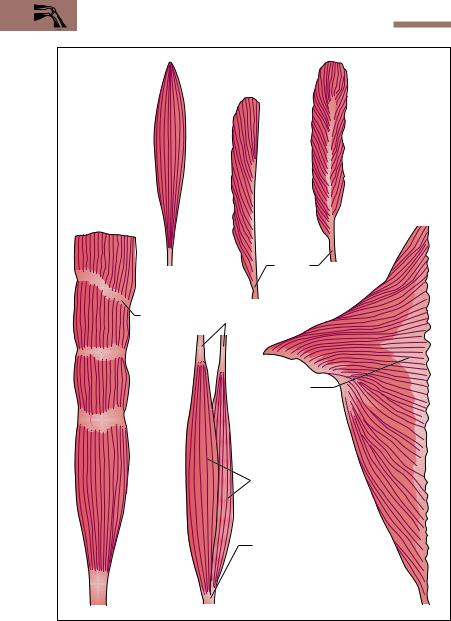
Muscles are also classified according to the way the muscle fibers are inserted into the tendons (e. g., pennate muscles from penna = feather) (Fig. 4.6). A muscle with parallel fibers can achieve considerable height of lift with relatively little force, but because of the small total cross-section of its muscle fibers (physiological cross-section), their lifting strength is rather small. The fibers of a unipennate muscle are inserted on only one side of the tendon of origin and insertion. This results in a large physiological cross-section and considerable muscular strength. Because the fibers of such a muscle are short, the height of lift is small. In a bipennate muscle the muscle fibers originate from a bifurcated tendon and run alongside both sides of the tendon of insertion. The physiological crosssection and so its ability to develop power is here even greater than in a unipennate muscle.
The fine structure of a muscle is determined not only by its striated muscle fibers but also by its connective tissue structures, which form the boundaries between the individual components of each skeletal muscle and are the conduit for the vessels and nerves supplying the muscle fibers (see Fig. 3.12a−e). Loose alveolar tissue in the form of endomysium forms a sheath around the individual fibers, which in their turn are grouped together by denser white connective tissue (perimysium). Several primary bundles are pulled together by another strong connective tissue sheath, the epimysium, into the secondary bundles (fleshy fibers) that are visible to the naked eye. A stout connective tissue sheath, the muscle fascia, envelops the whole muscle. Loose areolar tissue (epimysium) separates the muscle from the fascia. Several individual muscles can be enclosed by a common fascia.
126 4 The Locomotor System (Musculoskeletal System)
Unipennate muscle (e.g., m. semi-
Muscle with parallel membranosus) fibers (e.g., m.
palmaris longus)
Bipennate muscle (e.g., m. tibialis anterior)
Insertion ends of tendons
Tendinous |
Tendon of origin |
intersection |
|
Aponeurosis  (tendon sheet)
Muscle bellies
|
Insertion end of tendon |
|
Muscle with |
Two-headed |
|
several bellies |
Flat muscle |
|
(e.g., m. rectus |
muscle (e.g., m. |
|
abdominis) |
biceps brachii) |
(e.g., m. trapezius) |
Fig. 4.6 Various types of muscle. (After Platzer)

General Anatomy of the Locomotor System 127
The Tendons
The tendons attaching the muscles to the bones are composed of bundles of collagen fibers of great tensile strength. During muscle contractions they transfer force from the muscle to the skeleton. The attachments of the tendons to bone (tendon attachments) are of considerable functional significance, since the elasticity of the tendons at these points must be adapted to that of the bones. Tendons are classified according to their shape. When the tendons are very short and invisible to the naked eye, the muscle is said to have a fleshy muscular attachment, as, for example, in the case of the pectoralis major muscle. By contrast, the tendons of the muscles of the foot and hand are very long and thin. Flat tendon sheets (Fig. 4.6), such as those of the oblique abdominal muscles, are called aponeuroses. Tendons may run in the same direction as the muscles and act directly on the bones (sometimes called lever tendons), while others change their direction by running around a bone, which exerts pressure on them as they run over it. Because the bone acts as a pulley for these tendons, they are sometimes called pulley tendons and the bone is known as the fulcrum. An example of this is the tendon of insertion of the peroneus longus muscle, which runs around the side of the cuboid bone toward its insertion on the sole of the foot.
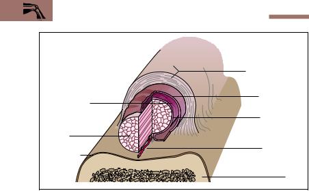
Auxiliary Structures of Muscles and Tendons
The function of muscle fasciae, tendon sheaths, bursae, and sesamoid bones is to reduce friction during muscular work, so that there is a minimum of reduction in force. Fasciae allow individual muscles or muscle groups to glide over each other. If tendons run next to a bone or run over a bony protrusion, they are protected by guiding channels that improve their ability to glide. The structure of the wall of such a synovial tendon sheath (vagina synovialis) is similar to that of a joint capsule and the fluid in the cavity around which the inner and outer synovial layers glide is similar to synovial fluid (Fig. 4.7). Where a muscle runs directly over a bone, it is protected by a synovial bursa, which is also filled with synovial fluid and which acts like a water cushion, distributing pressure evenly (Fig. 4.8). Bursae occur most often at the origins and insertions of
128 4 The Locomotor System (Musculoskeletal System)
|
Outer fibrous layer |
|
|
Outer fibrous layer |
|
|
(fibrous |
|
|
(fibrous membrane) |
|
|
Outer synovial |
|
Synovial cavity |
Outer synovial |
|
layer |
||
Synovial cavity |
||
|
layer |
|
|
Inner synovial |
|
|
Inner synovial |
|
|
layer |
|
|
layer |
|
Tendon |
|
|
Tendon |
|
|
|
Vascular fold |
|
Periosteum |
Vascular fold |
|
(mesotendon) |
||
|
Bone |
|
|
Bone |
Fig. 4.7 Structure of a tendon sheath (vagina synovialis tendinis). (After Frick)
The inner layer of the synovial membrane is firmly adherent to the tendon, the outer layer to the fibrous inner membrane of the tendon sheath. The cavity around which the two layers glide is filled with synovial fluid. The whole structure reduces friction over bone.
muscles, but they may also be found near joints. Occasionally they are continuous with the joint capsule, and are then considered extensions or recesses of the capsule. Sesamoid bones (ossa sesamoidea)are bones embedded in tendons. Their functional significance is that they extend the effective lever arm of a muscle and so save muscular effort. The largest sesamoid bone in humans is the kneecap (patella). Tendon sheaths and bursae can become inflamed by chronic irritation (tenosynovitis and bursitis).

Special Anatomy of the Locomotor System 129
Subcutaneous bursa |
Clavicle |
Acromion |
|
Subacromial bursa (bursa |
|
under acromion) |
|
Subdeltoid bursa (bursa |
|
under the deltoid |
|
muscle) |
|
|
1st rib |
Subtendinous bursa of m. |
|
subscapularis |
|
Bursa of m. pectoralis major |
|
Deltoid muscle |
|
M. teres major |
|
Latissimus dorsi |
|
muscle |
|
Upper arm bone (humerus) |
|
Fig. 4.8 Synovial bursae of the shoulder girdle. Right shoulder seen from in front. The muscles have been partially removed.
Special Anatomy of the Locomotor System
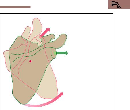
The fold-out at the end of the book shows the human skeleton in different views.
The Skeleton of the Trunk
With the transition to upright posture, the shape of the human body changed considerably. The trunk, rising vertically over the lower limbs, supports the head and the upper limbs. In this way, the lower limbs become the organs of locomotion, while the upper limbs become impor-

130 4 The Locomotor System (Musculoskeletal System)
tant “tools” for grasping and feeling. As the trunk becomes erect, the vertebral column develops the curvatures typical for the human body and the hipbones widen and combine with the sacrum to form the pelvis as a solid part of the trunk (Fig. 4.9).
The skeleton of the trunk includes the spinal column (vertebral column) and the chest cage (thorax), which includes the ribs (costae), the breastbone (sternum), and the thoracic vertebrae (Fig. 4.9). The spinal column contains the spinal canal (vertebral canal) with the spinal cord. The ribs run forward (ventrally) from the spinal column and form the bony structure of the chest cavity (thoracic cavity), which below is continuous with the abdominal cavity (see p. 349). The thorax and abdomen are enclosed by the bones and muscles of the trunk and abdominal wall. Anatomically, the wall of the trunk can be divided into ventral, lateral, and dorsal thoracic and abdominal walls. The back is that part of the dorsal wall that extends from the base of the skull to the tip of the coccyx. The lower part of the trunk, made up of parts of the abdominal wall and of the lower extremities, can be considered separately as the pelvic girdle. While the diaphragm divides chest and abdominal cavities, the muscles of the pelvic floor separate the abdomen from lower structures.
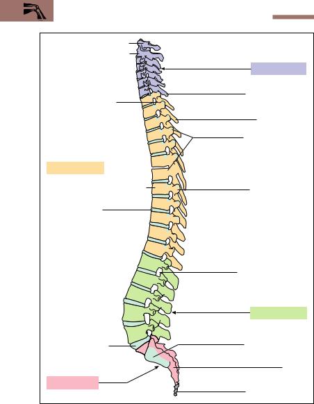
The Spinal Column
The spinal column forms the axis of the human skeleton (Fig. 4.10). It consists of 33−34 vertebrae, the intervertebral disks, and a number of ligaments. The vertebrae consist of 7 cervical vertebrae (neck), 12 thoracic vertebrae (chest), 5 lumbar vertebrae (loins), 5 sacral vertebrae (sacrum) and 4−5 coccygeal vertebrae (coccyx). The indivicual vertebrae are often designated by a shorthand notation as follows: “C5” for the fifth cervical vertebra (for example), and similarly T2, L1, S3, Co1, etc. The 24 vertebrae above the sacrum are called true or presacral vertebrae, forming the true spinal column In the adult they attain a length of 55−63 cm (ca. 35 % of the body’s height). The sacral and coccygeal vertebrae are fused and form the sacrum and the coccyx (tailbone).
In the erect posture, the spinal column in the adult forms a double-S curvature in the sagittal plane, with two anteriorly oriented convexities (cervical and lumbar lordoses) and two anteriorly oriented concavities (thoracic and sacral kyphoses) (Fig. 4.10). This arrangement creates a flexible rod with elastic springiness that is especially fitted to cushion axial
Special Anatomy of the Locomotor System 131
Head (caput) |
|
Shoulder girdle with |
|
clavicle and |
|
shoulder blade (scapula) |
Cervical spine |
|
|
|
Chest cage (thorax) |
Shoulder joint |
with ribs (costae) and |
|
breastbone (sternum) |
Upper arm (brachium) |
|
|
Thoracic spine |
Elbow joint |
Lumbar spine |
|
|
Forearm |
Hip bone |
(os coxae) |
|
(antebrachium) |
Sacrum |
|
|
Hip joint |
(Os sacrum) |
Wrist joint |
|
Hand (manus) |
|
|
Symphysis pubis |
Thigh bone (femur) |
|
Knee joint |
|
Lower leg (crus) |
|
Ankle joint |
|
Foot (pes) |
|
Fig. 4.9 Overview of the human skeleton side by side with the surface of the human body. Cartilaginous parts are shown in light blue
132 4 The Locomotor System (Musculoskeletal System)
Atlas (1st cervical vertebra)
Axis (2nd cervical vertebra) 

Vertebral synovial joint (zygapophyseal joint)
Thoracic kyphosis 


Vertebral body (corpus vertebri)
Intervertebral disk
Disk between 5th lumbar vertebra and
sacrum (lumbosacral disk, sacral promontory)
Sacral kyphosis
Cervical lordosis
Vertebra prominens
Spinous process
Joint surfaces for ribs (articular facets)
Transverse process
Intervertebral foramen
Lumbar lordosis
Articular facet of the iliosacral joint
Sacrum
Tailbone (os coccyx)
Fig. 4.10 Left lateral view of the spinal column

Special Anatomy of the Locomotor System 133
(vertical) loads, e. g., during running or jumping. The degree of spinal curvature varies in each individual. Lateral curvatures are pathological and are termed scoliosis.
The structural principle of the spinal column recalls the structure of a bowstring. In the area of the trunk, the thoracic curvature represents the bow, while the abdominal muscles represent the tensed string. In the area of the cervical and lumbar lordoses, tension is achieved by the muscles and ligaments of the back. These tension systems may become unbalanced; for example, if the abdominal muscles are poorly developed, the lumbar lordosis can become exaggerated (hyperlordosis).
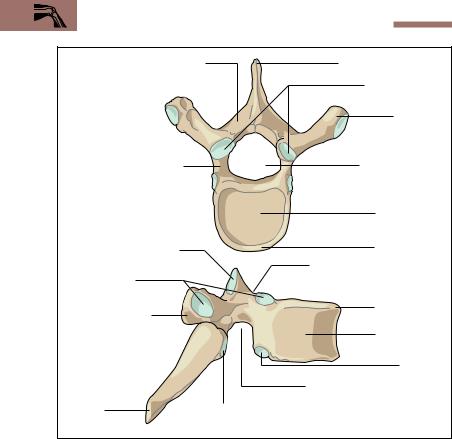
Structure of a Vertebra
Vertebrae have a common basic shape that changes in the different segments of the spinal column to adapt to their various static needs. Every vertebra—with the exception of the first cervical (atlas, C1)—has a body (corpus vertebrae), an arch, a spine, two transverse processes, and four articular facets (Fig. 4.11a, b). The vertebral body and arch enclose the vertebral foramen. All the vertebral foramina together form the vertebral canal, which houses the spinal cord. Corresponding to the increasing load, the size of the vertebrae increases from above down. The body and transverse processes of the thoracic vertebrae bear joint facets for the ribs. Every vertebral arch at its origin from the vertebral body is marked above and below by a notch (incisura vertebralis, inferior and superior vertebral notch). The notches of two adjoining vertebrae form the intervertebral foramen (Fig. 4.10), which transmits the spinal nerves.
Atlas and Axis: Atlanto-Occipital and Atlanto-Axial Joints
The first and second cervical vertebrae have special functions (Fig. 4.12). The atlas, which supports the head, has no vertebral body and is shaped like a ring. The two upper joint facets form the atlanto-occipital joint with the joint facets (condyles) of the occiput. This is a condylar joint and allows lateral inclination as well as forward and backward movement. The body of the axis carries on its upper surface a process shaped like a tooth (dens axis) that has a joint surface anteriorly. This joint connects
134 4 The Locomotor System (Musculoskeletal System)
Vertebral arch (arcus vertebrae) |
Spinous process (spine) |
|
Superior articular |
|
processes |
|
Transverse |
|
process |
Pedicle and location of the |
Vertebral foramen |
intervertebral foramen |
|
|
Vertebral body |
a |
|
Superior articular process
Articular facets for 6th rib
Transverse process
b |
|
Spinous |
Inferior articular process |
process (spine) |
|
Epiphyseal ring
Inferior vertebral notch for the intervertebral foramen
 Epiphyseal ring
Vertebral body
Facet for head of 7th rib
Superior vertebral notch for the intervertebral foramen
Fig. 4.11 Sixth thoracic vertebra as an example of the basic shape of a vertebra a Viewed from above; b viewed from the right. (After Frick)
Cartilaginous articular facets are shown in light blue
the axis with the atlas. Together the atlas and axis form the atlanto-axial joint, which allows the head to rotate in both directions (total extent of rotation about 50°). The transverse processes of the cervical vertebrae each enclose a hole (foramen transversarium) through which the vertebral artery runs toward the head on both sides. The 7th cervical vertebra has a particularly large spinous process (vertebra prominens), which is the first one from above palpable and visible through the skin.

Special Anatomy of the Locomotor System 135
|
Transverse ligament |
|
|
Articular facet |
of atlas |
Odontoid process of axis |
|
|
Anterior arch of |
||
for the occiput |
|
||
|
atlas |
||
Atlas |
|
||
|
|
||
(1st cervical vertebra) |
Transverse |
||
|
|
process |
|
Articular facet |
|
Holes in the trans- |
|
between atlas and axis |
|||
verse processes for |
|||
|
|
||
Axis |
|
the vertebralartery |
|
|
(foramen transversarium) |
||
(2nd cervical vertebra) |
|||
|
|||
Vertebral |
|
Posterior arch of atlas |
|
body of axis |
|
||
Spinous process (spine) |
|
||
|
|
||
Fig. 4.12 Posterior view of the 1st and 2nd cervical vertebrae (atlas and axis). The two vertebrae have been slightly separated to provide a better view. The arrows show the major directions of motion
Joints and Ligaments of the Vertebral Column
The spinal column is composed of independently mobile segments that are connected with each other by mobile and immobile joints. An independently mobile segment is a functional unit. It consists of the bones of two neighboring vertebrae with their connecting disk, the synovial vertebral joints of the vertebral arch (zygapophyseal joints, juncturae zygapophysiales), the ligaments, and the corresponding muscles (Fig. 4.13a). The intervertebral disk is of pivotal importance in the independently mobile segment. It is composed of an outer dense fibrous tissue ring (annulus fibrosus) and a central gelatinous core (nucleus pulposus) (Fig. 4.13b). The disks are attached to the neighboring vertebral bodies by synchondroses, and their position is further secured by the anterior and posterior longitudinal ligaments. Additionally, the spines, the transverse processes, and the vertebral arches are connected to each other by a strong ligamentous system. The articular facets of the articular processes (see Figs. 4.10 and 4.13a, b) are flat and are classified as synovial joints. The variations in the directions of their facets determine the mobility of each vertebral segment.
136 4 The Locomotor System (Musculoskeletal System)
Intervertebral foramen |
Anterior longitudinal |
Outer fibrous |
||
|
|
|
||
|
|
|
ligament |
ring (annulus |
|
|
Interspinous |
Nucleus |
fibrosus) |
|
|
|
||
Intervertebral |
ligament |
pulposus |
|
|
disk |
|
|
Posterior |
|
|
|
|
|
|
|
|
|
longitudinal |
|
|
|
|
ligament |
|
|
|
|
Ligamenta |
Vertebral |
|
|
|
flava |
foramen |
|
|
|
Inter- |
|
|
|
|
tranverse |
|
|
|
|
ligaments |
Interspinous |
|
|
|
|
|
|
|
Synovial vertebral joint |
|
ligaments |
a |
|
of the vertebral arch |
b |
|
(articulatio zygapophysialis) |
|
|||
Fig. 4.13 a, |
b Independently mobile |
segments and ligaments |
of the vertebral |
|
column
aLateral view of an independently mobile segment of the lumbar region. The parts of the mobile segment are highlighted in color (muscles and ligaments are shown in part only)
bLumbar vertebra with disk seen from above. The course of the individual ligaments is shown in red
Function of the Intervertebral Disk. The function of intervertebral disks can be compared to the function of automobile shock absorbers. When a load is imposed on the disks (when erect), they are pressed down; during prolonged relief from load (when recumbent), they again assume their original shape. They resemble a “water pillow”, distributing a central load evenly from the nucleus pulposus to the adjoining annulus fibrosus. In a ruptured disk the annulus fibrosus ruptures, and parts of the nucleus pulposus protrude. Compression of an emerging spinal nerve by the protruded tissue can lead to pain or weakness, e. g., of the lower extremity.
Special Anatomy of the Locomotor System 137
0° |
3 |
0–40 |
|
|
|
|
|
0° |
|
|
|
|
|
° |
|||
|
|
|
|
|
|
|||
|
|
° |
|
|
|
5 |
|
|
|
|
|
|
|
3 |
|
|
|
|
|
|
|
– |
|
|
|
|
|
|
|
|
0 |
|
|
|
|
|
|
|
|
3 |
|
|
|
|
|
|
|
|
|
|
|
|
9 |
|
|
|
|
|
|
|
|
0 |
|
|
|
|
|
|
|
|
– |
|
|
|
|
|
|
|
|
1 |
|
|
|
|
|
|
|
|
0 |
|
|
|
|
|
|
|
|
0 |
|
|
|
|
|
|
|
|
° |
|
|
30° |
|
|
|
|
|
|
|
|
0° |
|
|
|
|
|
|
|
|
30° |
|
|
|
|
|
|
Lateral flexion |
Rotation |
|
|
Flexion and extension |
||||
Fig. 4.14 Mobility of the vertebral column. The extent of mobility from zero position (0°) is given in degrees
Movements of the Vertebral Column
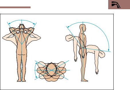
The free mobility of the vertebral column results from the sum of individual movements in several independent regions. The degree of mobility differs in the various segments. The following main movements are observed:
Forward and backward bending (flexion and extension) in the sagittal plane
Lateral bending (lateral flexion) in the coronal plane
Rotation (torsion) around a vertical axis
The cervical spine is the most mobile. The thoracic segment of the spinal column allows primarily rotation, while in the lumbar region the main possible movements are flexion and extension (Fig. 4.14). The degree of movement depends largely on the extensibility of the muscles, the ligaments and also body build.
138 4 The Locomotor System (Musculoskeletal System)
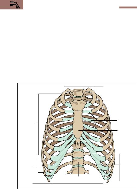
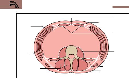
The Rib Cage (Thorax)
The bones of the thorax enclose the thoracic cavity, which has an inlet and an outlet (apertura superior thoracis, apertura inferior thoracis). The thorax protects the organs of the thoracic cavity and is formed by the breastbone (sternum), the ribs (costae), and the thoracic spine (Fig. 4.15). The sternum is a flat bone and consists of the manubrium (handle), body (corpus) and a variously shaped xiphoid process (Fig. 4.16). Normally the thoracic cage is composed of 12 pairs of ribs, of which the first seven pairs, also called true ribs, reach the sternum. Of the remaining five pairs of ribs, the 8th, 9th, and 10th are a part of the structure of the costal margin. The remaining two pairs of ribs (floating ribs) usually end free in the
1st to 7th ribs (true ribs)
8th, 9th, and 10th ribs
Thoracic outlet (apertura thoracis inferior)
Thoracic inlet (apertura thoracis superior)
Articular facet for the left clavicle
Rib cartilage
Breastbone
(sternum)
12th thoracic vertebra |
11th and 12th ribs
Fig. 4.15 Rip cage (thorax) seen from in front. (After Feneis)

Special Anatomy of the Locomotor System 139
lateral abdominal muscles. The bony parts of the ribs are joined to the sternum by their cartilaginous parts, the rib cartilages, in a synchondrosis. The cartilages of the 1st to 7th ribs generally articulate with the body of the sternum by synovial joints (Fig. 4.16).
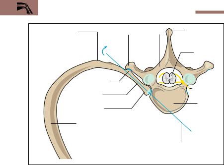
Each rib is divided into a head (caput costae), a neck (collum costae), and a body (corpus costae). Between the body and the neck of a rib there is a small process, the tubercle of the rib (tuberculum costae), where the rib is angled sharply forward (Fig. 4.17). The ribs form the costo-vertebral joints with the vertebrae. With the exception of the 11th and 12th ribs all ribs have two articular facets, one on the tubercle, the other on the head of the rib. These facets join the ribs with the transverse processes and the vertebral body (Fig. 4.17). The movements of both joints must work in tandem.
|
Clavicle |
|
|
Sternoclavicular |
|
|
joint |
|
External |
Manubrium |
|
(manubrium sterni) |
||
intercostal |
||
|
||
muscles |
Sternal angle |
|
|
Joint cavity |
|
|
Body of sternum |
|
Internal |
(corpus sterni) |
|
Rib cartilage |
||
intercostal |
||
Bony portion |
||
muscles |
||
|
of rib |
|
|
Xiphoid process |
|
|
of sternum |
Fig. 4.16 Sternum with 1st to 7th ribs and left sternoclavicular joint seen from in front. The 1st to 7th ribs are connected to the sternum by synovial joints (double blue line). The intercostal muscles are illustrated on the right side
140 4 The Locomotor System (Musculoskeletal System)
Costal angle |
Transverse Vertebral arch |
Vertebral spine |
|
process |
|
|
|
Spinal cord |
|
of rib |
|
|
costae) |
Spinal nerve |
|
|
|
|
rib |
|
|
costae) |
Vertebral |
|
|
|
|
rib |
body |
|
costae) |
|
|
rib |
|
|
costae) |
|
|
|
Axis of motion in |
|
|
costovertebral joints |
Fig. 4.17 Thoracic vertebra and costovertebral joints of a right rib seen from above.
Joint facets are shown as blue double lines
The ribs are moved by the internal and external intercostal muscles located in the intercostal spaces between the ribs (Fig. 4.16). They serve respiration by expanding and contracting the thorax. Additional muscles, the auxiliary respiratory muscles, take part in the movements of the thorax.
The cartilaginous portion of the ribs can lose its elasticity even early in life through calcification, thus restricting the mobility of the thorax. The sternum is situated directly under the skin and, being a flat bone, contains red marrow, which is thus accessible to sternal puncture for diagnostic purposes.
The Muscles of the Trunk
The movements of the trunk are effected by large muscle groups that act primarily on the vertebral column. The chest and back especially are also
Special Anatomy of the Locomotor System 141
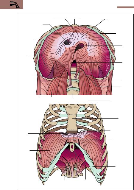
covered by muscles of the shoulder girdle and the upper extremity in addition to the true muscles of the trunk (Figs. 4.18a, b and 4.19). These gradually extended their origins to the trunk in the course of phylogenetic development. The muscles of the trunk are arranged in individual segments like the skeleton. With the exception of a few muscles (e. g., the intercostal muscles), the segments are often not maintained but rather fuse with neighboring segments into larger muscles. The musculature of the trunk is divided into the muscles of the back, the chest, and the abdomen together with the diaphragm and the pelvic muscles (Figs. 4.18a, b and 4.19).
The Back
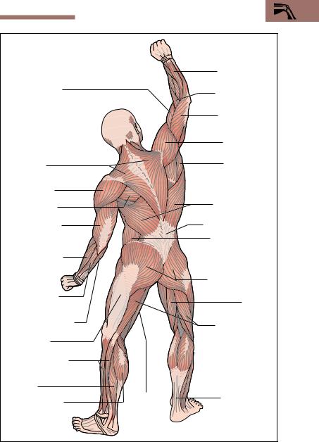
The muscles of the back proper are applied directly to the skeleton and run in two great muscular columns on each side of the vertebral column from the neck to the pelvis. This group of muscles has the collective name erector spinae (see Fig. 4.21). It is the actual segmental musculature of the back and it may be contrasted with the muscles covering it, which encroach on the back from the upper limb and are usually classified with the muscles of the upper extremity. These encroaching muscles are the muscles of the shoulder girdle, including the trapezius, the greater and lesser rhomboid muscles (rhomboideus major and minor), the levator scapulae (elevator of the shoulder), and one of the muscles of the free upper extremity, the latissimus dorsi (broad muscle of the back) (Figs. 4.18a, b and 4.19). The trapezius muscle originates from the occipital bone and the spines of the vertebrae. It is inserted into the clavicle, the acromion, and the spine of the scapula, and is one of the most important muscles in the movements of the shoulder blade. The latissimus dorsi muscle takes its origin chiefly from the spinous processes and the iliac crest, and is inserted below the lesser tubercle of the humerus. Its chief action is on the shoulder joint, in that it pulls the arm into the body (adduction), pulls it backward (extension), and rotates it inward (medial rotation).
The Chest Wall
The muscles of the chest wall are arranged in three layers. According to the course and position of the muscles, they are divided into external, in-

142 4 The Locomotor System (Musculoskeletal System)
ternal, and innermost (mm. intercostales externi, interni, and intimi) (see Fig. 4.16). They are the actual muscles of respiration and are responsible for the movement of the chest wall in inspiration and expiration. They, too, are covered by the superficial muscles. The scalene muscles continue the intercostal muscles cephalad. They originate from the cervical vertebrae and are inserted into the first three ribs. They are the most important muscles in quiet respiration, as they lift the thorax. They are completely covered by other muscles, e. g., the pectoralis major and the serratus anterior (Fig. 4.18b).
Another muscle encroaching from the upper limb is the serratus anterior (Fig. 4.18b), the serrations of which originate from the 1st to the 9th ribs, and which is inserted into the medial border of the scapula. It pulls the scapula forward, and its inferior portion (pars inferior) can rotate the inferior angle of the scapula forward (Fig. 4.20). This movement allows the arm to be elevated above the horizontal (elevation). When the shoulder girdle is fixed, the serratus anterior can lift the ribs, and so become an auxiliary muscle of respiration.
The pectoralis major is another muscle encroaching on the trunk from the upper limb. It is a muscle of the shoulder and originates chiefly from the clavicle, the sternum, and the ribs. It is inserted into the greater tubercle of the humerus (Figs. 4.18b and 4.19). It is a strong muscle, used to adduct the arm and to rotate it medially. It, too, can function as an auxiliary muscle in respiration by lifting the thorax when the shoulder girdle is fixed.
Another muscle that can lift the thorax is the sternomastoid (sternocleidomastoid). Like the trapezius, it is a muscle encroaching on the trunk. It originates from two heads attached to the clavicle and the sternum, and is inserted into the mastoid process of the occipital bone. The sternomastoid muscle acts principally on the joints of the head and the cervical vertebrae. When both sternomastoids contract, the head is thrown back toward the back of the neck; with unilateral contraction, the muscle inclines the head toward the same side and turns it to the opposite side. When the head is fixed, the sternomastoid is also a very important auxiliary respiratory muscle.
Fig. 4.18 a, b View of the superficial musculature from the back and side (a) and from the front and side (b). From a “Somso” cast
Special Anatomy of the Locomotor System 143
M. biceps brachii.
(biceps muscle of the arm; two heads)
M. trapezius
Deltoid muscle
M. infraspinatus (infraspinous muscle)
M. triceps brachii (triceps muscle of the arm; three heads)
M. brachioradialis
Mm. extensores carpi radialis longus and brevis (short and long extensor muscles of the wrist
M. flexor carpi ulnaris
(ulnar flexor muscle of wrist)
Iliotibial tract
Long and short peroneal mmuscles. (mm. fibulares longus and brevis)
M. soleus
M. gastrocnemius
M. extensor digitorum (long extensor of the fingers)
M. brachioradialis
M. triceps brachii
Deltoid muscle
M. teres major
M. latissimus dorsi
Thoracolumbar fascia
External abdominal oblique muscle
M. gluteus maximus
M. semimembranosus
M. biceps femoris
M. Semi- |
Achilles tendon |
tendinosus |
(combined |
|
tendon of |
|
soleus and mm. |
|
gastrocnemius) |
144 4 The Locomotor System (Musculoskeletal System)
|
Muscles of the thenar eminence |
M. flexor carpi radialis |
M. brachioradialis |
|
|
M. palmaris longus |
|
|
M. biceps brachii |
M. pronator teres |
Muscles of facial expression |
|
|
M. triceps brachii |
|
M. teres major |
M. latissimus dorsi |
M. serratus anterior |
M. masseter
Sternomastoid muscle
(sternocleidomastoid muscle) M. trapezius
M. pectoralis major |
Deltoid muscle |
|
|
||
External abdominal oblique |
|
|
muscle (m. obliquus |
M. biceps brachii |
|
abdominis externus) |
|
|
Rectus sheath |
M. brachialis |
|
M. pronator teres |
||
M. rectus abdominis |
||
|
||
|
M. brachioradialis |
|
M. rectus femoris |
M. flexor carpi |
|
|
radialis |
|
M. quadriceps |
Mm. extensor carpi |
|
femoris |
radialis longus and |
|
|
brevis |
|
M. semitendinosus |
|
|
M. gracilis |
M. tensor fasciae latae |
|
|
M. adductor longus |
|
M. tibialis |
M. quadriceps femoris |
|
anterior |
||
(M. sartorius) |
||
M. extensor |
||
M. triceps surae |
||
hallucis longus |
||
(extensor muscle |
(mm. gastrocnemius |
|
of the big toe) |
and soleus) |
Special Anatomy of the Locomotor System 145
Deltoid muscle
M. pectoralis major
M. iliopsoas
(mm. iliacus and psoas major)
Mm. soleus and gastrocnemius (M. triceps surae)
M. latissimus dorsi
M. triceps brachii
M. rectus abdominis
M. deltoid
Muscles of the back of the neck
M. triceps brachii
Segmental back muscles (erector spinae muscles)
M. gluteus maximus
M. triceps brachii
Extensor muscles (extensors) of the forearm
M. quadriceps femoris
Extensor muscles (extensors)of the forearm
M. triceps brachii
Extensor muscles (extensors) of the leg
M.quadriceps femoris
M. iliopsoas (mm. iliacus and
psoas major)
M. quadriceps femoris
Extensor muscles (extensors) of the leg
Extensor muscles (extensors) of the |
forearm |
Mm. soleus and gastro- |
cnemius (M. triceps surae) |
M. quadriceps femoris
Mm. soleus and gastrocnemius (M. triceps surae)
Segmental back muscles (erector spinae muscles)
M. latissimus dorsi
M. gluteus maximus
Fig. 4.19 Muscle manikins. To illustrate how the action of certain muscles results from their origin, insertion, and course
146 4 The Locomotor System (Musculoskeletal System)
Extensor muscles (extensors) of the forearm
M. triceps brachii
Deltoid muscle
Inferior portion of m. serratus anterior
Segmental back muscles (erector spinae muscles)
M. gluteus maximus
Mm. soleus and gastrocnemius (M. triceps surae)
Mm. gluteus medius and gluteus minimus
M. gluteus maximus
Mm. soleus and gastrocnemius (M. triceps surae)
M. biceps brachii
M. brachialis
(flexor muscle of the arm)
Mm. soleus and gastrocnemius (M. triceps surae)
M. quadriceps femoris
Segmental back muscles (erector spinae muscles)
Inferior portion of m. serratus anterior
Muscles of the back of the neck
M. triceps brachii
Extensor muscles (extensors) of the forearm
M. iliopsoas (mm. iliacus and psoas major)
Adductor muscles
Forearm flexor muscles
M. trapezius
M. latissimus dorsi
Adductor muscles
Fig. 4.19 (continued)
Special Anatomy of the Locomotor System 147
Elevation of the arm above the horizontal by rotating the joint surface upward and forward
Normal position of the scapula. The joint surface is oriented forward and to the side
Rotation
Action of the lower portion of the serratus anterior (serrations originating from the 5th to 9th ribs): the lower angle of the scapula is rotated forward
Fig. 4.20 Action of the inferior part of the serratus anterior in lifting the upper arm above 90° (elevation). The normal position of the scapula and its joint cavity is edged in green; the rotated scapula during elevation is edged in red. (After Faller)
The Abdominal Wall
The muscles of the abdominal wall are divided according to their location into straight (anterior), oblique (lateral) and deep abdominal muscles. The oblique muscles are arranged in three layers and their aponeuroses (flat tendons) anteriorly form a sheath (rectus sheath) that encloses the rectus abdominis (straight muscle) and serves it as a guiding channel (Figs. 4.18b, 4.21). The rectus abdominis has several tendons separating multiple bellies and runs from the superior border of the pubis, lateral to the symphysis, upward to the insertion of the ribs into the sternum. Of the oblique muscles, the external oblique runs superficially from the
148 4 The Locomotor System (Musculoskeletal System)
Lateral oblique muscles: |
|
|
|
Linea alba |
||
|
|
|
|
|
|
Posterior and anterior |
External |
|
|
|
|
|
laminae of rectus |
oblique muscle |
|
|
|
|
sheath |
|
|
|
|
|
|
|
M. rectus |
Internal |
|
|
|
|
|
abdominis |
|
|
|
|
|
|
|
oblique |
|
|
|
|
|
Deep |
muscle |
|
|
|
|
|
abdominal |
|
|
|
|
|
|
muscles: |
M. trans- |
|
|
|
|
M. psoas |
|
versus |
|
|
|
|
|
major |
|
|
|
|
|
|
M. quadratus |
|
|
|
|
|
|
lumborum |
Segmental back |
|
|
|
|
M. latissimus dorsi |
|
|
|
|
|
Deep and superficial |
||
muscles (erector |
|
|
|
|
||
spinae) |
|
|
|
|
layers of thoracolumbar fascia |
|
Fig. 4.21 |
Schematic |
cross-section |
through |
the |
trunk, |
showing the abdominal |
muscles. (After Faller)
lateral portions of the lower ribs obliquely forward to the iliac crest and to the rectus sheath. The internal oblique runs in the layer deep to this, its fibers running almost at right angles to the external oblique. Its aponeurosis also fuses with the rectus sheath. The innermost layer is formed by the transversus abdominis. The psoas major and the quadratus lumborum muscles, together with the lumbar spine, form the posterior abdominal wall (Fig. 4.21).
The muscles of the abdominal wall, together with their aponeuroses, form a functional entity, in that they exert their pull in different directions, forming an oblique support. The arrangement of the abdominal musculature allows rotation, flexion, and lateral inclination of the trunk. At the same time, the abdominal muscles act to exert pressure on the abdominal contents, and so assist the emptying of the urinary bladder and the rectum. They also take part in expiration by raising the diaphragm.

Special Anatomy of the Locomotor System 149
The Diaphragm
The dome-shaped diaphragm is the most important muscle of respiration and separates the abdominal from the thoracic cavity in the form of a musculotendinous partition. The muscle arises in a ring from the inferior thoracic outlet (Fig. 4.22). Its muscle fibers arch upward and radiate into a central tendon (centrum tendineum, trefoil tendon). According to its various origins, the diaphragm is divided into a costal part (pars costalis), a lumbar (vertebral) part (pars lumbalis), and a sternal part (pars sternalis). The inferior vena cava runs through an opening in the central tendon; the aorta and the esophagus enter the abdominal cavity from the thorax through two slitlike openings (e. g., aortic hiatus) in the lumbar part.
In life, the shape and position of the diaphragm change with breathing (Fig. 4.23), posture, and body position and the degree of fullness of the inner organs. When the diaphragm contracts, the thoracic cavity enlarges and thereby supports inspiration. In the relaxed state, with simultaneous contraction of the abdominal muscles, the diaphragm rises, which leads to expiration. Each displacement of the diaphragm has an effect on the abdominal organs. For instance, the inferior border of the liver descends with inspiration and rises again on expiration. In the erect posture, the left dome of the diaphragm projects to the upper border of the 5th rib during maximal expiration, while the right dome rises a little higher, to the level of the 4th intercostal space. During maximal inspiration the two domes descend by about 3−6 cm (Fig. 4.23).
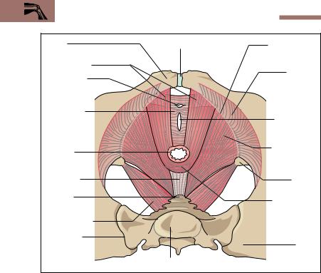
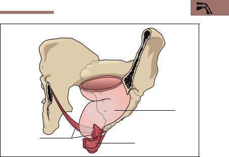
The Pelvic Floor
The pelvic outlet, and therefore the abdominal cavity, is incompletely closed by muscles and connective tissue structures. Together they form the pelvic floor, which plays a significant role in the position of the pelvic and abdominal organs. The mechanical strength of the fibromuscular pelvic floor is limited by the emergence through it of the intestinal as well as the urinary and genital tracts (Fig. 4.24).
The pelvic floor consists in part of a funnel-shaped muscle, the levator ani, which, together with its connective tissue sheath (fascia), forms the pelvic diaphragm; and in part of a muscle crossed by connective tissue bands, the deep transverse perineal muscle, which, together with
150 4 The Locomotor System (Musculoskeletal System)
Sternal part of diaphragm |
|
Xiphoid process |
Central tendon of |
Vena caval hiatus |
|
of sternum |
diaphragm |
|
|
|
|
(foramen venae |
|
|
|
cavae) |
|
|
Costal arch |
|
|
|
|
Diaphragm |
|
|
|
|
|
|
Esophageal |
Costal |
|
|
hiatus |
|
|
|
|
part of |
|
|
|
dia- |
|
|
|
phragm |
|
|
|
|
|
|
Aortic |
|
|
|
hiatus |
Verte- |
|
|
|
brocostal |
|
|
|
trigone |
|
|
12th rib |
|
|
|
Lumbar |
a |
|
|
part of |
|
|
diaphragm |
|
|
|
|
|
M. quadratus |
Lumbar spine |
M. psoas major |
|
lumborum |
|||
|
|
|
Sternum |
Vena |
|
|
Central tendon |
caval |
|
|
of diaphragm |
hiatus |
|
|
Sternal part of |
|
|
|
|
|
|
|
diaphragm |
Costal part |
|
|
|
of dia- |
|
|
|
phragm |
|
|
|
|
|
|
Costal arch |
|
Lumbar part of |
Xiphoid |
|
b |
diaphragm |
process |
|
Fig. 4.22 a, b Diaphragm, viewed from below (a) and from in front (b). Parts of the thoracic cage have been removed. (After Benninghoff)
Special Anatomy of the Locomotor System 151
|
1st rib |
Position of |
|
ribs after |
|
deep |
|
inspiration |
|
4th |
|
intercostal |
|
space |
|
Right |
5th rib |
dome of |
|
diaphragm |
|
|
Position of |
|
diaphragm |
Position of diaphragm |
after |
at maximal inspiration |
expiration |
Fig. 4.23 Position of ribs and diaphragm at maximal inspiration and expiration.
With maximal inspiration the thoracic cage widens and the diaphragm flattens. At maximal expiration the diaphragm arches back into the thorax and the thorax narrows. (After Frick et al)
its fascia, forms the urogenital diaphragm (Fig. 4.24). The levator ani muscle is attached to the internal surface of the true pelvis in a semicircular fashion and has an anteriorly directed opening, the tendinous arch of the levator ani. It transmits the urethra, the rectum, and in the female also the vagina. The tendinous arch of the levator ani is closed by the urogenital diaphragm, which extends in the shap of a trapezium between the pubic rami (Fig. 4.24).
152 4 The Locomotor System (Musculoskeletal System)
Pubis |
Symphysis pubis |
Tendinous |
|
|
arch (origin of |
M. levator ani |
|
m. levator ani) |
|
Internal |
|
Opening for |
|
|
|
obturator m. |
|
urethra |
|
|
|
(m. obturator |
|
Urogenital |
|
internus) |
|
|
|
diaphragm |
|
|
below the |
|
Opening for |
levator hiatus |
|
vagina |
Rectum |
|
Outer part of |
|
m. levator ani |
|
|
|
|
Anococcygeal |
|
|
ligament |
|
Ischial |
|
|
spine |
Coccyx |
|
Inner part of |
|
|
|
|
|
levator ani |
M. coccygeus |
|
muscle (m. |
|
puborectalis) |
|
|
|
|
Sacroiliac |
|
|
joint |
|
Ilium |
|
Sacrum |
|
Fig. 4.24 Muscles of the female perineum from above. (After Faller)
The inner part of the levator ani (puborectalis m.), together with the external anal sphincter (sphincter ani externus m.), forms a very effective (voluntary) closing mechanism (sphincter) of the rectum (Fig. 4. 25). The sphincter urethrae muscle allows the urethra to be closed voluntarily, damming urinary flow.
If the muscles of the pelvic floor are overdistended, e. g., during parturition, the internal genital organs of a woman can prolapse. Similarly, tears in the course of parturition (perineal tears) may damage the levator ani or the sphincter ani externus to the point of impairing control of rectal content (rectal incontinence).
Special Anatomy of the Locomotor System 153
Rectum
Inner part of m.
levator ani External anal sphincter (m. sphincter ani externus)
Fig. 4.25 Position of the rectum in relation to the pelvic floor. Left anterior view. Parts of the pelvic skeleton have been removed. (After Leonhardt)
The Upper Extremity
One of the essential tasks of the two upper extremities in man is to support grasping and touching. The basis for these activities is an extensive mobility of the upper limb, giving the hand as much room to move as possible. The upper extremity includes the shoulder girdle and the free upper limb. (A complete illustration of all bones and comprehensive labeling of all structures is provided in the pull-out illustration at the end of the book.)
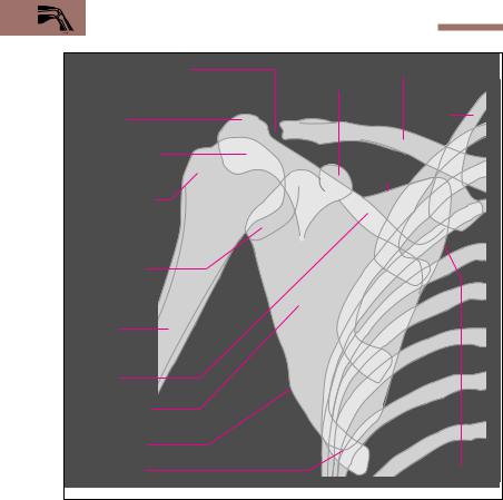
The Shoulder Girdle—Bones, Joints, Muscles
The shoulder girdle includes the clavicle and the shoulder blade (scapula). It forms the base for the upper extremity and, contrary to the pelvic girdle, is not firmly anchored to the trunk (Fig. 4.26). It is linked to the
154 4 The Locomotor System (Musculoskeletal System)
Acromioclavicular joint |
Coracoid |
Clavicle |
|
process |
|
Acromion |
|
1st rib |
|
|
|
Head of humerus |
|
Superior |
(caput humeri) |
|
|
|
|
border |
Greater tubercle |
|
|
Glenoid cavity |
|
|
Shaft of |
|
|
humerus |
|
|
Spine of |
|
|
scapula |
|
|
Shoulder blade |
|
|
(scapula) |
|
|
Lateral border |
|
|
Inferior angle |
|
Medial border |
Fig. 4.26 Simplified tracing of a radiographic image of the right shoulder seen from in front. In the radiographic image the joint cavity appears to be wider because the joint cartilage is not seen.
trunk by a joint, the sternoclavicular joint (Fig. 4.27), which, by virtue of its range of motion, functionally represents a ball-and-socket joint. The scapula is guided along the thorax by a muscular sling.
The clavicle is an S-shaped bone connecting the sternum with the scapula. It is attached to the trunk by strong ligaments to the 1st rib, the sternum and the coracoid process. The clavicle is joined to the scapula by the acromioclavicular joint. The scapula is a triangular flat
Special Anatomy of the Locomotor System 155
Supraspinous fossa |
Thoracic spine |
Spine of scapula |
|
(fossa supraspinata) |
|
|
|
|
|
Acromion |
|
Acromio- |
1st rib |
Coracoid |
|
process |
|||
clavicular joint |
|||
|
|||
|
|
||
Superior |
|
Clavicle |
|
|
|
||
border of scapula |
Breastbone (sternum) |
Sternoclavicular joint |
|
|
|
Fig. 4.27 Shoulder girdle seen from above
bone with a shallow facet (glenoid cavity) for the shoulder joint at its upper lateral end. The posterior surface features the spine, which runs obliquely upward, and the outer end bears the acromion process, which can easily be palpated externally (Fig. 4.26 and 4.27). The acromion, the coracoid process, and the strong ligament connecting them (coracoacromial ligament) form a roof over the shoulder joint (fornix humeri) (see Fig. 4.30b).
The axillary and vertebral borders (margo lateralis and margo medialis) of the scapula meet at the inferior angle of the scapula (Fig. 4.26). The scapula is moved on the trunk by a muscular sling. The muscles of the shoulder girdle include the trapezius, the serratus anterior, the levator scapulae, and the major and minor rhomboid muscles (Figs. 4.18a, b and 4.19). (The three last-named muscles lie deep and are therefore not illustrated.)

156 4 The Locomotor System (Musculoskeletal System)
The Free Upper Limb—Bones, Joints, Muscles
The free upper limb begins at the shoulder joint and includes the humerus, the two bones of the forearm, radius and ulna, and the hand (manus), composed of wrist (carpus), metacarpus, and fingers (digits). Toward the fingers, the number of bones, and therefore the possible number of joints, increases. The important joints of the free upper limb are the shoulder joint, the elbow joint, the wrist joint, the carpometacarpal joint, the saddle joint of the thumb, and the proximal, middle, and distal joints of the fingers.
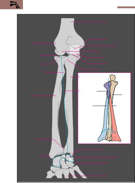
The Bones of the Upper Arm, the Forearm, and the Hand
Upper Arm (Brachium). The humerus is a long bone, and it has a shaft (corpus humeri) as well as proximal and distal ends (epiphyses). The end nearest the body is formed by the head of the humerus, which is connected to the glenoid cavity (Fig. 4.26). At the transition to the humeral shaft there are two solid tubercles, which provide attachment to the muscles acting on the shoulder joint. In front lies the lesser tubercle, which distally (away from the trunk) is continued in the crest of the lesser tubercle (crista tuberculi minoris); laterally lies the greater tubercle, which is continued in the crest of the greater tubercle (crista tuberculi majoris). Between the two tubercles lies a groove (bicipital groove) for the tendon of the long head of the biceps (see Fig. 4.30b).
The distal end of the shaft of the humerus ends in the spherical capitulum humeri, which forms the joint with the radius, and in a markedly larger bobbin-shaped roll, the trochlea humeri, which forms the joint with the ulna (Fig. 4.28a, b). The medial and lateral sides of the distal end are marked by large bony protrusions, the medial and lateral epicondyles. Behind the larger medial epicondyle runs the ulnar nerve to the forearm and hand. Pressing this nerve against the bone elicits a brief painful sensation that radiates into the fingers (“funny bone”).
Forearm (Antebrachium). The skeleton of the forearm consists of the radius and the ulna. A membrane, the interosseus membrane (see Fig. 4.32), connects the shafts of both bones and ensures their cohesion. It also distributes any pull and pressure exerted by one bone to

Special Anatomy of the Locomotor System 157
the other and serves as origin for the forearm muscles. The ulna features at its proximal end a hook-shaped process, the olecranon (Fig. 4.28a, b). With this process the ulna grasps the trochlea of the humerus and so forms the hinge joint between the humerus and the ulna. Distally the ulna narrows and contributes only a small surface to the wrist joint. By contrast, the radius is thinner proximally and broader and more strongly developed distally. At its proximal end it bears a small flat head (caput radii), the cartilaginous facet of which forms joints with the humerus (elbow joint, humeroradial joint) and the ulna (superior radioulnar joint, proximal radioulnar joint). The distal end of the radius, like the ulna, bears a small process, the condyloid process. Both condyloid processes are easily palpable through the skin (Fig. 4.28a).
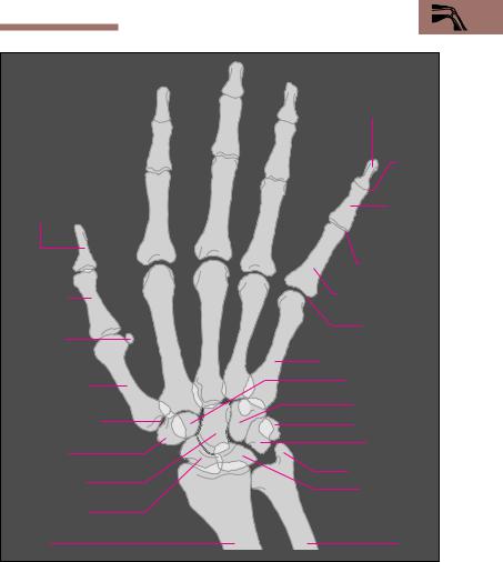
The Hand (Manus). The wrist consists of eight bones arranged in two rows, a proximal and a distal row (Fig. 4.29). The proximal row consists of the radially situated scaphoid (navicular) bone, the middle lunate bone, the ulnar triquetrum, and the pisiform bone (pea-shaped bone). The distal carpal bones include (from radial to ulnar) the trapezium, the trapezoid bone, the centrally situated capitate bone, and the hamate bone.
Of the five metacarpal bones, only the first metacarpal forms a mobile joint, the saddle joint of the thumb, with the trapezium. All the other metacarpals are joined to the bones of the wrist by plane joints with strong ligaments that limit their mobility. The metacarpal bones are continued in the bones of the fingers (phalanges), each finger consisting of a proximal, middle, and distal phalanx. The thumb has only two phalanges, lacking a middle phalanx (Fig. 4.29). The metacarpals are joined to the proximal phalanges and the proximal, middle, and distal phalanges to each other, by synovial joints. These are the metacarpophalangeal and the proximal and distal interphalangeal joints (Fig. 4.29).
The Shoulder Joint (Articulatio Humeri)
The shoulder joint is the most mobile ball-and-socket joint in the human skeleton. Because of the difference in the size of the head of the humerus and the glenoid cavity of the scapula, the joint provides little bony
158 4 The Locomotor System (Musculoskeletal System)
|
|
Shaft of humerus |
|
|
(corpus humeri) |
|
|
Medial epicondyle |
Lateral |
|
Olecranon |
epicondyle |
|
|
|
|
Capitulum of humerus |
Humeroradial |
|
Trochlea of humerus |
joint |
|
|
|
|
|
Head of radius |
|
Humeroulnar joint |
(caput radii) |
|
Proximal radioulnar joint |
Radial tuberosity |
|
|
|
|
|
|
Ulna |
|
|
Axis of |
|
Radius |
pronation/ |
Ulna |
|
supination |
|
|
Radius in |
Radius in |
|
supination |
pronation |
Distal |
|
|
radioulnar |
b |
|
joint |
|
|
|
|
Ulnar styloid process |
Radial styloid |
|
Wrist joint |
process |
|
|
|
|
|
|
|
Middle wrist joint |
a |
|
Metacarpal bones |
|
|
|
Fig. 4.28 a, b |
|
|
Special Anatomy of the Locomotor System 159
|
Distal phalanx |
|
|
of little finger |
|
|
Terminal |
|
|
interphal- |
|
|
angeal |
|
|
joint |
|
Distal phalanx of |
Middle |
|
thumb |
||
phalanx |
||
|
||
|
of little |
|
|
finger |
|
|
Middle inter- |
|
Proximal |
phalangeal joint |
|
Proximal phalanx |
||
phalanx of |
||
of little finger |
||
thumb |
||
|
||
|
Metacarpo- |
|
Sesamoid |
phalangeal joint |
|
bone |
5th metacarpal bone |
|
|
||
1st metacarpal |
Trapezoid bone |
|
|
||
bone |
Hamate bone |
|
|
||
Synovial cavity of |
Pisiform bone |
|
saddle joint of thumb |
||
Triquetrum |
||
Trapezium |
||
|
||
Capitate bone |
Styloid process |
|
Lunate bone |
||
|
||
Scaphoid bone |
|
|
Radius |
Ulna |
Fig. 4.29 Simplified tracing of a radiographic image of the right hand in dorsal view
Fig. 4.28 a, b Simplified tracing of a radiographic image of the right forearm (the blue lines indicate the lines of the joints of the wrist)
a In supination. Ulna and radius are parallel b Radius in supination and pronation

160 4 The Locomotor System (Musculoskeletal System)
guidance. It is secured primarily by muscles and to a lesser extent by ligaments. For this reason, dislocations (subluxations) of the shoulder joint are especially common (about 45 % of all dislocations affect the shoulder joint).
The glenoid cavity is bordered by a fibrocartilaginous lip (labrum glenoidale), about 5 mm in breadth, that increases the joint contact and so the stress-absorbing surface. The joint capsule is wide and lax, and with the arm dependent forms a recess on the medial side (Fig. 4.30a). This extra fold ensures that there is no resistance especially during abduction.
Muscles and Movements. The coracoid process and the acromion, together with the coracoacromial ligament (fornix humeri, Fig. 4.30b), limit upward mobility and at the same time secure the head of the humerus in the glenoid cavity. When the arm is lifted above the horizontal, the scapula, and so the glenoid cavity, must rotate. The movements at the shoulder joint take place in three axes, as in every ball- and-socket joint. The muscular action occurs chiefly in the musculotendinous cuff (rotator cuff, i.e., supraspinatus, infraspinatus, subscapularis, and teres minor muscles) and the deltoid muscle, the most important abductor of the upper arm (Figs. 4.18a, b and 4.19). In addition, the anterior part of the deltoid (pars clavicularis) flexes the arm and the posterior part (pars scapularis) extends it (Fig. 4.31).
The muscles of the musculotendinous cuff all have their origin on the scapula and are inserted into the greater or lesser tubercle of the humerus. While the teres minor and the infraspinatus muscles rotate the upper arm outward (external rotation), the subscapularis muscle is an important internal rotator. Finally, the supraspinatus muscle takes part in abduction, especially its initiation. The pectoralis major, the latissimus dorsi, and the teres major muscles adduct the arm (pull it toward the trunk) and to varying extents rotate it externally or internally (Figs. 4.18a, b and 4.31).
The Elbow Joint (Articulatio Cubiti)
The elbow joint is a hinge joint and a compound joint with three joint elements in the lax joint capsule. It is composed of three separate joints, the humeroradial joint, the humeroulnar joint, and the proximal radioulnar
Special Anatomy of the Locomotor System 161
Head of humerus |
|
Joint |
|
|
capsule |
Glenoid lip |
|
|
(labrum glenoidale) |
|
|
Joint cavity |
|
|
Glenoid cavity |
|
|
Glenoid lip |
|
|
|
|
Tendon |
Pouch of glenoid |
|
of long |
|
head of |
|
capsule |
|
|
|
biceps |
|
|
|
|
a |
|
(caput |
|
longum) |
|
|
|
|
|
Shaft of humerus |
|
Insertion of subcapularis |
|
|
muscle |
Acromion |
|
|
Coracoacromial |
|
|
ligament |
Coracoa- |
|
|
|
|
Coracoid |
cromial |
|
process |
joint |
|
|
Joint capsule |
|
|
Pouch of glenoid |
|
|
capsule |
|
Tendon of long head |
|
|
of biceps (caput longum) |
|
b |
Shaft of humerus |
|
Fig. 4.30 a, b Right shoulder joint a Coronal section, anterior view
b Joint capsule and acromioclavicular joint, anterior view (After Platzer)
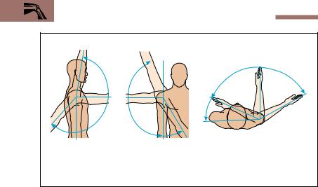
162 4 The Locomotor System (Musculoskeletal System)
Elevation |
Elevation |
|
|
150–170° |
150–170° |
0° |
|
|
|
|
|
90° |
|
70° |
60° |
|
|
||
|
|
|
|
40° |
|
20–40° |
|
0° |
0° |
|
|
Flexion and |
Abduction |
Adduction |
Rotation |
extension |
|
|
|
Fig. 4.31 Movements of the shoulder joint
joint. While the joint between the humerus and ulna (humeroulnar joint) acts as a typical hinge joint, allowing only flexion and extension, the head of the radius has joints with the humerus (humeroradial joint) as well as the ulna (proximal radioulnar joint) (Figs. 4.28a, 4.32).
Muscles and Movements. The proximal radioulnar joint, in combination with the distal radioulnar joint, allows the hand to be turned forward and backward (pronation and supination) (Figs. 4.28b, 4.33). As the hand turns, the radius pivots in place at the elbow joint, while pivoting around the ulna at its distal (hand) end. The position in which the forearm bones are parallel and the palm faces upward, is called supination. When the radius crosses the ulna, the back of the hand faces upward. This position is called pronation. The head of the radius is fixed by two collateral ligaments aided by an annular ligament that surrounds the radial head (Fig. 4.32a−c).
Muscles originating from the humerus or the shoulder girdle and inserted into the ulna include the triceps (m. triceps brachii), which runs along the posterior side of the upper arm and extends the elbow, and the
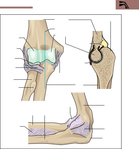
Special Anatomy of the Locomotor System 163
|
Humerus |
Olecranon |
|
Plane of section c |
|
Capitulum |
Trochlea of humerus |
|
|
|
|
of humerus |
Joint capsule |
|
Lateral |
|
|
|
|
|
collateral |
|
|
ligament |
|
|
Head of |
|
|
radius |
Humeroulnar |
|
Annular |
joint |
|
|
|
|
ligament |
|
|
(of radius) |
Medial collateral |
|
Radius |
ligament |
|
|
|
|
Radial |
Ulna |
|
tuberosity |
|
c |
(bicipital |
|
|
tuberosity) |
|
|
a |
|
|
|
Joint capsule |
Humerus |
Radius |
Ulna |
|
Interosseus |
|
Medial |
membrane |
|
collateral |
|
|
ligament |
|
|
Olecranon |
b |
|
|
Fig. 4.32 a−c Right elbow joint a Ligaments, anterior view
b Ligaments and joint capsule, medial view
c Longitudinal section through the humeroulnar joint (After Platzer)
biceps (m. biceps brachii), which runs along the front of the upper arm and flexes the elbow. Both the biceps and the triceps also act on the shoulder joint, because one head of each originates from the scapula (Figs. 4.18a, b and 4.19). Four muscles primarily take part in pronation
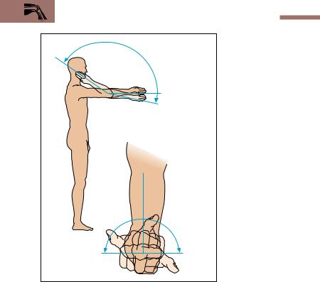
164 4 The Locomotor System (Musculoskeletal System)
|
Fig. 4.33 Movements at |
150° |
the elbow joint. Pronation |
and supination additionally |
|
|
involve the distal radioulnar |
|
joint |
0°
10° Flexion and extension
80–90° |
80–90° |
Supination |
Pronation |
and supination, two internal rotator muscles (m. pronator teres and m. pronator quadratus) and two external rotator muscles (mm. supinator and biceps brachii).
Beside its function as flexor at the elbow, the biceps, with its insertion in a roughened area of the radius (radial tuberosity) (Fig. 4.32), is the strongest supinator. This external rotation is especially strong when the elbow is flexed and at the same time the biceps tendon unwinds from a coiled position around the radius.

Special Anatomy of the Locomotor System 165
The Joints of the Hand
The proximal wrist joint (radiocarpal joint) between the radius and ulna on one side, and the wrist on the other, is a typical condyloid joint. The middle carpal joint is formed by the proximal and distal rows of carpal bones and forms an S-shaped joint cavity with interdigitation of the bones (Fig. 4.28a). The trapezium and the first metacarpal bone form an extraordinarily mobile joint, the saddle joint of the thumb (carpometacarpal joint of the thumb).
Muscles and Movements. Because of its saddle shape, the joint allows four movements: abduction, adduction, flexion, and extension of the thumb. The joint also allows a composite rotational movement—opposi- tion—in which the thumb touches the index finger. In all nine muscles are involved in movements of the thumb. In movements of the wrist both wrist joints act in concert, though the radiocarpal joint makes the largest contribution. Flexion (palmar flexion) and extension occur around an axis running transversely to the radius and ulna. Lateral movements of the hand (adduction and abduction) occur around a second axis that runs vertically through a carpal bone (the capitate). Because of the interdigitation at the midcarpal joint, these movements occur exclusively at the radiocarpal joint.
With the exception of the short muscles of the hand, the muscles acting on the carpal joints and the saddle joint of the thumb originate predominantly from the forearm. Their tendons run in protective tendon sheaths to the wrist or the metacarpal bones, where they act exclusively on the wrist, or to the fingers, which they move simultaneously with the joints of the hand. Eighteen short muscles of the hand and 15 forearm muscles take part in the small movements of grasping and touching. The forearm muscles are divided by their position. The radius and ulna and the interosseus membrane between them separate the flexors anteriorly from the extensors posteriorly. A separate radial muscle group lies between these two muscle groups. Flexors and extensors are both divided into superficial and deep groups (Figs. 4.18a, b and 4.19).

166 4 The Locomotor System (Musculoskeletal System)
The Lower Extremity
The lower extremities in the human are exclusively organs of support and locomotion. They can be divided into a pelvic girdle and a free lower limb. In contrast to the shoulder girdle, the pelvic girdle is solidly connected to the axial skeleton.
The Pelvic Girdle and the Pelvis—Bones, Joints, Muscles
The components of the pelvic girdle are the two hip bones (innominate bones, ossa coxae) that in combination with the sacrum form the bony pelvis. The Sacrum and innominate bones are connected to each other by the sacroiliac joints, synovial joints that, by virtue of their strong ligaments, are almost immobile. Anteriorly, the two innominate bones are connected by the symphysis pubis (Fig. 4.34a, b). The sacroiliac joints and symphysis pubis connect the bony parts of the pelvis into a stable ring and allow only very limited movement.
The bony pelvis is composed of the two innominate bones and the sacrum. The innominate bone is composed of three parts, the ilium, the ischium, and the pubis (Fig. 4.35a, b). The bony parts fuse at the acetabulum (articular facet of the hip joint) about the 14th to 16th years of life to form a single bone, the innominate bone. The sacrum and the innominate bone are connected on each side by two strong ligaments, which exert a strong stabilizing effect on the pelvis. The stronger band (sacrotuberal ligament) runs from the ischial tuberosity to the sacrum, while the second, weaker band (sacrospinal ligament) connects the ischial spine with the sacrum.
The pelvis is divided into the false pelvis and the true pelvis. The line between them (linea terminalis, iliopectineal line) is situated in the plane of the pelvic brim. It is formed by a bony line (the arcuate line), the upper border of the symphysis pubis, and the disk between the 5th lumbar vertebra and the sacrum (sacral promontory) (Figs. 4.34a, b and 4.36). In the erect posture the plane of the pelvic inlet forms a 60° angle to the horizontal (Fig. 4.36). The false pelvis is formed laterally by the iliac fossae and posteriorly by the sacrum. The upper border of the iliac fossa is termed the iliac crest. Anteriorly it ends in the anterior superior iliac spine, from which the inguinal ligament stretches to the pubic tubercle
(Fig. 4.36).
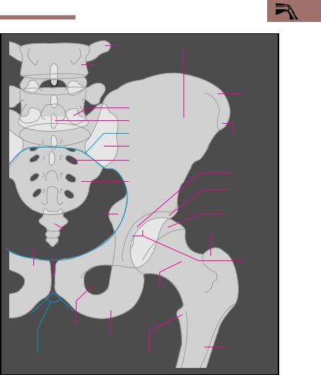
Special Anatomy of the Locomotor System 167
|
|
Transverse |
Ilium |
|
|
|
process |
|
|
|
4th lumbar vertebra |
|
|
|
|
|
|
|
Iliac crest |
|
|
Articular facet |
|
|
|
|
Spinous process |
|
|
|
|
Linea terminalis |
Anteror superior |
|
|
|
|
|
|
|
|
Sacroiliac joint |
iliac spine |
|
|
|
Sacral |
|
|
|
|
foramen |
|
Acetabulum |
|
|
Sacrum |
|
|
|
|
|
|
|
|
|
|
|
Acetabular lip |
|
|
|
|
(labrum acetabulare) |
|
Ischial |
|
|
Head of femur |
|
spine |
|
|
|
|
|
|
(caput femoris) |
|
|
|
|
|
|
|
Coccyx |
|
|
Greater trochanter |
|
|
|
|
|
|
Pubis Symphysis |
|
|
|
|
pubis |
|
|
Hip joint |
|
|
|
|
|
|
|
Neck of |
|
|
|
|
femur |
|
|
|
Obturator |
Ischium |
|
|
|
foramen |
|
|
|
|
|
|
|
of femur |
|
Pubic angle |
Lesser trochanter |
|
|
a |
(between pubic rami) |
|
|
|
Fig. 4.34 a Simplified tracing of a radiographic image of the pelvis and left hip joint
(articulatio coxae) in anterior view
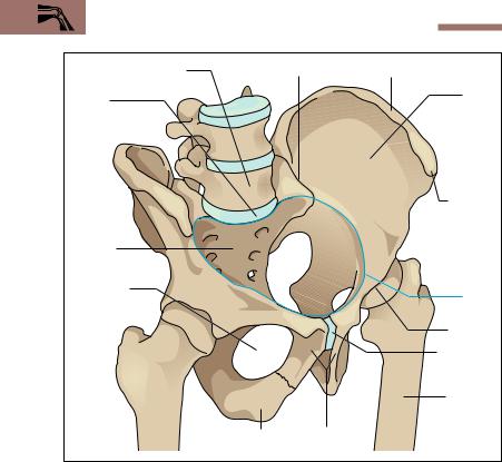
168 4 The Locomotor System (Musculoskeletal System)
5th lumbar vertebra |
Sacroiliac joint |
Iliac crest |
||
Sacral |
|
|
|
Ilium |
|
|
|
|
|
promontory |
|
|
|
|
|
|
|
|
Anterior |
|
|
|
|
superior |
|
|
|
|
iliac spine |
Sacrum |
|
|
|
|
Obturator |
|
|
|
Linea |
foramen |
|
|
|
|
|
|
|
terminalis |
|
|
|
|
|
|
|
|
|
|
Hip joint |
|
|
|
|
Symphysis |
|
|
|
|
pubis |
|
|
|
|
Shaft of |
|
|
|
|
femur |
|
|
Ischium |
Pubis |
|
Fig. 4.34 b |
Bony pelvis in right anterior view (After Schwegler) |
|||
The true pelvis is formed by the two pubic bones and the two ischial bones. Between the upper and lower branches (superior and inferior rami) of the pubis and the bordering ischium there is an oval opening (obturator foramen), closed by a connective tissue membrane (obturator membrane) (Fig. 4.34a, b). The pelvic outlet is bordered by the coccyx, the ischial spine, and the ischial tuberosity. Pelvic inlet, pelvic canal, and pelvic outlet of the true pelvis form the birth canal and as such have great practical significance.
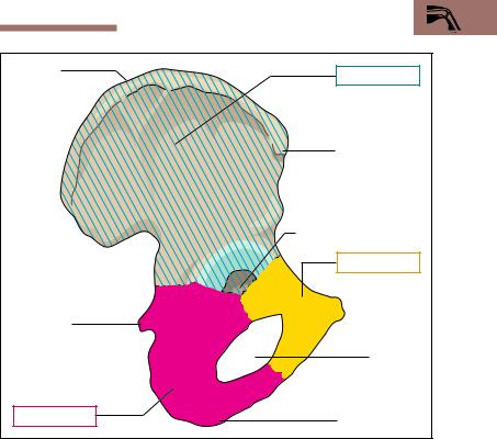
Special Anatomy of the Locomotor System 169
Iliac crest |
Ilium |
|
|
|
|
|
|
Anterior superior |
|
|
iliac spine |
|
|
Acetabulum |
|
|
Pubis |
Ischial spine |
|
|
|
|
Obturator |
|
|
foramen |
Ischium |
Ischial tuberosity |
|
|
|
|
Fig. 4.35 |
Right hip bone (innominate bone, os coxae) in lateral view |
|
The shape of the pelvis shows marked sexual differences: in the female the two iliac fossae extend more laterally. The transition from the false to the true female pelvis is oval in its transverse dimension, while in the male it is smaller and posteriorly narrowed markedly by the more prominent sacral promontory. The angle between the two inferior pubic rami is greater in the female pelvis than in the male (Fig. 4.37). Finally, the obturator foramen in the female pelvis is transversely oval, while that of the male is almost round. The pelvic outlet, determined by the
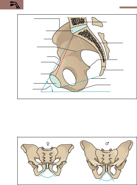
170 4 The Locomotor System (Musculoskeletal System)
Iliac crest |
5th lumbar vertebra |
|
|
||
Sacral promontory |
|
|
Anterior superior |
|
|
iliac spine |
Sacrum |
|
Arcuate line |
||
|
||
Plane of pelvic inlet |
|
|
True conjugate |
Ischial spine |
|
|
||
(narrowest part |
|
|
of true pelvis) |
Coccyx |
|
|
||
Pubic tubercle |
|
|
Symphysis |
Plane of pelvic outlet |
|
|
||
pubis |
Ischial tuberosity |
|
Angle of |
|
|
inclination |
|
|
of pelvis |
|
Fig. 4.36 Right half of pelvis, internal view (sagittal section). The angle of pelvic inclination between the plane of the pelvic inlet and the horizontal is about 60° in the erect posture. (After Faller)
90°
70°
Fig. 4.37 Female and male pelvis. Female pubic angle = 90°; male pubic angle = 70°. (After Schwegler)
Special Anatomy of the Locomotor System 171
ischial tuberosity and the ischial spine, is also wider in the female. The musculature of the pelvis and the pelvic floor is described with that of the trunk (see above Muscles of the Trunk: The Pelvic Floor).
The Free Lower Limb—Bones, Joints, Muscles
The free lower limb is connected to the pelvic girdle by the hip joint. It consists of the thigh bone (femur), the (lower) leg (crus) including tibia and fibula, and the foot (pes), which includes the ankle (tarsus), metatarsals, and toes (digits). The movements between femur and lower leg occur at the knee joint (genu), which is a principal factor in the movement sequence of walking. The tibia and fibula are connected to the foot by the ankle joint. Together with the mid-tarsal (talocalcaneonavicular) joint and the other joints of the foot, this freely mobile joint enables the foot to roll during walking.
Bones of the Thigh, Lower Leg, and Foot
The Thigh (Femur). The thigh bone (femur) is the longest and strongest bone of the human skeleton. Proximally, the shaft of the femur
(diaphysis) continues into the femoral neck (collum femoris), which is directed obliquely upward and which ends in the head of the femur (caput femoris). In the adult the angle of the neck to the shaft (collodiaphyseal angle) is about 125−126°. In the newborn the angle is markedly greater (ca. 150°), while in the elderly it may be smaller than 126° (Fig. 4.38a−c). At the junction between the shaft and neck there are two processes, the robust externally directed greater trochanter, and the somewhat smaller internally directed lesser trochanter (Fig. 4.34a, b). Both trochanters are the site of muscle insertions. Anteriorly and laterally, the femoral shaft is smooth, while along its posterior aspect runs a rough line (linea aspera) with an inner and an outer lip for muscle insertions.
Distally, the femur expands to form the two femoral condyles, extensively covered by hyaline cartilage. Anteriorly, they are extended into a cartilaginous groove, the patellar surface, in which the kneecap (patella) glides downward during flexion of the knee (Figs. 4.39 and 4.40a, b). Posteriorly, the two condyles are separated by a broad groove, the intercondylar notch (intercondylar fossa).
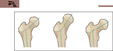
172 4 The Locomotor System (Musculoskeletal System)
125° |
140° |
115° |
a |
b |
c |
Fig. 4.38 a−c Angle of the neck of the femur (collodiaphyseal angle). Collodiaphyseal angle in the adult (a), in a 3-year old child (b), and in the elderly (c). (After Frick)
Lower Leg. The skeleton of the lower leg is formed by the tibia and the fibula. The tibia is the stronger bone and the true supporting pillar and connection between the femur and the foot. Proximally, the two bones are connected by the strong ligamentous tibiofibular joint (Fig. 4.40). Distally, the two bones are connected by a fibrous joint (syndesmosis). The mortise formed by the medial and lateral malleoli of these bones, together with the trochlea of the talus, makes up the ankle joint (Fig. 4.41).
Proximally, the tibia expands and features on its anterior surface the tibial tuberosity, into which the quadriceps femoris is inserted. The superior articular surface (Fig. 4.40), formed by the medial and lateral condyles, forms the knee joint with the femur. Like the radius and ulna, the tibia and fibula are connected by an interosseus membrane, which gives origin to some muscles in the leg.
Foot (Pes). The foot, like the hand, is divided into three groups arranged end to end (Fig. 4.41): the ankle (tarsus), the intermediate group (metatarsus), and the toes (digits). There are seven tarsal bones, of which the talus, the navicular, and the three cuneiform bones lie on the medial side, while the calcaneum (calcaneus, os calcis), and the cuboid bone are lateral. The calcaneum is the largest bone of the foot, its posteriorly directed calcaneal tubercle (tuberculum calcanei) forming the bony base
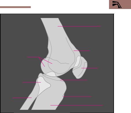
Special Anatomy of the Locomotor System 173
|
|
Femur |
|
|
|
Patellar surface |
|
Femoral condyles |
of femur |
||
|
|||
|
|
Kneecap |
|
|
|
(patella) |
|
Head of fibula |
Superior articular |
||
surface of tibia |
|||
|
|
||
Fibula |
|
Tibial tubercle |
|
|
|
||
|
|
Tibia |
|
Fig. 4.39 |
Simplified tracing of a lateral radiograph of the knee joint |
||
of the heel. The five metatarsal bones are long bones, like the metacarpal bones of the hand. Each is divided into a base, a shaft, and a head. The first (medial) metatarsal bone is the thickest and strongest metatarsal bone. The 2nd to 5th toes consist of proximal, middle and distal phalanges, while the 1st toe (big toe, hallux), only has a proximal and a distal phalanx (Fig. 4.41).
Inspection of the skeleton of the foot reveals that, while the talus lies above the calcaneum behind, the two bones lie side by side in the middle and in front. This arrangement creates medially a marked longitudinal arch, and at the level of the cuneiform bones and the metatarsal bones a
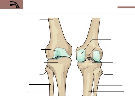
174 4 The Locomotor System (Musculoskeletal System)
Femoral shaft |
Femoral shaft |
|
Medial condyle |
Patellar surface |
Intercondylar fossa |
|
|
of femur |
Lateral condyle |
|
|
Head of tibia = |
|
superior articular |
Intercondylar |
surface |
eminence |
Head of fibula |
Head of fibula |
Fibula |
Fibula |
|
|
Tibia |
Tibia |
a |
b |
Fig. 4.40 a, b Components of the knee joint, anterior (a) and posterior (b) views
transverse arch running from medial to lateral (Fig. 4.42a, b). Muscles and tendons serve to keep these arches intact. The calcaneal tubercle and the heads of the 1st to 5th metatarsals may be regarded as the bases on which all of the body weight rests on level ground.
The Hip Joint (Articulatio Coxae)
The hip joint is a ball-and-socket joint in which the head of the femur moves in the acetabulum (Figs. 4.34a, b and 4.43a). The acetabulum is formed by all three components of the hip bone, (ilium, ischium and pubic bone, see Fig. 4.35). It grasps almost half the ball of the femur, giving the hip joint a solid bony channel. The joint contact is augmented by a fibrocartilaginous lip, (acetabular labrum), which extends the joint beyond its equator by forming a ring around the acetabulum. In this way the joint maintains solidity and stability in every position. The capsule of
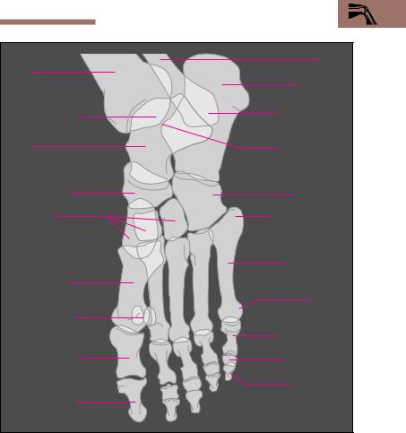
Special Anatomy of the Locomotor System 175
|
Fibula |
Tibia |
|
|
Calcaneum |
Medial malleolus |
Lateral malleolus |
|
|
Talus |
Trochlea of talus |
Navicular bone |
Cuboid bone |
Cuneiform |
Base of metatarsal |
bones |
bone |
|
5th metatarsal |
1st metatarsal |
bone |
|
|
bone |
Head of |
|
|
Sesamoid bones |
metatarsal bone |
|
|
|
Proximal phalanx |
Proximal phalanx |
Middle phalanx |
of big toe (hallux) |
|
|
Distal phalanx |
Distal phalanx of |
|
big toe (hallux) |
|
Fig. 4.41 Simplified tracing of a radiographic image of a plantarflexed foot in lateral view
the joint is reinforced by three strong ligaments running from the three parts of the hip bone: the iliofemoral, the pubofemoral, and the ischiofemoral ligaments (Fig. 4.43b, c). The iliofemoral ligament is the strongest ligament of the body, with a tensile strength of 350 kg.
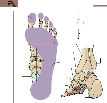
176 4 The Locomotor System (Musculoskeletal System)
Distal |
Longitudinal arch |
|
|
|
phalanx |
|
|
||
|
|
|
||
|
Transverse arch |
|
|
|
Proximal |
|
|
|
|
phalanx |
of little toe |
|
|
|
|
|
|
||
|
Middle phalanx |
Distal phalanx |
||
|
Proximal phalanx |
|
|
|
1st |
Tibia |
Malleolar |
Fibula |
|
metatarsal |
mortise |
|||
|
|
|||
bone |
|
|
|
|
|
Medial |
|
Lateral |
|
Cuneiform |
malleolus |
|
||
|
|
malleolus |
||
bones |
|
|
|
|
|
Cuboid bone |
|
|
|
Navicular |
Navicular |
|
|
|
bone |
|
|
||
|
bone |
|
Talus |
|
Talus |
Medial |
|
|
|
cuneiform |
|
|
||
|
bone |
|
|
|
|
1st meta- |
|
|
|
|
tarsal bone |
|
|
|
Calcaneum |
|
|
|
|
a |
Calcaneum |
|
||
b |
|
|
||
Fig. 4.42 a, b Longitudinal and transverse arches of the right foot a Skeleton of foot from above, with superimposed footprint
b Skeleton of foot with bones of the lower leg, medioposterior view
The ligaments run a characteristic spiral course around the neck of the femur. In the erect posture they are tense, while they relax during flexion of the hip joint, allowing considerable mobility during flexion.
Muscles and Movements. Because a ball-and-socket joint has three main axes of movement, the leg can be moved around a transverse axis anteriorly and posteriorly (flexion and extension), a sagittal axis away from and toward the trunk (abduction and adduction), and a ver-
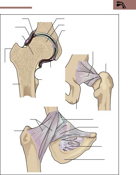
Special Anatomy of the Locomotor System 177
Acetabular lip |
Acetabulum |
|
(labrum) |
Head of femur |
|
Articular |
|
|
|
|
|
capsule |
Ligament of head of femur |
|
Femoral neck |
|
|
|
|
Ischiofemoral ligament |
|
|
Greater |
Greater |
|
trachanter |
|
|
|
trach- |
Labrum |
|
anter |
acetabuli |
|
Shaft of |
Lesser |
|
trochanter |
|
|
femur |
|
|
|
|
|
a |
|
|
|
Ischium |
c |
|
|
|
Iliofemoral ligament |
|
Articular capsule |
Greater |
|
Pubofemoral |
|
ligament |
|
trochanter |
|
|
|
|
Obturator |
|
|
membrane |
|
|
Ischium |
b |
|
|
Fig. 4.43 a−c Right hip joint
a coronal section; b, c ligaments: anterior (b) and posterior (c) views
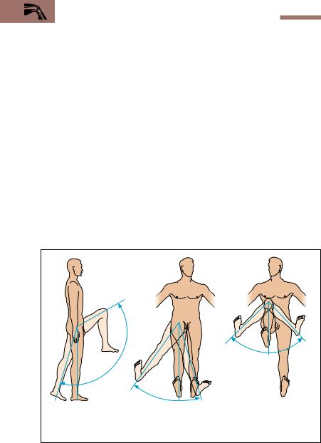
178 4 The Locomotor System (Musculoskeletal System)
tical axis around which it is rotated (internal and external rotation) (Fig. 4.44).
The most important hip flexors muscles include the deep iliopsoas, the sartorius, and the rectus femoris muscles. The extensors at the hip are the gluteus maximus muscle and the muscles running from the ischial tuberosity to the lower leg (ischiocrural muscles) (Figs. 4.18a, b and 4.19): the biceps femoris, the semitendinosus, and semimembranosus muscles). The gluteus medius and the gluteus minimus muscles function both as abductors and as internal and external rotators.
On the medial side of the thigh, between the extensors and the flexors runs a group of five muscles that adduct the thigh toward the trunk (adductors, e. g., m. adductor magnus). They take their origin from the pelvis and are inserted into the medial lip of the linea aspera of the femur. The external rotators arise from the posterior aspect of the pelvis and run to the femur, e. g., the gluteus maximus. Internal rotators are the anterior parts of the gluteus medius and gluteus minimus. During movement of the hip, the leg may be moved freely on the hip or the leg may be fixed (standing) and the hip moved against it. In walking these functions alternate.
|
|
1 |
|
|
|
|
|
|
3 |
|
|
|
|
|
|
0 |
|
|
|
|
|
|
– |
|
|
|
|
|
|
1 |
|
|
|
|
|
|
4 |
|
|
|
|
|
|
0 |
|
|
|
|
|
|
° |
30°–45° |
|
|
40°–50° |
|
|
|
|
|
||
|
|
|
Internal |
0° |
0° |
External |
|
|
|
rotation |
|
|
rotation |
|
|
30°–45° |
|
|
|
|
15° |
0° |
|
0° 20°–30° |
|
|
|
Extension and flexion |
|
Abduction and Adduction |
Rotation |
|||
Fig. 4.44 Mobility of the hip joint

Special Anatomy of the Locomotor System 179
The Knee Joint (Articulatio Genus)
The knee joint is the largest joint of the human body. It is a compound joint, formed by the combined action of the femur, tibia, patella and two fibrocartilaginous disks, (menisci) (Figs. 4.45 and 4.46c). The joint consists of the joint between the condyles of the femur and the tibia and the joint between the femur and the patella. The two menisci adapt the joint surfaces of the femur and the tibia to each other and increase the surface for the transmission of force. When the knee joint is flexed, the femur executes a combined gliding and rolling motion over the tibial articular surface, during which the menisci are shifted more posteriorly the further the knee is flexed. The movements of the knee joint are guided by two sets of ligaments, the medial and lateral collateral ligaments and the anterior and posterior cruciate ligaments (Fig. 4.46).
While the collateral ligaments primarily stabilize the extended leg, the cruciate ligaments take over that function in the flexed knee joint. Because of the uneven curvature of the femoral condyles, the collateral ligaments are fully tensed only during extension of the knee joint; they are relaxed during flexion. With the knee flexed, the cruciate ligaments limit internal and external rotation of the lower leg, internal rotation being more severely limited than external rotation by the coiling of the cruciate ligaments.
Muscles and Movements. Muscles inserted into the medial surface of the tibia (e. g., mm. semitendinosus and semimembranosus) are internal rotators. The biceps femoris is inserted into the head of the fibula, and is the only external rotator of the lower leg (Figs. 4.18a, b and 4.19). All three muscles are flexors of the knee joint, as is the sartorius. The most important extensor of the knee joint is the quadriceps femoris, inserted by the patellar ligament into the tibial tuberosity. The patella, the largest sesamoid bone in the human body, is embedded in the patellar ligament (Fig. 4.45). It is triangular in shape and articulates with the anterior surface of the distal femur (patellar surface). As the knee flexes, the patella moves downward. Because there is considerable transfer of force in the joint between femur and patella, especially during flexion, this is the most highly stressed joint in the body, and is the earliest to show degenerative changes in its cartilage.
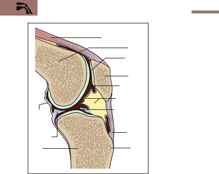
180 4 The Locomotor System (Musculoskeletal System)
|
Fig. 4.45 Sagittal section |
|
|
of knee joint |
|
|
Quadriceps femoris |
|
|
Femur |
|
|
Femoro- |
|
|
patellar |
|
|
articulation |
|
|
Patella |
|
|
Joint cavity |
|
|
Joint capsule |
|
Posterior |
Joint surfaces |
|
between |
||
part of |
||
femur and |
||
joint |
||
tibia |
||
capsule |
||
Patellar |
||
Meniscus |
||
ligament |
||
|
||
Tibia |
Tibial |
|
|
tuberosity |
The Ankle Joint (Articulatio Talocruralis) and the Intertarsal Joint (Articulationes Subtalaris and Talocalcaneonavicularis)
The movements of the foot against the lower leg occur in two joints: the ankle joint and the talocalcaneonavicular joint.
Ankle Joint. The ankle joint is formed by the tibia, fibula, and talus. The distal ends of the tibia and fibula form a mortise made up of the medial and lateral malleoli, which grasp the trochlea of the talus (Fig. 4.47a−c). The joint is a hinge joint, the axis of which runs through the trochlea of
Fig. 4.46 a−c Ligaments of the right knee joint |
|
a Anterior view (patella and patellar ligament are reflected downward) |
|
b Posterior view |
|
cSuperior articular surface of the tibia seen from above (the femur has been removed and the ligaments transsected)
Special Anatomy of the Locomotor System 181
Posterior |
|
Patellar surface of femur |
|
|
cruciate ligament |
|
|
|
|
Lateral |
|
|
|
|
condyle |
|
|
|
|
Anterior |
|
|
|
|
cruciate |
|
Medial condyle |
|
|
ligament |
|
|
||
Lateral |
|
|
|
|
collateral |
|
|
|
|
ligament |
|
Medial collateral ligament |
|
|
Superior arti- |
|
|
||
|
|
|
||
cular surface |
|
|
|
|
of tibia |
|
|
Medial |
|
Head |
|
|
condyle |
|
|
|
Inter |
||
of fibula |
|
|
||
|
|
condylar |
||
|
|
|
||
Patellar |
|
|
fossa |
|
ligament |
|
|
Lateral |
|
Patella |
|
|
condyle |
|
|
|
Lateral |
||
|
|
|
||
|
|
|
collateral |
|
|
|
|
ligament |
|
Fibula |
|
|
Lateral |
|
|
|
|
meniscus |
|
|
|
Medial |
Tibio- |
|
a |
|
collateral |
||
|
fibular |
|||
|
|
ligament |
joint |
|
|
|
|
||
|
|
Medial |
Head |
|
|
|
meniscus |
of fibula |
|
Anterior |
|
Hyaline |
Posterior |
|
cruciate |
Posterior |
cartilage of |
||
cruciate |
||||
ligament |
cruciate |
superior articular |
||
ligament |
||||
|
ligament |
surface of tibia |
||
|
|
|||
Head of |
|
Tibia |
Fibula |
|
fibula |
|
|
|
|
Lateral |
|
b |
|
|
collateral |
|
|
|
|
ligament |
|
Medial collateral ligament |
|
|
Lateral |
|
Medial meniscus |
|
|
meniscus |
|
|
|
|
c |
|
|
|

182 4 The Locomotor System (Musculoskeletal System)
the talus and connects both malleoli. The movements at this joint are called dorsiflexion (lifting of the point of the foot) and plantarflexion (lowering of the point of the foot toward the sole = plantar side of the foot). When the foot is fixed, the lower leg moves backward and forward. A strong set of ligaments stabilizes the ankle joint. It consists of three lateral ligaments and the medial deltoid ligament. The tibia and fibula are tightly joined together by a syndesmotic joint at the level of the malleoli.
Intertarsal Joint. In the intertarsal joint, the talus articulates with the calcaneus and the navicular bone. This joint is, in fact, composed of two joints that are anatomically entirely separate: posteriorly the subtalar joint, and anteriorly the talocalcaneonavicular joint. In the subtalar joint, the talus articulates with the calcaneus; in the talocalcaneonavicular joint, the ball-shaped head of the talus articulates with the calcaneus and the navicular bone. (Fig. 4.47a−c) The axis of movement in the talocalcaneonavicular joint runs from the center of the navicular obliquely downward, laterally, and backward through the calcaneum. The movements in this joint are lateral movements called supination (lifting of the inner = medial border of the foot) and pronation (lifting of the outer = lateral border of the foot) (Fig. 4.48c).
Muscles. The muscles running from the lower leg to the foot act on the ankle joint and the talocalcaneonavicular joint. They are divided onto three groups according to their position on the leg: the posterior calf muscles (mm.triceps surae, tibialis posterior, flexor digitorum longus, and flexor hallucis longus) act as supinators, plantar flexors (point of the foot turned down), and flexors of the toes (mm. flexor digitorum longus and flexor hallucis longus only). The muscles of the anterior compartment of the lower leg (mm. tibialis anterior, extensor digitorum longus, and extensor hallucis longus) act as dorsiflexors (lifting of the point of the foot) and supinators of the foot (m. tibialis anterior only). The muscles of the lateral compartment of the calf (mm. peronei longus and brevis) are primarily pronators and assist plantar flexion. The strongest group are the superficial muscles of the calf (m. triceps surae), usually considered to be composed of two muscles, the soleus
and the gastrocnemius muscles (Fig. 4.18a, b and 4.19). They join to form the Achilles tendon, which is inserted into the calcaneal tubercle. If the Achilles tendon is torn, the triceps surae cannot act and it be-
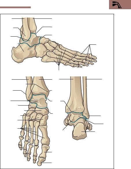
Special Anatomy of the Locomotor System 183
Fibula
Lateral malleolus
Talus
Calcaneum
a
Fibula
Malleolar mortise
Lateral malleolus
Talus
Calcaneum
Navcicular bone
Cuboid bone
Cuneiform bones
Metatarsal bones
b
Tibia
Ankle joint
Talocalcaneonavicular joint
Phalanges
Metatarsal bones |
|
|
Tibia |
Fibula |
|
Medial |
|
|
malleolus |
|
|
Ankle |
|
|
joint |
|
|
Talocal- |
Lateral |
|
caneo- |
||
malleolus |
||
navicular |
||
|
||
joint |
Talus |
|
|
||
Subtalaris joint |
Calcaneum |
|
c |
|
Proximal phalanx of the big toe (hallux)
Distal phalanx of the big toe
Fig. 4.47 a−c Ankle joint and talocalcaneonavicular joint (the joints are shown as blue lines)
a Lateral view b Anterior view c Posterior view

184 4 The Locomotor System (Musculoskeletal System)
Dorsi- |
|
Plantar |
0° |
0° 30° |
|
flexion |
0° |
flexion |
|||
50° |
|
||||
20–30° |
|
40–50° |
|
|
|
|
|
Plantar |
Dorsi- |
||
|
|
|
|||
|
|
|
flexion |
flexion |
|
|
|
|
b |
|
a |
30° |
60° |
|
||
|
|
|
|
0° |
0° |
|
c Pronation |
Supination |
Fig. 4.48 a−c Movements of the ankle and talocalcaneonavicular joints a Ankle joint (free leg)
b Ankle joint (fixed leg)
c Talocalcaneonavicular joint
comes impossible to stand on tip-toe; supination is also severely impaired.
Head and Neck
The Neck (Collum)
The neck joins the head to the trunk and contains the vascular and nervous pathways (blood and lymph vessels as well as nerve fibers) that run from the head to the trunk and the upper extremity. The boundary between the trunk and the neck lies at the clavicles in front and at the spine of the 7th cervical vertebra in the back. Cephalad, the neck extends to a line joining the lower border of the mandible, the two mastoid processes, and the external occipital protuberance. The neck may be

Special Anatomy of the Locomotor System 185
divided into the posterior cervical refion (back of the neck, nucha) and the neck proper (cervix, collum).
The organs of the neck lie in front of the vertebral column. They include the throat (pharynx), the cervical part of the esophagus, the larynx, and the cervical part of the air passages (trachea). The thyroid gland occupies the lateral portion of the compartment containing the organs. The lateral parts also contain the vascular and nervous pathways (common carotid artery and its branches, internal jugular vein, lymphatics of the neck, vagus nerve, sympathetic trunk).
The Neck Muscles. The space containing the organs is enclosed on all sides by connective tissue sheaths (fascias) (Figs. 4.49, 4.50). Posteriorly, the muscular covering of the neck is formed by the segmental neck muscles, which are to a large extent covered by the trapezius muscle, and laterally by the sternocleidomastoid muscle. In front of and at the side of the cervical spine lie the derivatives of the trunk muscles (scalene muscles), and the prevertebral muscles (e. g., mm. longus capitis and longus colli). The organs of the neck are covered in front by the infrahyoid muscles. The most superficial muscle is the platysma, which is classified as one of the muscles of facial expression.
The Head (Caput)
The bony basis of the head is the skull (cranium). It serves partly as a bony capsule for the brain and the sense organs, and partly as support for the face; it also contains the upper ends of the digestive and respiratory tracts. It consists of a brain case (neurocranium) and a facial skeleton
(splanchnocranium, viscerocranium), both sharing the base of the skull, which descends obliquely backward (see Figs. 4.54, 4.55). Externally, the boundary between the two parts can be seen to run from the root of the nose, through the upper rims of the orbits, to the external canals of the ears (external auditory meati). The internal aspect of the base of the skull separates the neurocranium from the facial skeleton and so serves as floor for the neurocranium, while anteriorly it becomes the roof of the facial skeleton. The posterior half is connected to the vertebral column by the atlanto-occipital joint and provides the origin for the neck muscles.
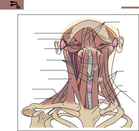
186 4 The Locomotor System (Musculoskeletal System)
|
Suprahyoid |
|
|
muscles |
|
Mandible |
|
|
Hyoid bone |
|
|
Thyroid cartilage |
|
|
Infrahyoid muscles |
|
|
Thyroid gland |
Scalene |
|
|
||
|
muscles |
|
Sternocleidmastoid |
|
|
muscle |
Sternum |
|
Clavicle |
||
1st rib |
||
|
||
Trapezius |
|
|
muscle |
|
Fig. 4.49 Muscles of the neck, ventral view (the platysma and fasciae of the neck and on the left side the muscles of the shoulder girdle have been removed)
General Aspects of the Skull
Both parts of the skull consist of separate bones. With the exception of the mandible, the ossicles of the ear, and the hyoid bone, they are connected by sutures or cartilaginous joints (synchndroses) or by bony joints (synostoses).
The Neurocranium. The neurocranium extends from the upper borders of the orbits (supraorbital margins) to the superior nuchal line. It includes the frontal bone, the two parietal bones, parts of the two tem-
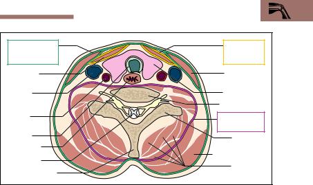
Special Anatomy of the Locomotor System 187
Superficial |
Pretracheal |
|
fascia of the |
fascial layer |
|
neck |
of the neck |
|
Infrahyoid |
Sternocleido- |
|
muscles |
mastoid muscle |
|
Internal |
Thyroid gland |
|
jugular vein |
||
Vertebral body |
||
Common |
||
|
||
carotid |
Prevertebral |
|
artery |
||
fascia |
||
|
||
Trachea |
Transverse |
|
Esophagus |
process |
|
Spinal cord |
Trapezius muscle |
|
Segmental |
||
Spinous process |
||
neck muscles |
Fig. 4.50 Horizontal section of the neck at the level of the thyroid gland (the various fasciae of the neck are marked in color)
poral bones, and the uppermost part of the occipital bone. In front, the two parietal bones are joined to the frontal bone by the coronal suture (Fig. 4.51). Posteriorly, the lambdoid suture lies between the parietal bones and the occiput. The sagittal suture runs between the two parietal bones from the center of the coronal suture to the lambdoid suture. Laterally, the temporal bone is connected to the parietal bone posteriorly and the greater wing of the sphenoid bone (ala major ossis sphenoidalis), anteriorly by the squamous suture. The part of the skull visible externally is the cranial vault; the internal part, not visible from the outside, is the internal base of the skull. The latter is the internal boundary between the neurocranium and the facial skeleton.
The Fontanelles. In the newborn, broad gaps filled with connective tissue, the fontanelles, remain between the bones of the cranial vault (Fig. 4.52). The anterior, larger fontanellele is defined by the two developing halves of the frontal bone and the two developing parietal bones. It does not close completely until the 36th month of life. The posterior, triangular fontanellele lies between the two developing parietal bones and the developing occipital bone. It closes first, about the 3rd
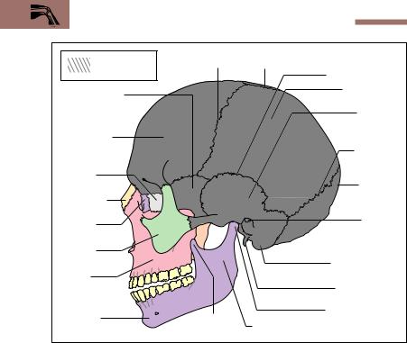
188 4 The Locomotor System (Musculoskeletal System)
|
Coronal |
Sagittal |
|
Neurocranium |
suture |
suture |
|
|
|
Squamous suture |
|
Greater wing of |
|
Parietal bone |
|
|
|
||
sphenoid bone |
|
Temporal |
|
|
|
||
|
|
bone |
|
Frontal bone |
|
|
|
|
|
Lambdoid |
|
|
|
suture |
|
Ethmoid |
|
Occipital |
|
bone |
|
||
Nasal bone |
|
bone |
|
|
|
||
Lacrimal |
|
External |
|
|
auditory meatus |
||
bone |
|
||
|
|
||
Zygoma |
|
|
|
|
|
Mastoid process |
|
Maxilla |
|
|
|
|
|
Styloid process |
|
Mandible |
Coronoid |
Head of mandible |
|
Ramus of mandible |
|||
|
process |
Fig. 4.51 Lateral view of adult skull
month of life. The placement of the fontanelles makes it possible to determine the position of the child’s head during birth.
The Facial Skeleton (Splanchnocranium, Viscerocranium). The parts of the facial skeleton are the upper jaw (maxilla), the zygoma, the nasal bones, the lacrimal bone, the palatine bone, the vomer, and the lower jaw
(mandible) (Fig. 4.51). (The palatine bone is shown in Fig. 4.54; the vomer lies in the nasal cavity and cannot be seen in the figure.) The upper jaw takes part in the structure of the orbit, the nasal cavity, and the roof of the mouth (hard palate. In a dental process, the alveolar arch, it contains the roots of the upper row of teeth. It encloses the largest of the four nasal sinuses, the maxillary sinus.
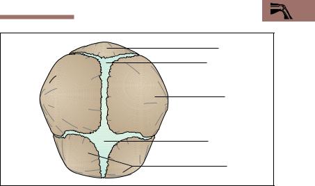
Special Anatomy of the Locomotor System 189
Occipital bone
Posterior fontanelle
Parietal bone
Anterior fontanelle
Frontal bone
Fig. 4.52 Skull of a newborn seen from above
The lower jaw moves on the rest of the skull at the temporomandibular joint (see Fig. 4.58). It consists of a body, two condylar processes bearing the heads of the mandible, and two rami with the muscle-bearing processes for the attachment of the temporalis muscles (coronoid processes) (Fig. 4.51) branching from them. The lower dentition is also rooted in an alveolar arch, which contains the sockets for the teeth (alveoli). A canal for the nerve and vessels to the teeth (canalis mandibulae) opens on the inner side of the mandibular ramus. An opening in each side allows passage of the nerves and vessels supplying the skin of the chin.
Internal and External Aspects of the Base of the Skull. The base of the skull is the floor of the cranial cavity on which the brain rests (Fig. 4.54). The external surface of the base is the part visible from the outside, looking at the skull from below. The internal surface (Fig. 4.55) is the aspect not visible from the outside.
The external surface of the base of the skull (Fig. 4.54) extends from the external occipital protuberance to the incisor teeth in the upper jaw. The lateral boundaries are formed from imaginary lines drawn between
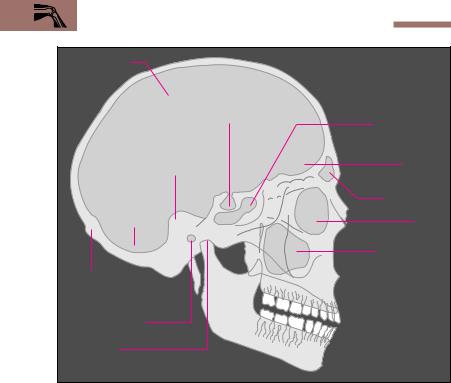
190 4 The Locomotor System (Musculoskeletal System)
Cranial cavity
Pituitary fossa
|
Sphenoid sinus |
|
Petrous part of |
|
|
the temporal |
Anterior |
|
bone |
||
cranial fossa |
||
|
||
|
Frontal sinus |
|
Posterior |
|
|
cranial fossa |
Orbit |
|
|
Maxillary sinus |
External occipital protuberance
External auditory meatus
Temporomandibular joint
Fig. 4.53 Simplified tracing of a lateral radiograph of the skull
the alveolar arches and the mastoid processes. The anterior part forms the roof of the oral cavity and the floor of the nasal cavity. At the posterior end of the hard palate lies the entrance to the nasal cavity (choanae). At the posterior end of the base of the skull lies a large opening, the foramen magnum, and next to it on each side sit the occipital joint facets
(occipital condyles) that, in conjunction with the atlas, form the atlantooccipital joint. The articular fossae for the temporomandibular joints can be found immediately in front of the mastoid processes.
Anteriorly, the internal surface of the base of the skull (Fig. 4.55) is formed by the frontal bone, which also forms the roof of the two orbits. In between lie parts of the ethmoid bone, with its cribriform plate (lamina
Special Anatomy of the Locomotor System |
191 |
|
Palate (hard palate) |
|
|
Wings of left sphenoid bone |
|
|
|
Anterior |
|
Vomer |
section of |
|
the external |
||
|
||
Choanae |
aspect of the |
|
Zygoma |
base of the |
|
skull |
||
Inferior |
||
|
||
orbital fissure |
Middle |
|
Foramen ovale |
section of |
|
the external |
||
|
||
Foramen lacerum |
aspect of the |
|
base of the |
||
|
||
External |
skull |
|
auditory meatus |
|
|
Mastoid process |
|
|
Stylomastoid |
Posterior |
|
foramen |
||
section of |
||
Jugular foramen |
||
the external |
||
Foramen magnum |
aspect of the |
|
base of the |
||
|
||
Occipital condyles |
skull |
|
|
External occipital protuberance
Fig. 4.54 Skull seen from below (external surface of base of skull). The mandible has been removed
cribrosa), which transmits the olfactory nerves to the nasal mucosa. The ethmoid bone takes part in the formation of the nasal cavity and the internal boundary of the orbits. In its center projects a crest like a cock’s comb (crista galli), which gives attachment to the falx cerebri. The frontal bone and the ethmoid, combined with the lesser wing of the sphenoid, form the two anterior cranial fossae (fossae craniis anterior).
Behind these bones lies the sphenoid bone, followed by the occipital bone. On each side lie the temporal bones. The sphenoid bone is the center of the base of the skull. It is composed of the body of the sphenoid surrounding the sphenoid sinus and forming the sella turcica, the floor of which is occupied by the pituitary gland (hypophysis). The two greater
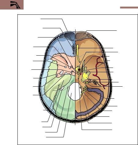
192 4 The Locomotor System (Musculoskeletal System)
Cock’s comb |
|
|
|
(crista galli) |
|
|
|
Cribriform plate |
|
|
Frontal sinus |
|
|
|
|
Sella turcica |
|
|
|
Superior |
A |
|
Olfactory nerve |
orbital fissure |
|
|
|
|
|
Optic nerve |
|
Optic canal |
|
|
|
|
|
Oculomotor nerve |
|
|
|
|
|
Foramen |
|
|
Middle meningeal |
rotundum |
|
B |
artery |
|
|
Trochlear nerve |
|
Carotid |
|
|
|
|
|
Internal carotid |
|
canal |
|
|
|
Foramen |
|
|
artery |
|
|
Trigeminal nerve |
|
ovale |
|
|
|
|
|
Facial nerve |
|
|
|
|
|
Foramen |
|
|
Acoustic |
spinosum |
|
|
(vestibulo- |
Foramen |
|
|
cochlear) nerve |
|
|
|
|
lacerum |
|
|
Glossopharyn- |
(occluded by |
C |
|
geal nerve |
cartilage) |
|
Vagus nerve |
|
|
|
||
|
|
|
|
Internal auditory |
|
|
Accessory nerve |
meatus |
|
|
|
|
|
|
|
Petrous part of |
|
|
Hypoglossal nerve |
|
|
|
|
temporal bone |
|
|
Transverse sinus |
|
|
|
|
Jugular foramen |
|
|
Abducent nerve |
Hypoglossal canal |
|
|
|
|
|
|
|
Foramen magnum |
|
|
|
Fig. 4.55 Internal surface of base of skull (on the left side the structures covering the bone have been removed). A Anterior; B middle, and C posterior cranial fossa
wings of the sphenoid form a part of the middle cranial fossa (fossa cranii media) and below give rise to the pterygoid processes (Fig. 4.54). The posterior part of the middle cranial fossa is formed by the anterior superior wall of the petrous bone (pyramid of the petrous bone), which houses the middle and inner ear. The petrous bone is a part of the temporal

Special Anatomy of the Locomotor System 193
bone. The floor of the unpaired posterior cranial fossa (fossa cranii posterior) is formed almost entirely by the occipital bone. Through its middle passes the foramen magnum, through which the brain is connected with the spinal cord. Anteriorly and laterally, the petrous bone takes part in the formation of the posterior cranial fossa.
Foramina (holes) and canals traverse all the cranial fossae downward, transmitting the cranial nerves and vessels (Fig. 4.55). Between the lesser and greater wings of the sphenoid bone lies the superior orbital fissure, which leads into the orbit. Nerves that run through it into the orbit include the first division of the trigeminal nerve (ophthalmic nerve) and cranial nerves III, IV, and VI (oculomotor, trochlear, and abducens) to the external muscles of the eye. The optic nerve and optic artery pass through a separate canal, the optic foramen. The second division of the trigeminal nerve (maxillary nerve) leaves the skull through the foramen rotundum in the greater wing of the sphenoid, while the third division (mandibular nerve) passes through the foramen ovale of the greater wing of the sphenoid. Behind it lies a small foramen, the foramen spinosum, which transmits the artery supplying the dura mater (middle meningeal artery).
Inside the petrous pyramid runs the internal auditory meatus (Fig. 4.55), through which cranial nerves VII and VIII (facial and acoustic or vestibulocochlear nerves) enter the petrous bone. On each side of the sella turcica lies a separate canal, the carotid canal, through which the internal carotid artery enters the skull. Between the temporal and occipital bones there is an opening (jugular foramen) where cranial nerves IX (glossopharyngeal nerve) X (vagus nerve), and XI (accessory nerve) pass through the base of the skull. Additionally, the venous blood from the brain drains through the jugular foramen into the internal jugular vein. Cranial nerve XII (hypoglossal nerve) leaves the skull through an opening (hypoglossal canal) immediately next to the foramen magnum.
The Muscles of the Skull
Two sets of muscles can be distinguished on the facial skeleton: the chewing muscles (muscles of mastication) and the muscles of facial expression. The muscles of mastication take their origin from the base and the lateral wall of the skull and are inserted into the mandible. The muscles of facial expression are cutaneous muscles (see below). They are of significance in facial expressions, have a protective function, and

194 4 The Locomotor System (Musculoskeletal System)
take part in the ingestion of nutrients. Both groups of muscles move and put stress on the bones of the skull and influence their shape and structure.
Muscles of Mastication. Muscles with a variety of origins and functions take part in the act of chewing (Fig. 4.56a, b). Muscles of mastication in the more restricted sense include the masseter, the lateral and medial pterygoid, and the temporalis muscles. They are all innervated by the third division of the trigeminal nerve. Of all these muscles, the temporalis muscle is the largest and strongest, developing almost 50 % of the chewing power. The lateral pterygoid muscle occupies a special place among the chewing muscles, in that it guides the movement of opening the mandible by pulling the mandible forward. It is aided by the suprahyoid muscles, which, in addition to gravity, also take part in opening the mouth.
Muscles of Facial Expression. The muscles of expression are mostly made up of thin sheets of muscle fibers lying immediately below the subcutaneous tissue (Fig. 4.57). Unlike the skeletal muscles, they have no fascial sheaths and extend from bones to skin; hence they can move the skin. The original arrangement of the muscles of expression around the orbits, the nose, the mouth, and the pinna of the ear remains in its original state only around the eyes and the mouth. The muscles of facial expression also include the occipitofrontalis muscle (m. epicranius), which consists of a frontal and occipital belly. Both parts are connected by a strong aponeurosis, the galea aponeurotica, which covers almost the whole neurocranium like a bathing cap. All the muscles of facial expression are supplied by the facial nerve (cranial nerve VII).
The Jaw Joint (Temporomandibular Joint)
The temporomandibular joints are hinge joints. Their movements in the human serve not only nutritional intake but also the articulation of speech, facial expression, and singing. The temporomandibular joint connects the mandible with the temporal bone (Fig. 4.58). Between the ellipsoid joint surface of the head of the mandible and the mandibular fossa of the temporal bone lies an articular disk, which divides the temporomandibular joint into two completely separate chambers. The
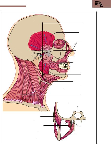
Special Anatomy of the Locomotor System 195
|
Plane of b |
|
|
M. temporalis |
|
|
Muscles of |
|
|
expression |
|
|
Lateral (external) |
|
|
pterygoid muscle |
|
|
Temporomandibular |
|
|
joint |
|
|
M. masseter |
|
|
Suprahyoid muscles |
|
|
Hyoid bone |
|
|
Sternocleidomastoid muscle |
|
|
M. trapezius |
|
|
Greater wing of sphenoid |
|
a |
Sphenoid sinus |
|
|
||
M. temporalis |
|
|
Zygoma |
|
|
Lateral (external) |
Medial and |
|
pterygoid muscle |
lateral pterygoid |
|
M. masseter |
plate |
|
Medial pterygoid |
||
Mandible |
||
muscle |
||
b |
|
Fig. 4.56 a, b Lateral view (a) and coronal section (left side only) (b) of the muscles of mastication (highlighted in red)
196 4 The Locomotor System (Musculoskeletal System)
Epicranial aponeurosis |
M. orbicularis oculi |
M. depressor |
(galea aponeurotica) |
|
supercilii |
Frontal belly of m. |
|
|
occipitofrontalis |
|
M. corrugator |
|
|
|
Temporoparietal |
|
supercilii |
|
|
|
muscle |
|
|
|
|
Mm. levator |
|
|
labii superioris |
|
|
et alae nasae |
Auricular muscles
M. levator labii |
Nasal muscles |
|
|
||
superioris |
M. levator |
|
|
||
|
anguli oris |
|
M. zygomaticus minor |
M. orbicularis |
|
|
||
M. zygomaticus major |
oris |
|
|
||
M. buccinator |
M. depressor |
|
labii inferioris |
||
|
||
M. risorius |
|
|
M. depressor anguli oris |
M. mentalis |
Fig. 4.57 Overview of the muscles of facial expression (superficial muscles colored light, deep musckes dark)
movements of the temporomandibular joint take place around one axis that runs from external and anterior to internal and posterior, and one that runs obliquely up and down. The human temporomandibular joint carries out two main functions during the act of mastication: (1) elevation (adduction) and depression (abduction) of the mandible and (2) grinding motions. Every time the mouth opens, the head of the mandible and the articular disk are pulled forward on the articular tubercle. If the forward pull occurs on only one side the result is a grinding motion.
|
|
Summary |
197 |
Joint cartilage |
Glenoid cavity |
Postglenoid |
|
|
|
(mandibular fossa) |
tubercle |
Brain |
|
|
|
Articular disk |
Lateral pterygoid |
||
|
muscle |
||
|
|
|
|
Mastoid process |
|
|
|
with air cells |
|
|
|
|
|
Pterygoid process |
|
Auditory meatus |
|
|
|
Head of mandible |
|
|
|
Joint cavity |
Coronoid process |
||
|
|
||
Mandible |
|
|
|
Fig. 4.58 |
Anatomy of the temporomandibular joint. (After Faller) |
|
|
Summary The Locomotor System (Musculoskeletal System)
Locomotor System
General Anatomy of the Locomotor System
The skeletal system and the muscles together constitute the locomotor system, in which the bones and the joints comprise the passive motor system and the striated muscles the active motor system.
The Bones
Bones may be long bones (pipe bones), flat bones (e. g., skull), short (e. g., wrist and ankle bones), irregular (e. g., vertebrae), and bones containing air-filled cavities (e. g., frontal bone), as well as sesamoid bones (e. g., kneecap).
The Joints
Joints may be movable or immovable. The immovable joints (synarthroses) include syndesmoses, synchondroses, and synostoses. In

198 4 The Locomotor System (Musculoskeletal System)
movable joints (synovial joints, diarthroses), the joint surfaces are covered with cartilage and are separated by a joint space. Inside the joint, the articular cavity is filled with synovial fluid and enveloped in a joint capsule.
Joints may be classified according to the shape of the joint surfaces
as:
Ball-and-socket joints (hip joint, shoulder joint)
Condylar joints (wrist joint)
Trochlear joints
−Hinge joints (elbow joint)
−Pivot joints (proximal radioulnar joint)
Saddle joints (saddle joint of the thumb)
Plane joints (articular joints of the vertebrae)
Joints with mobility limited by strong ligaments (sacroiliac joints)
Skeletal Muscle
As a rule, a skeletal muscle consists of one or more heads, a muscle belly, and a proximal tendon of origin as well as a distal tendon of insertion connecting the muscle with a bone. A muscle may have multiple bellies if it has tendons between them, and it may be monarthric (uniarticular, monarticular) or polyarthric (multiarticular) depending on whether it runs over one or more joints.
The length and force of lift of a muscle depend on the way the muscle fibers are arranged on the tendons, that is, from the angle at which they are inserted. Because of their long fibers, muscles with parallel fibers achieve a great height of lift but relatively little force of lift, since their physiological cross-section is small. Unipennate, and especially bipennate, muscles have a large physiological cross-section and so develop considerable strength, but because of their short muscle fibers their height of lift is small.
Tendons transfer the force of a muscle contraction from the muscle to the skeleton. There are very short tendons (“fleshy” origin; e. g., pectoralis major), long, narrow tendons (muscles of the hand and foot), and flat tendons (“aponeuroses”; e. g., oblique abdominal muscles). The tendons may exert pull or pressure. In the latter case, they wind around a bone that acts as a fulcrum (e. g., tendon of insertion of the peroneus longus).
Summary 199
Muscle fasciae, tendon sheaths, bursae, and sesamoid bones are auxiliary structures for muscles and tendons. Their function is to reduce friction during muscular work and so reduce effort.
Special Anatomy of the Locomotor System
The Skeleton of the Trunk
The skeleton of the trunk includes the vertebral column and the chest cage (thorax), which includes the ribs, the breastbone (sternum), and the thoracic spine.
The Spinal Column
The spinal column consists of 33−34 vertebrae: 7 cervical, 12 thoracic, 5 lumbar, 5 sacral (fused in the sacrum), and 4−5 coccygeal (fused in the coccyx). It forms two S-shaped curves (cervical and lumbar lordoses, thoracic and sacral kyphoses); its spinal canal contains the spinal cord.
The basic vertebra consists of a vertebral body, a vertebral arch, a spinous process, two transverse processes and four articular processes. The vertebral body and the vertebral arch enclose the vertebral foramen. Together, the vertebral foramina form the spinal canal. The spinal nerves leave the spinal canal through the intervertebral foramina between two adjacent vertebrae.
Two adjoining vertebrae form one independently mobile segment, consisting of the two vertebrae, the intervertebral disk between them, the synovial joints of the vertebral arch, the ligaments, and the corresponding muscles.
The atlas supports the head and with the occipital condyles forms the atlanto-occipital joint (inclines the head laterally, forward and backward). With the axis it forms the atlanto-axial joint (rotation of the head).
The Chest Cage
The chest cage is composed of 12 pairs of ribs. The first seven of these (true ribs) are connected to the sternum directly. The 8th to 10th pairs of ribs take part in the formation of the costal margin, while the 11th and 12th ribs end unattached in the lateral abdominal wall. Each rib in the 1st to 10th pairs is connected to the vertebrae by two costovertebral joints. The movements of the ribs (expansion and contraction of
200 4 The Locomotor System (Musculoskeletal System)
the thorax) subserve respiration (muscles of respiration, including intercostal muscles).
The Muscles of the Trunk
The muscles of the trunk include back, thoracic, and abdominal muscles as well as diaphragm
The Back
Segmental muscles of the back: m. erector spinae
Muscles encroaching on the back: mm. trapezius, rhomboideus major and minor, together with levator scapulae (shoulder girdle muscle) and latissimus dorsi (muscle of the upper extremity)
The Thoracic Wall
Intercostal muscles (external, internal, intimi)—inspiration, expiration; scalenus muscles—inspiration
Muscles encroaching on the trunk: m. serratus anterior—abduc- tion of the arm, auxiliary muscle of respiration; m. pectoralis major—adduction, internal rotation of the upper arm, auxiliary muscle of respiration; sternocleidomastoid muscle, also auxiliary muscle of respiration.
The Abdominal Wall
Muscles: m. rectus abdominis, external and internal oblique muscles, mm. transversus abdominis, psoas major, and quadratus lumborum— rotation, flexion, lateral inclination of the trunk, compression of the abdomen.
The Diaphragm
The most important muscle of respiration (contraction—inspiration, relaxation—expiration).
The Pelvic Floor
Levator ani muscle—forms the pelvic diaphragm; m. transversus perinei profundus—forms the urogenital diaphragm; medial part of the levator ani (puborectalis), m. sphincter ani externus—voluntary constriction of the anus; m. sphincter urethrae—voluntary constriction of the urethra.
Summary 201
The Upper Extremity
The Shoulder Girdle
Bones: clavicle and shoulder blade (scapula)
Joints: sternoclavicular joint, acromioclavicular joint between clavicle and shoulder blade
Muscles: mm. trapezius, serratus anterior, levator scapulae, romboideus major and minor.
Free Upper Limb
Bones: (1) Upper arm (brachium)—humerus. (2) Forearm (ante- brachium)—radius and ulna. (3) Hand (manus)—wrist (carpus) with eight bones (navicular or scaphoid, lunate, triquetrum, pisiform, trapezium, trapezoid, capitate, hamate bones); metacarpus with 5 metacarpal bones; 5 fingers, each with 3 phalanges, but only two for the thumb.
Shoulder joint: ball-and-socket joint between head of humerus and glenoid cavity of the shoulder blade.
Muscles: muscles of the rotator cuff (m. supraspinatus—abduc- tion, mm. infraspinatus and teres minor—external rotation; subscapular—internal rotation of the arm), deltoid muscle (abduction, extension, and flexion of the arm), mm. pectoralis major, latissimus dorsi, and teres major (adduction, external and internal rotation).
Elbow joint (articulatio cubiti): compound hinge joint composed of three separate joints. (1) humeroradial joint, (2) humeroulnar joint, (3) proximal radioulnar joint.
Muscles: m. triceps brachii (extension at the elbow joint); m. brachialis (flexion), biceps brachii (flexion, supination); m. supinator (supination); mm. pronator teres and pronator quadratus (pronation).
Joints of the hand: (1) proximal wrist joint (radiocarpal joint—be- tween ulna and radius as well as the proximal row of carpal bones); (2) middle carpal joint (articulatio mediocarpea—between the distal and proximal row of carpal bones); (3) saddle joint of the thumb (articulatio carpometacarpea pollicis).
Muscles: 18 short muscles of the hand, together with 15 forearm muscles (flexors, extensors, radial group). 9 muscles take part just in the movements of the thumb.

202 4 The Locomotor System (Musculoskeletal System)
The Lower Extremity
The Pelvic Girdle
Bones: the two hip bones (ossa coxae; each composed of ilium, ischium, pubic bone) and the sacrum together form the bony pelvic girdle.
Joints: sacroiliac (between sacrum and ilium)
Muscles: The muscles of the pelvis are part of the musculature of the trunk (see Pelvic Floor).
Free Lower Limb
Bones: (1) Thigh, femur (thighbone). (2) Patella (kneecap). (3) Lower leg (crus)—tibia and fibula. (4) Foot (pes)—ankle (tarsus) composed of 7 bones (talus, navicular, 3 cuneiform bones, calcaneum, cuboid bone); metatarsus with 5 metatarsal bones; 5 toes (digits) with 3 phalanges each, except 2 phalanges for the big toe (hallux).
Hip joint: ball-and-socket joint between the head of the femur (caput femoris) and the acetabulum of the hip bone (os coxae).
Muscles: mm. iliopsoas (flexion), sartorius (flexion), rectus femoris (flexion), gluteus maximus (extension); posterior (ischiocrural) group (mm. biceps femoris, semitendinosus, semimembranosus—extension); mm. gluteus medius and gluteus minimus (abduction, internal and external rotation); adductor group (5 muscles, e. g., m. adductor magnus—adduc- tion).
Knee joint (articulatio genus): compound joint. (1) Between femoral condyle and tibia, (2) between patellar surface of femur and patella.
Muscles: mm. semimembranosus and semitendinosus (internal rotation, flexion), biceps femoris (external rotation, flexion), sartorius (flexion), quadriceps femoris (extension).
Tibiofibular joint: syndesmotic joint.
Ankle joint (articulatio talocruralis): the distal ends of the tibia and fibula grasp the talus and trochlea of the talus in a malleolar mortise, forming a hinge joint.
Muscles: mm. gastrocnemius and soleus (triceps surae), tibialis posterior, flexor digitorum longus, flexor hallucis longus, peroneus longus and brevis (plantar flexion); flexor digitorum lon-

Summary 203
gus and flexor hallucis longus (plantar flexion of the toes); tibialis anterior, extensor digitorum longus, and flexor hallucis longus (dorsiflexion).
Talocalcaneonavicular joint: two separate joints coupled functionally. (1) Calcaneum and talus. (2) Talus and navicular bone.
Muscles: mm. soleus and gastrocnemius (triceps surae muscle), tibialis anterior and posterior, flexor digitorum longus, flexor hallucis longus (supination); peroneus longus and brevis, extensor digitorum longus (pronation).
Head and Neck
The Neck
The neck (collum) joins the trunk to the head; the posterior region is called the nape of the neck or the back of the neck (nucha), the anterior the neck proper (cervix). The organs of the neck (throat [pharynx], cervical portion of the esophagus, larynx, cervical part of the air passages [trachea]), lie in front of the vertebral column, and are covered by the fasciae and muscles of the neck (muscles: posteriorly, segmental muscles of the neck, m. trapezius; laterally, sternocleidomastoids; anterior and lateral to the vertebral column, muscles derived from the muscles of the trunk [mm. scaleni, prevertebral muscles such as mm. longus capiti and longus colli]; anterior to the organs of the neck, infrahyoid muscles and platysma).
The Head
The bony foundation of the head is the skull, which consists of the brain case (neurocranium) and a facial skeleton (splanchnocranium) (boundary: line from the root of the nose—upper orbital rims—exter- nal auditory meati).
Bones of the neurocranium: (1) Cranial vault: frontal bone, paired parietal bones, parts of both temporal bones, uppermost part of the occipital bone. (2) Internal aspect of the base of the skull: frontal bone, ethmoid bone with the cribriform plate, crista galli, sphenoid bone, occipital bone, temporal bones.
In the newborn, gaps filled with connective tissue occupy the space between bones (fontanelles). They close between the 3rd and 36th month of life and are then replaced by bony sutures (coronal, lambdoid, sagittal, squamous sutures).
204 4 The Locomotor System (Musculoskeletal System)
Bones of the facial skeleton: upper jaw (maxilla, with a process for the teeth [alveolar arch]), zygoma, nasal bone, lacrimal bone, palatine bone, vomer, mandible (with alveolar arch).
Base of skull: bony base of the cranial cavity, on which the brain rests. The foramen magnum is seen if the skull is viewed from below (inferior aspect of base of skull). This is where the vertebral column is connected to the skull by the atlanto-occipital joint. The superior aspect of the base of the skull is part of the neurocranium (see above). It forms the boundary between neurocranium and facial skeleton.
Muscles of the skull: (1) Mandibular muscles (masticatory muscles): masseter, lateral and medial pterygoid, temporalis, syprahyoid muscles. (2) Muscles of facial expression: 21 muscles. They have no fascia and extend from skin to bone (cutaneous muscles).
Temporomandibular joint (TMJ): hinge joint between mandible and temporal bone.