


534 13 The Central and Peripheral Nervous Systems
Classification of the Nervous System
The nervous system connects the organism with its environment (somatic nervous system) and its internal organs (autonomic or vegetative nervous system). The characteristics of the somatic nervous system are conscious sensation, voluntary movement, and rapid processing of information. The vegetative nervous system, on the other hand, is responsible for maintaining a constant internal milieu (homeostasis) and for the autonomous regulation of organ functioning in response to environmental demands.
The somatic and the autonomic nervous systems include sensory (afferent) as well as motor (efferent) connections. Afferent conduction occurs when impulses are conducted from the periphery (e. g., sensations from skin or internal organs) to the center (brain and spinal cord). In efferent conduction, impulses are conducted from the center to the periphery (e. g., skeletal muscle, smooth muscle, gland cells) (Fig. 13.1).
By its spatial distribution the nervous system is divided into a central nervous system (CNS) and a peripheral nervous system. The CNS includes the brain and spinal cord. The peripheral nervous system includes all somatic and autonomic nerves, including the collections of nerve cells called ganglia.
Role of the Nervous System
The central and peripheral nervous systems coordinate the performance of organ systems directly (through nerves) or indirectly (through endocrine glands). They regulate the activities of the locomotor apparatus, the respiratory, circulatory, digestive, and urogenital systems, and the system of endocrine glands. Integration and evaluation of incoming impulses occur in the central nervous system, while the peripheral nervous system conducts impulses originating in the CNS to the periphery of the body and conducts impulses from the periphery to the CNS. (For a discussion of the generation, conduction, and transmission of nerve impulses, see Chapter 3: The Nerve Impulse [Action Potential], and Chapter 3: The Synapse.) Beyond these functions, the so-called “higher functions” of the nervous system (memory, ability to learn and think, judgment, language) are tied to the activity of the central nervous system.

Development of the Nervous System 535
Joints, skin, |
|
Skeletal |
|
muscles |
|
skeletal |
|
|
muscle |
|
|
Somatic |
|
Motor |
Afferent pathways |
CNS |
Efferent pathways |
Visceral |
|
Autonomic |
|
|
Glands, smooth |
Inner organs |
|
muscle, cardiac |
|
muscle |
|
|
|
Fig. 13.1 Diagram showing the connections between the central and peripheral nervous systems. CNS = brain and spinal cord
Development of the Nervous System
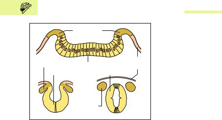
The components of the central and peripheral nervous system develop from the outer germ layer, the ectoderm. The first rudiment appears about the 18th day of embryonic development as a platelike thickening of the ectoderm of the embryonic disk (neural plate) (Chapter 12: Derivatives of the Germ Layers). Within the neural plate, between two lateral folds (neural folds), develops a depression—the neural groove—which subsequently closes to form the neural tube and migrates inward. Parts of the neural folds that do not take part in the formation of the neural tube form the neural crests (Fig. 13.2a−c).
The neural tube becomes the central nervous system (brain and spinal cord), while the neural crests form the peripheral nervous system (peripheral nerves and ganglia).
|
Central Nervous System |
537 |
|
Forebrain |
|
Cephalic flexure |
|
(prosen- |
|
|
|
cephalon) |
|
|
|
Midbrain |
|
Pontine flexure |
|
|
|
|
|
(mesencephalon) |
|
Cervical flexure |
|
|
|
||
Hindbrain |
Notochord |
|
|
(rhomben- |
(chorda dorsalis) |
|
|
cephalon) |
|
|
|
a |
Spinal cord |
b |
Spinal cord |
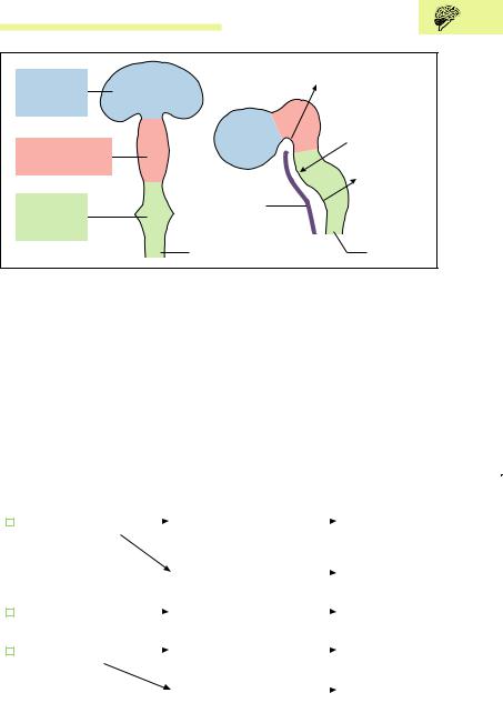
Fig. 13.3 a, b Development of the brain (stage of three primary brain vesicles).
(After Faller)
a View from above
bLateral view. The three primary embryonic brain vesicles form at the anterior end of the neural tube; the posterior end becomes the spinal cord
brain) formed from the anterior end of the neural tube (Fig. 13.3a, b). The posterior part of the neural tube becomes the spinal cord. The wall of the neural tube develops into the gray and white matter of brain and spinal cord, while its lumen develops into a system of cavities in the brain (ventricles) and a narrow canal (central canal) in the spinal cord.
The derivatives of the primary brain vesicles are as follows:
|
Forebrain |
|
Cerebrum |
|
Lateral ventricles |
||||
L |
|
|
|||||||
|
|
||||||||
|
(prosencephalon) |
(telencephalon) with the |
|
||||||
|
|
|
|
|
cerebral hemispheres |
|
|||
|
|
|
|
|
Diencephalon |
|
|
Third ventricle |
|
|
|
|
|
|
|
|
|||
|
|
|
|
|
(interbrain) |
|
|||
|
Midbrain |
|
|
Midbrain |
|
|
Aqueduct |
||
L |
|
|
|
|
|
|
|||
|
|
|
|
||||||
|
(mesencephalon) |
|
|
|
|
|
|||
|
Hindbrain |
|
Pons and |
|
Fourth ventricle |
||||
L |
|
|
|
|
|||||
|
|
||||||||
|
(rhomben- |
cerebellum |
(superior part) |
||||||
|
cephalon) |
Medulla |
|
|
Fourth ventricle |
||||
|
|
|
|
|
|
||||
|
|
|
|
|
|
||||
|
|
|
|
|
oblongata (bulb) |
(inferior part) |
|||

538 13 The Central and Peripheral Nervous Systems
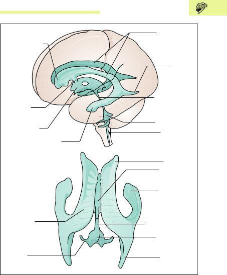
The ventricular system (see Cerebrospinal Fluid [CSF] and the Ventricular System below) consists of four cavities inside the brain, namely, the two lateral ventricles, the third ventricle of the midbrain, and the fourth ventricle of the hindbrain. They are interconnected (e. g., the third and fourth ventricle by the aqueduct = aqueduct of Sylvius) and are filled with cerebrospinal fluid.
The Brain (Encephalon)
The brain of a newborn weighs about 400 g and grows in the course of the first 9 months of life to about 800 g. It reaches close to its final weight of about 1310 g at about 5−7 years of age and is fully formed at 10 years. Data concerning brain weight vary remarkably (1100−1600 g). The brain of a man weighs on average 1375 g, that of a woman about 1245 g. The lesser weight in the woman is ascribed to the weaker development of her locomotor system and its consequently reduced representation in the CNS. However, when the actual brain weight is compared to body weight (brain/body weight ratio), women show on an average 22 g of brain weight per kg of body weight, while in men the figure is 20 g.
The striking example of the blue whale shows that the absolute weight of the brain is of no significance. A whale weighing about 74 000 kg has a brain weight of 7 kg. If, now, we again compare brain weight to body weight, 1 kg body weight corresponds to 0.1 g brain weight. If a similar body/brain weight ratio were applied to a human brain, it would weigh on average 7 g.
Cerebrum or Forebrain (Telencephalon)
Cerebral Hemispheres
The cerebrum is the highest integration center of the CNS and for this reason is the most markedly differentiated segment of the human brain. Essentially it is composed of two structures: the two cerebral hemispheres and several bilateral gray nuclei (basal ganglia). The latter have a partial role in motor activity, especially the initiation and performance of slow movements. They lie deep inside the hemispheres and cannot be seen
Central Nervous System 539
Pineal gland (epiphysis) |
Cerebral hemisphere |
|
Connecting duct |
(telencephalon) |
|
|
||
third to fourth |
Corpus callosum |
|
ventricle (cerebral |
||
|
||
aqueduct) |
|
|
|
Diencephalon |
|
Cerebellum |
(interbrain) |
|
|
||
Midbrain |
|
|
(mesen- |
|
|
cephalon) |
|
|
Pons |
|
|
Bulb |
|
|
(medulla |
|
|
oblongata) |
|
|
Pituitary gland |
|
|
(hypophysis) |
|
|
Spinal cord |
|
|
(medulla spinalis) |
|
|
Brainstem |
|
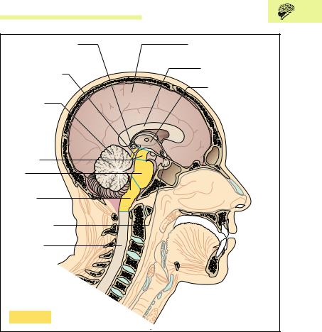
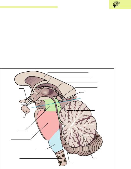
Fig. 13.4 Organization of the brain. Sagittal section through the head of an adult man; view of the left half from the medial side. The midbrain, pons, and medulla oblongata together form the brainstem
until the brain is cut. The two voluminous cerebral hemispheres are separated by a deep division, the longitudinal fissure (Fig. 13.7) and comprise the major part of the visible substance of the brain.
540 13 The Central and Peripheral Nervous Systems
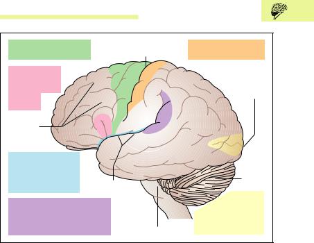
Lobes of the Brain (Cerebral Lobes)
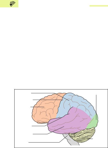
The surface of the brain is formed by convolutions (gyri) separated by fissures (sulci). The two lateral sulci and the central sulcus may be considered to divide each hemisphere into four lobes (Fig. 13.5):
Frontal lobe
Parietal lobe
Temporal lobe
Occipital lobe
The frontal lobe lies in front of the central sulcus, the parietal lobe behind it. The temporal lobe lies below the lateral sulcus, and an imaginary line drawn down from the parieto-occipital sulcus separates the parietal lobe from the occipital lobe. Deep inside the lateral sulcus lies the insula (island), covered by the frontal, parietal, and temporal lobes. The insula is often regarded as a fifth lobe. It has no known function in the human brain.
Parietal lobe |
Occipital lobe |
Central fissure (sulcus centralis)
Frontal lobe
Lateral sulcus
Temporal lobe
Cerebellum
Brainstem
Fig. 13.5 Lobes of the cerebrum. Seen from the left side. (After Frick et al)
Central Nervous System 541
Each lobe in turn has its specific convolutions and fissures. For instance, the frontal lobe comprises the precentral gyrus, which lies immediately in front of the central fissure. It is the motor center that sends impulses to the voluntary muscles. The point situated closest to the forehead, the frontal pole, is the seat of personality.
Gray and White Matter
In a brain cut in the horizontal or coronal plane, the cerebral hemisphere can be seen to consist of an outer gray cortical layer; the cerebral cortex, about 2−5 mm thick, mainly composed of cell bodies; and an inner white core composed mainly of nerve fibers (myelinated axons) (Figs. 13.6a, b and 13.7a, b). The cerebral cortex has a total surface of about 2,200 cm2, with about 10 000−20 000 nerve cells per mm3. This may be contrasted with the white matter, which houses about 300 000−400 000 km of nerve fibers.
Medullary Tracts and Internal Capsule
Fibers interconnecting the hemispheres are called commissural fibers. One example is the vast corpus callosum (Figs. 13.4 and 13.6a), a fiber system containing about 200 million nerve fibers. Nerve fibers confined to one hemisphere are called association fibers. These run within the same hemisphere from lobe to lobe or fissure to fissure. Finally, there are fibers that run from the cerebral cortex to other parts of the CNS, called projection fibers. The majority of these form the internal capsule (Figs. 13.6a, b and 13.7a, b). This structure consists of an anterior limb (crus anterius), a posterior limb (crus posterius), and a segment between them, the genu (knee).
Basal Ganglia
Several basal ganglia are located next to the genu of the internal capsule (Figs. 13.6a, 13.7a, and 13.8), e. g., the globus pallidus and the putamen. The globus pallidus and the putamen together are also known as the lentiform nucleus. The anterior limb of the internal capsule is bounded by the caudate nucleus and the lentiform nucleus, which together form the corpus striatum (striate body). The posterior limb of the
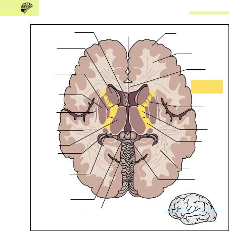
542 13 The Central and Peripheral Nervous Systems
Anterior horn of |
Fornix |
Cerebral gray matter |
lateral ventricle |
|
(cerebral cortex) |
Head of |
|
White matter |
caudate |
|
|
nucleus |
|
|
Globus |
|
Corpus |
|
callosum |
|
pallidus |
|
Internal |
|
|
|
Putamen |
|
capsule |
|
|
|
Claustrum |
|
Anterior |
(barrier) |
|
crus (crus |
|
|
anterius) |
Acoustic |
|
Genu |
|
(knee) |
|
radiation |
|
|
|
Posterior |
|
Thalamus |
|
|
|
crus (crus |
|
|
|
|
Occipital |
|
posterius) |
horn of |
|
|
lateral ventricle |
|
Optic radiation |
Tail of caudate |
|
|
|
|
|
nucleus |
|
Pineal gland |
|
|
(epiphysis) |
Third ventricle |
|
|
Quadrigeminal plate |
Plane of cut |
|
(lamina tecti) |
|
|
|
|
|
a |
|
b |
Fig. 13.6 a Horizontal section through the cerebrum and diecephalon. The internal capsule is shown in yellow. b Plane of the horizontal section
internal capsule is bounded medially by the thalamus. This is an important nuclear area of the diencephalon and is not considered part of the basal ganglia.
The basal ganglia have an important function in the so-called extrapyramidal motor system (see later in this chapter). The current view is that their function is to regulate the extent and direction of voluntary movements. The symptoms of lesions of the basal ganglia include disturbances of muscle tone and involuntary movements.
Central Nervous System 543
|
Longitudinal fissure |
|
|
|
(fissura longitudinalis) |
Gray matter of |
|
|
|
||
Choroid plexus in |
|
cerebral cortex |
|
|
|
||
central part of |
|
White matter |
|
lateral ventricle |
|
||
|
|
||
Caudate |
|
Corpus |
|
|
callosum |
||
nucleus |
|
|
|
|
|
Internal |
|
Thalamus |
|
capsule |
|
|
|
||
Globus |
|
|
|
pallidus |
|
Lateral |
|
|
|
fissure |
|
|
|
(sulcus |
|
Tail of the |
|
lateralis) |
|
|
|
||
caudate |
|
Insula |
|
nucleus |
|
|
|
Third ventricle |
Plane of cut |
Claustrum |
|
a |
(barrier) |
||
|
|||
Mammillary body |
|
Putamen |
|
|
|
Choroid plexus in |
|
|
|
temporal horn of |
|
|
|
lateral ventricle |
|
|
b |
|
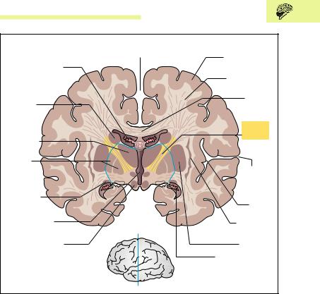
Fig. 13.7 a Coronal section through the cerebrum and diencephalon. The broken blue line shows the junction of the diencephalon and the telencephalon. The internal capsule is highlighted in yellow. b Plane of the coronal cut
Lateral Ventricles
The lateral ventricles are situated in the two cerebral hemispheres. They are a part of a system of cavities (ventricles) that contain the cerebrospinal fluid (CSF). The CSF is formed by an arterial plexus (choroid plexus) that extends into both lateral ventricles. The lateral ventricles possess a frontal horn, a body, an occipital horn, and a temporal horn (see Cerebrospinal Fluid [CSF] and the Ventricular System below and Fig. 13.25a, b). Each
544 13 The Central and Peripheral Nervous Systems
Thalamus |
|
Globus |
Putamen |
pallidus |
|
|
Caudate nucleus |
|
Amygdala (corpus |
|
amygdaloideum) |
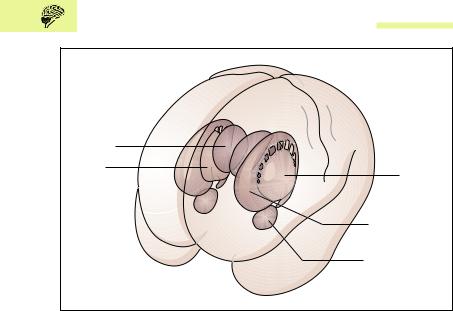
Fig. 13.8 Location of the basal ganglia seen from the front and left. The thalamus is shown for orientation, as it is not one of the basal ganglia. Putamen and globus pallidus = lentiform body. Caudate nucleus and lentiform body = corpus striatum. (After Duus)
lateral ventricle is connected with the third ventricle of the diencephalon by an opening, the interventricular foramen.
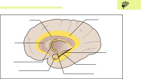
Limbic System
The term “limbic system” derives from the anatomical relation to the corpus callosum, which it surrounds like a hem (limbus) (Fig. 13.9). In the course of evolution, the limbic system was formed from an old part of the cerebrum that consisted mainly of the paleocortex and the archicortex (archipallium). The paleocortex and archicortex are often lumped together under the term allocortex, which takes up the major part of the cortex in lower mammals, for example. In humans these older parts of the brain are overgrown by the newer, strongly developed neocortex (isocortex).
The paleocortex consists essentially of the olfactory brain. The archicortex includes among other structures the amygdala (almonds) the
Central Nervous System 545
Indusium griseum |
Cingular gyrus |
|
(supracallosal gyrus) |
|
|
Corpus |
|
|
callosum |
|
|
|
Dentate |
|
|
gyrus |
|
Fornix |
Pes hippocampi |
|
|
||
Amygdala |
(foot of hippocampus) |
|
Mammillary body |
||
(corpus amygdaloideum) |
Fig. 13.9 The limbic system. Right cerebral hemisphere, medial view. The limbic system is highlighted in yellow
hippocampus, the dentate gyrus, the cingulate gyrus, and the indusium griseum (supracallosal gyrus). From the hippocampus, a fiber tract (fornix) arches to areas of the hypothlamus, especially the mammillary bodies. From the mammillary bodies, in turn, pathways run to the reticular formation in the brainstem (see p. 555). The hippocampus and the cingulate gyrus are the central structures of the limbic system. The hippocampus plays an important role in aggression and in behavioral motivation, as well as in learning processes, particularly the formation of explicit memories, i.e., those that are accessible to deliberate, conscious recall. Short-term and long-term memory are components of explicit memory.
The limbic system evaluates experience affectively and triggers emotional reactions. Stimulation of these regions triggers rage, for example, but also pleasurable reactions. Thus, it is hardly surprising that many of their pathways end in the hypothalamus (see Diencephalon), which is the major coordinating and reflex center for many sensations such as smell and taste. Since the hypothalamus also regulates the autonomic nervous system (see p. 605), it makes sense that emotional reactions lead to autonomic disturbances (rise in blood pressure, blushing, becoming pale, etc.), while autonomic disturbances express themselves in emotions and psychosomatic illnesses.

546 13 The Central and Peripheral Nervous Systems
Functional Cortical Areas
The cerebral cortex is especially highly developed in humans (Fig. 13.10). It is responsible for the characteristics that distinguish human beings from animals. This includes, for instance, the ability to use the hand for skillful and difficult movements, highly developed language, logical thinking, personality, and conscience. All this has become known because these characteristics are lost or are severely diminished when certain cortical areas are damaged.
Of the two hemispheres, the left is dominant in 80−90 % of cases, mostly demonstrated by right-handedness (projection of the left hemisphere on the right half of the body by crossing of the nerve fibers). The two cerebral hemispheres are also known to differ in their intellectual abilities. For instance, the ability to read, speak, and write is especially marked in the left hemisphere. On the other hand, aptitudes such as memory, language comprehension, visualization of spatial relationships, and musical comprehension dominate in the right hemisphere.
The cerebral cortex in lower animals is small and almost exclusively responsible for the processing of olfactory stimuli (archicortex and paleocortex), which are among the most important sensations for these animals. The thalamus processes all stimuli from the sensory nerves and special senses, while the basal ganglia are the motor centers.
In the course of evolution the cerebral cortex has increased in size (neocortex) and has taken over other functions. For instance, the postcentral gyrus, situated behind the central fissure, has become the most important center for conscious somatic perception (somatosensory area). Meanwhile, the phylogenetically older center, the thalamus, has become a transmission and relay station for all impulses from general and special senses flowing toward the cerebral cortex and into consciousness. From the development of the motor cortex (precentral gyrus) it follows that the human basal ganglia perform only gross motor activities (see p. 542).
As the neocortex increases in size and takes over more functions, the number of neurons also increases. They are arranged in six layers. In order to be able to greatly increase its surface without increasing its volume, the cerebral cortex develops folds, the characteristic gyri and sulci. In lower mammals, as for instance the rat, the cerebral surface remains smooth.

548 13 The Central and Peripheral Nervous Systems
nized. The transverse gyri deep in the lateral sulcus of the temporal lobe form the acoustic cortex (primary acoustic cortex). They are framed by the auditory association area (secondary acoustic center) (Fig. 13.10).
Sensory Aphasia
Lesions of the dominant auditory association area (Wernicke’s area) (Fig. 13.10) (for most people, even left-handers, the left hemisphere is the dominant side for speech) lead to receptive or sensory aphasia. Affected patients do hear sounds and noises, but they have no significance. The patients behave as if they are hearing a foreign language. Aphasia is defined as the inability to derive information from speech or writing or to understand it and subsequently pass it on.
Motor Aphasia
The motor speech center (Broca’s area) can be found on the inferior frontal gyrus (Fig. 13.10). With lesions of this area on the dominant side of an adult, the patient cannot speak even though the laryngeal muscles are not paralyzed (motor aphasia). Patients know what they want to say, but all they can produce are distorted sounds or words that are repeated over and over. If the lesion occurs in childhood, the child relearns speech by using the nondominant side.
Diencephalon
The diencephalon is an area of the brain that lies between the cerebral hemispheres and surrounds the third ventricle (Figs. 13.11, 13.6, and 13.7). It consists of the thalamus, which is the central relay station of the sensory pathways (pain, temperature, pressure, touch, as well as vision and hearing) and the hypothalamus below it.
The thalamus is the major subcortical relay station for all incoming impulses except those dealing with smell (the olfactory pathway). In the thalamus, these impulses are synaptically transmitted to neurons that mostly project to the cerebral cortex. Thus, the thalamus is sometimes called “the gateway to the cortex,” or even “the gateway to consciousness.”
 Plane of cut, see
 duct third to fourth
  ventricle (cerebral
 aqueduct)
550 13 The Central and Peripheral Nervous Systems
Midbrain (Mesencephalon)
The midbrain is the smallest part of the brain, lying between the diencephalon and the pons (Figs. 13.11 and 13.12). The area above the aqueduct is the roof of the midbrain (tectum), composed of four projections resembling hills, the lamina tecti. The two upper hills form the superior colliculi, the four lower hills the inferior colliculi. Together, the four colliculi are known as the corpora quadrigemina. They give rise to optic and acoustic reflex pathways to the spinal cord.
A number of bundles of fibers run to the tegmentum (cover), which lies under the tectum. This structure also includes the red nucleus (nucleus ruber) and the nuclei of the extrinsic eye muscle nerves, cranial nerves III (oculomotor nerve) and IV (trochlear nerve). At the base of the midbrain run a pair of massive bundles of fibers, the crura cerebri, made up of descending projection fibers (e. g., pyramidal tract) from the internal capsule. The last structure of the midbrain that needs to be mentioned is the substantia nigra (black substance), which, together with the crura cerebri and the tegmentum, forms the cerebral peduncles (pedunculi cerebri). The red nucleus and the substantia nigra form the basal ganglia of the midbrain. An expanding mass above the tentorium, such as a hematoma in or adjacent to the brain, can cause acute wedging (herniation) of the midbrain in the tentorial notch, leading rapidly to a decline of consciousness, coma, and death.
Pons (Bridge) and Cerebellum
The pons and cerebellum together form the metencephalon part of the hindbrain (rhombencephalon) (Fig. 13.11). The cerebellum lies in the posterior cranial fossa under the occipital lobe of the cerebrum, separated from it by the tentorium (tent) cerebelli. Its anterior surface forms the roof of the fourth ventricle. It is connected to the midbrain, the pons, and the medulla oblongata by the cerebellar peduncles. The function of the cerebellum is to maintain equilibrium and muscle tone and to coordinate the activity of voluntary muscles (coordination of antagonistic muscle groups, e. g., flexor/extensor). It works with the basal ganglia (p. 542) in the programming of movement.
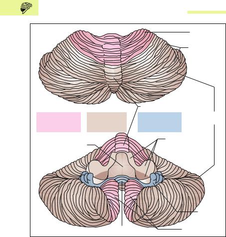
The cerebellum weighs about 130−140 g and consists of a central vermis (worm) and bilateral cerebellar hemispheres (Fig. 13.13a, b). The cere-
Central Nervous System 551
Parasympathetic accessory |
Aqueduct |
Superior colliculus |
oculomotor nuclei |
(aqueduct of Sylvius) |
|
(Edinger–Westphal nuclei) |
|
Medial lemniscus |
Area of nucleus of |
|
|
|
|
|
cranial nerve III |
|
|
(oculomotor nerve) |
|
Tectum |
Reticular formation |
|
|
|
|
|
Red nucleus |
|
Tegmentum |
(nucleus ruber) |
|
|
Substantia |
|
|
nigra (black |
|
|
substance) |
|
|
|
|
Cerebral |
|
|
peduncle |
Extra- |
|
(crus |
pyramidal |
|
cerebri) |
motor tracts |
|
|
Medial longitudinal |
Oculomotor nerve |
Pyramidal tract |
fasciculus |
(cranial nerve III) |
(tractus pyramidospinalis) |
Fig. 13.12 Transverse section through the midbrain at the level of the superior colliculus. The individual nerve fibers are arranged in somatotopic order in the pyramidal tract. The periaqueductal gray substance (substantia grisea centralis) is distributed around the cerebral aqueduct. For plane of section, see Fig. 13.11
bellar surface shows a large number of narrow, almost parallel folia (gyri) and sulci (fissures), much more marked than in the cerebrum. If the total surface of the cerebrum is compared to that of the cerebellum, the cerebellar surface is about 75 % that of the cerebrum, though with only one-tenth of its weight.
The phylogenetically oldest part of the cerebellum (archicerebellum) consists of the nodulus of the vermis and the bilateral flocculus (Fig. 13.13a, b). Together they form the flocculonodular lobe, which is responsible for the maintenance of equilibrium. The paleocerebellum is the second oldest part of the cerebellum, composed of the anterior lobes of the cerebellar hemispheres and part of the vermis. Its primary function is to regulate muscle tone. The phylogenetically youngest and largest part of the cerebellum is the neocerebellum, composed of the posterior lobes of the cerebellar hemispheres and the major part of the vermis. It is responsible for the coordination of voluntary muscles.
552 13 The Central and Peripheral Nervous Systems
|
|
|
Anterior lobe |
|
|
|
|
Posterior lobe |
|
a |
|
|
|
|
|
|
Vermis of cerebellum |
|
|
Paleocerebellum |
Neocerebellum |
Archicerebellum |
Cerebellar |
|
hemispheres |
||||
(old part of |
(new part of |
(primitive part |
||
|
||||
cerebellum) |
cerebellum) |
of cerebellum) |
|
|
|
|
Cerebellar |
|
|
Roof of fourth ventricle |
|
peduncles |
|
|
|
|
|
Flocculus |
|
b |
Nodulus |
Cerebellar tonsils |
||
|
||||
Fig. 13.13 a, b Cerebellum. (After Duus.) a Seen from above; b seen from below. The cerebellar peduncles have been divided
The cerebellar surface is made up of a gray cortex, below which is a core of white matter containing four important gray nuclei (cerebellar nuclei). The most important of these is the dentate nucleus. A median section of the cerebellum shows a treelike structure on the cut surface, called the arbor vitae (tree of life) (Fig. 13.11).

Central Nervous System 553
The tracts entering and leaving the cerebellum form the three cerebellar peduncles: the superior, middle, and inferior peduncles (Figs. 13.13 and 13.15). The pons lies between the midbrain and the medulla oblongata and is separated from the cerebellum above it by a cavity, the fourth ventricle. Several ascending and descending tracts run through the pons: it contains the nuclei of cranial nerves V, VI, and VII (trigeminal, abducens, and facial nerves) (Fig. 13.14).
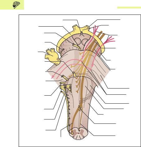
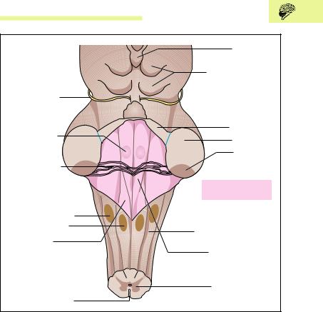
Medulla Oblongata
The medulla oblongata (myencephalon, medulla, bulb), about 4 cm long, ends at the transition between the brain and the spinal cord (Figs. 13.14 and 13.15) at the level of the foramen magnum of the skull. Anteriorly it
has a median groove |
(median |
sulcus, fissura mediana anterior) |
(Fig. 13.15), interrupted |
by the |
decussation of the pyramidal tracts |
(p. 566) (Fig. 13.14). The pyramidal tract, the most important tract of the voluntary motor system (see Voluntary Motor Tracts [Pyramidal Tracts] below) runs on each side of the groove, thickening below the pons into the pyramids. The olive, a folded gray nucleus, bulges beside it (Fig. 13.14).
The posterior surface of the medulla is partly covered by the cerebellum. If the cerebellum is removed, the floor of the fourth ventricle can be seen, known as the rhomboid fossa because of its shape (Fig. 13.15). The anterior portion of the rhomboid fossa forms part of the pons; the posterior part is part of the medulla. The bulges in the rhomboid fossa are formed by the nuclei of certain cranial nerves. On each side of the groove the medulla has two raised areas (nucleus cuneatus and nucleus gracilis), relay station in the ascending sensory tracts of the posterior spinal funiculus (fasciculi cuneatus and gracilis).
The medulla, like the pons and the midbrain, contains ascending and descending fibers and nuclei of cranial nerves (VIII to XII). It is also the seat of the respiratory and circulatory centers. If the intracranial pressure rises (e. g., due to hemorrhages or tumors), the medulla becomes compressed and this may lead to coma or death.


 (vestibulocochlear nerve)

556 13 The Central and Peripheral Nervous Systems
through the whole brainstem to the diencephalon. Its development is most marked in the tegmentum of the midbrain.
These nuclear areas receive impulses through the hypothalamic tracts and are also connected to the basal ganglia. Through descending tracts they connect with anterior horn cells and preganglionic neurons of the autonomic lateral horn (see p. 610). They also receive information from all important sensory and special sense organs (e. g., pain, temperature, pressure, touch, as well as vision and hearing), and pass this on to the thalamus, relay station of numerous sensory and motor tracts, and from there the information eventually reaches the cerebral cortex.
Among other functions, the reticular system plays an important role in the state of consciousness, that is, in alert wakefulness as well as the sleep−wake cycle. It is believed that wakefulness and/or sleep depend on the number of stimuli that reach the cerebral cortex via the reticular formation. If, for instance. the number of environmental stimuli decreases, attention flags and this leads to transition to a state of sleep. On the other hand, an increase in the number of stimuli reaching the cerebral cortex increases attention and this leads to transition to the waking state.
The Electroencephalogram (EEG)
Every stimulation of nerve cells in the brain elicits rhythmic potential oscillations of a few microvolts. Hence, as with the ECG, these oscillations can be conducted from the body surface (scalp) by metal electrodes and recorded as an electroencephalogram. The waves (α, , δ, and θ waves) of the EEG vary from region to region, especially in their height (amplitude) and frequency. Hence it is possible to make relatively crude interpretations concerning normal and pathological activities of the brain.
Sleep and Wakefulness
As in almost all living forms, an internal (biological) clock regulates the normal human sleep−wakefulness cycle. Such sleep−wake cycles are called circadian rhythms, because they correspond to about (= Latin circa) the duration of one day (= Latin dies). In actual life, these circadian

Central Nervous System 557
rhythms are adapted to the 24-hour rhythm of the day by external time divisions (work, leisure, and sleep phases, as well as light and dark periodicity).
While an awake organism is actively connected to the environment and reacts to external stimuli, contact with the surroundings is largely suspended during sleep. Nevertheless, the electroencephalogram demonstrates that sleep is not simply a state of cerebral rest, but simply an alternative state of consciousness. Depending on the depth of sleep, the EEG changes in characteristic ways. Several stages of sleep can be distinguished and these are repeated several times during the night, with a lightening of the depth of sleep toward morning.
REM Sleep
A special role is taken by the stage of sleep called REM (rapid eye movement) sleep, characterized by jerky eye movements and distinctly lower muscle tone. Such REM stages last on average 10−20 minutes and repeat about every 90 minutes. The experience of dreams frequently present during REM sleep mirrors the lively activity of the central nervous system during these stages. The percentage of REM sleep relative to total sleep is noticeably high in infants and small children (about 50 %). It is believed that this high proportion of REM sleep accompanies increased neuronal activity and is important for maturation of the brain. With increasing age, the relative proportion of REM sleep declines and amounts to about 20 % of total sleep in the adult.
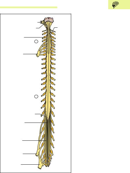
Spinal Cord (Medulla Spinalis)
The spinal cord is a cylindrical cord, about the thickness of a finger and about 40−45 cm long, that runs in the vertebral canal from the foramen magnum to the level of the 1st to 2nd lumbar vertebra. In it run the ascending and descending nerve tracts that connect the peripheral nerves with the brain. The peripheral nerves leave the spinal cord in 31 pairs of spinal nerves (Figs. 13.16 and 13.17). The spinal cord is thickened above and below, where there is a special accumulation of nerve cells (intumescentia cervicalis and lumbosacralis) (Fig. 13.16).

558 13 The Central and Peripheral Nervous Systems
These nerve cells, with their set of spinal nerves, supply the upper and lower limbs.
Spinal Nerves
Each pair of spinal nerves leaves the vertebral canal between two adjacent vertebrae through the intervertebral foramen (Fig. 3.17). The number of spinal nerve pairs corresponds to the number of vertebrae with one exception (in the cervical region eight pairs of spinal nerves, seven cervical vertebrae). There are eight spinal nerves from the cervical spine (C1− C8), 12 thoracic (T1−T12) from the thoracic spine, five lumbar (L1−L5) from the lumbar spine, five sacral (S1−S5) from the sacral spine, and 1 or 2 coccygeal from the coccyx (Co1−Co2). The first pair of spinal nerves (C1) leaves the vertebral canal between the base of the skull and the 1st cervical vertebra (atlas). Each part of the spinal cord giving rise to a pair of nerves is called a segment.
Spinal Nerve Roots
The spinal nerves are derived from anterior and posterior roots (radix anterior and posterior) (Fig. 13.18a) that arise from the spinal cord in the form of root filaments of spinal nerves (fila radicularia nervi spinalis) (Fig. 13.16) The posterior root is marked by a swelling (dorsal root ganglion) that contains the sensory nerve cells of the afferent nervous pathways from the periphery. The efferent nervous pathways from the spinal cord to the periphery run exclusively in the anterior roots (Fig. 13.18). Both roots come together just below the dorsal root ganglion and form a mixed spinal nerve (afferent and efferent nerve fibers), that, after a short course (1.5 cm), first divides into an anterior and a posterior branch (r. dorsalis and r. ventralis) (Figs. 13.18a and 13.29).
The roots of the upper spinal nerves run a more or less horizontal course and are relatively short. The further down the nerve, the more obliquely downward and the longer is its course in the vertebral canal before it emerges from the intervertebral foramen. During development, the spinal cord and spinal canal are equal in length, so that every spinal nerve leaves through the intervertebral foramen of its level. However, as development proceeds, the vertebral canal increases in length much more than the spinal cord, so that the lower end of the cord lies at an
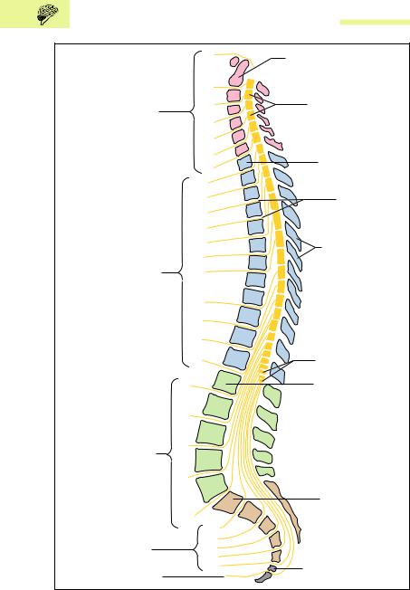


Central Nervous System 561
ever higher level in relation to the surrounding vertebrae. The lower the point of origin of the roots from the spinal cord, the longer is their course in the vertebral canal (Fig. 13.17). Below the end of the spinal cord (conus medullaris) at the level of the 1st or 2nd lumbar vertebra, the vertebral canal contains only nerve roots, which are called cauda equina (horse’s tail) because they resemble the tail of a horse (Fig. 13.16). These anatomical relationships make it possible, for instance, to obtain cerebrospinal fluid (CSF, see p. 578) without danger of damaging the spinal cord, either for diagnostic purposes (lumbar puncture) or to inject substances such as local anesthetics in order to attain anesthesia of the lower part of the body (spinal anesthesia).
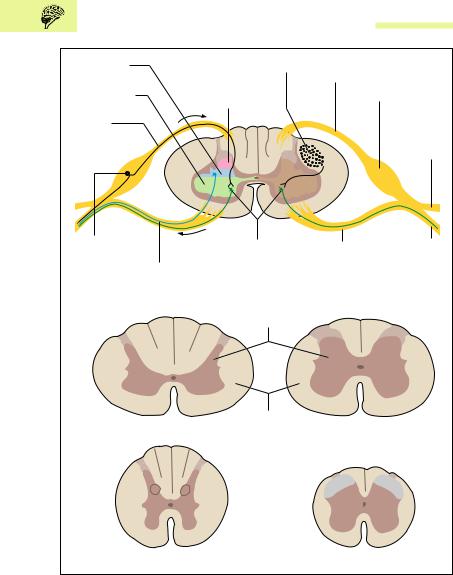
Gray and White Matter of the Spinal Cord
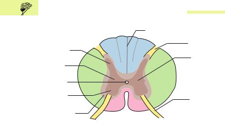
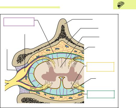
The spinal cord is divided into two symmetrical halves by the connective tissue dorsal median septum of the spinal cord (septum medianum posterium medullae spinalis) and a deep anterior groove (fissura mediana anterior medullae spinalis) (Fig. 13.19). In cross-section, two different areas that vary in different regions of the spinal cord can be distinguished: the gray substance (substantia grisea), shaped like a butterfly in cross-section and varying in size, and the white substance (substantia alba) surrounding it (Fig. 13.18). Exactly as in the cerebral hemispheres, the gray substance is composed primarily of cell bodies and the white substance of myelinated nerve fibers. The dorsal projections of the gray substance are called dorsal horns (cornua posteriora) and the anterior projections anterior horns (cornua anteriora). The thoracic region also contains a lateral horn (cornu laterale) lying between the dorsal horn and the anterior horn. In the middle of the gray substance runs the fluidfilled central canal.
Anterior horns: The anterior horns contain motor nerve cells, the axons of which leave the spinal cord through the anterior root and mainly supply striated muscle.
Posterior horns: The posterior horns contain sensory nerve cells, on which the afferent nerve fibers coming from the periphery end in synapses and are relayed.
Fig. 13.17 Lateral view of the vertebral canal with spinal cord and emerging spinal nerves. (After Kahle)
562 13 The Central and Peripheral Nervous Systems
Lateral horn |
Left pyramidal |
Posterior root |
(cornu laterale) |
tract |
(radix posterior) |
Anterior horn |
Posterior horn |
Spinal ganglion |
(cornu anterius) |
(cornu posterius) |
|
Afferent |
|
Posterior |
sensory nerve |
|
branch of |
fibers |
a |
spinal nerve |
|
(r. posterior) |
|
|
|
Pseudounipolar |
|
Anterior horn |
Anterior root |
Anterior |
nerve cell |
|
motor cells |
of spinal nerve |
branch of |
|
Efferent |
|
(radix anterior) |
spinal nerve |
a |
autonomic |
|
|
(r. anterior) |
nerve fiber |
|
|
|
Gray matter (substantia grisea)
|
White matter |
|
Cervical spinal cord |
(substantia alba) |
Lumbar spinal cord |
b Thoracic spinal cord Sacral spinal cord
Fig. 13.18 a, b Cross-section through the spinal cord
a With anterior and posterior roots; b at four different levels

Central Nervous System 563
Lateral horns: The lateral horns contain motor cells of the autonomic system, the axons of which also leave the spinal cord in the anterior roots and run, for example, to smooth muscle or glands.
Ascending and Descending Tracts of the Spinal Cord
The white substance of the spinal cord is composed mainly of ascending (afferent) and descending (efferent) myelinated nerve fibers. These are grouped according to their function in fiber bundles that may be welldefined (fascicles, fasciculi) or more diffuse (tracts, tractus), and that are as a rule named according to their origin and destination. Together the tracts and fascicles form cords (funiculi). The intersegmental fibers of the spinal cord itself run immediately adjacent to the gray substance (fasciculi proprii). They do not leave the spinal cord and subserve especially the spinal reflexes (see below). From the fasciculi proprii outward there follow dorsal, ventral, and lateral funiculi, the latter two sometimes being known as the anterolateral funiculus (Fig. 13.19).
Ascending Tracts
1.Tracts of the anterolateral funiculus: Afferent tracts to the thalamus for coarse touch and tactile sensation as well as pressure, pain, and temperature sensations of the extremities and the trunk (anterior and lateral spinothalamic tracts).
2.Tracts of the posterior funiculus (posterior columns): Afferent tracts to the thalamus for deep sensation (sensation of joint position and muscle tension = proprioception), vibration, light touch, and tactile discrimination on the limbs and trunk (fasciculus gracilis, fasciculus cuneatus). The fasciculus gracilis and fasciculus cuneatus project to the correspondingly named nuclei in the medulla, which, in turn, project to the thalamus by way of the medial lemniscus, an important fiber bundle in the brainstem (cf. Fig. 13.12).
3.Spinocerebellar tracts: Afferent tracts to the cerebellum for unconscious deep sensation (proprioception from muscles, tendons and joints (tractus spinocerebellaris ventralis and tractus spinocerebellaris dorsalis).
564 13 The Central and Peripheral Nervous Systems
|
|
|
|
|
|
|
|
|
|
|
|
|
|
|
|
|
|
|
|
Posterior median septum (septum |
|
||||
|
|
|
Dorsal funiculus |
|
||||||||
|
|
|
|
|
|
|
|
posterior medianum) |
|
|||
|
|
|
(funiculus posterior) |
|
|
|
|
|
|
|||
|
|
|
|
|
|
|
|
|
Posterior root |
|
||
|
|
|
|
|
|
|
|
|
|
|
|
|
|
|
Posterior horn |
|
|
|
|
|
|
(radix posterior) |
|
||
|
|
(cornu posterius) |
|
|
|
|
|
|
Gray matter |
|
||
|
|
|
|
|
|
|
|
|
|
|
|
|
|
|
Lateral horn |
|
|
|
|
|
(substantia grisea) |
|
|||
|
|
|
|
|
|
|
|
|
|
|||
|
|
(cornu laterale) |
|
|
|
|
|
|
|
|
||
|
|
|
|
|
|
|
Anterolateral |
|
||||
|
|
|
|
|
|
|
|
|
|
|
|
|
|
|
Central canal |
|
|
|
|
|
|
column (funi- |
|
||
|
|
(canalis centralis) |
|
|
|
|
|
|
culus lateralis) |
|
||
|
|
Anterior horn |
|
|
|
|
|
|
Anterior root |
|
||
|
|
(cornu anterius) |
|
|
|
|
|
|
|
|||
|
|
|
|
|
|
|
|
(radix anterior) |
|
|||
|
|
|
|
|
|
|
|
|
|
|
|
|
|
|
Intersegmental |
|
|
|
|
|
|
|
|
||
|
|
|
|
|
|
|
|
|
||||
|
|
|
|
|
|
|
|
|
||||
|
|
tract (fasciculi |
|
|
Anterior median fissure |
|
|
Anterior funiculus |
|
|||
|
|
|
|
|
||||||||
|
|
|
|
|
||||||||
|
|
proprii) |
|
(fissura mediana anterior) |
|
|
|
|||||
|
|
|
|
|
|
|
|
|
|
|
|
|
Fig. 13.19 Gray and white matter of the spinal cord. The posterior, lateral, and anterior funiculi together form the white matter. (After Faller)
Descending tracts:
1.Pyramidal tract: Efferent tracts from the motor cortex to the motor cells of the anterior horn for voluntary fine movements of the extremities and the trunk (corticospinal tracts).
2.Extrapyramidal tracts: Efferent tract from the brainstem to the motor cells of the anterior horn for involuntary movements, e. g., position and posture, automatic movements, associated movements (synkinesis) (reticulospinal tract, etc.).
Voluntary Motor Tracts (Pyramidal Tracts)
The corticospinal tract is the principal path for the nerves activating all voluntary muscular activity. Its large cell bodies lie in the precentral gyrus of the frontal lobe of the cerebrum (Figs. 13.10 and 13.20), where it originates. Because many of the cells have a pyramidal shape, the corticospinal tract is also called the pyramidal tract. From these cell bodies the axons leave the cerebral cortex and pass through the internal capsule, which is not really a capsule but the principal path for the ascending and descending tracts (see p. 563).
Central Nervous System 565
Precentral |
|
|
|
|
Tail of caudate nucleus |
||
|
Thalamus |
|
|
|
Internal capsule (capsula interna) |
||
|
Head of caudate nucleus |
||
Lentiform |
Midbrain |
|
|
nucleus (putamen |
Cranial part of |
||
and globus pallidus) |
(mesencephalon) |
||
|
|
pyramidal tract |
|
Corticospinal tract |
III |
(tractus corticonuclearis) |
|
(pyramidal tract to trunk |
|
||
IV |
|
||
and extremities) |
Cerebral peduncle |
||
|
|||
|
|
(crus cerebri) |
|
|
V |
Pons |
|
|
VI |
|
|
|
VII |
|
|
|
IX |
|
|
Pyramid |
X |
Bulb (medulla |
|
XII |
|||
Decussation of the |
XI |
oblongata) |
|
1st spinal |
|
||
pyramidal tracts |
|
||
nerve (C1) |
|
||
|
Lateral corticospinal |
||
Anterior corticospinal |
|
||
|
tract (crossed) |
||
tract (uncrossed) |
|
||
|
Motor end plate |
||
|
|
||
Upper motoneuron |
|
|
|
|
|
Striated |
|
|
|
muscle |
|
Lower motoneuron |
|
|
|
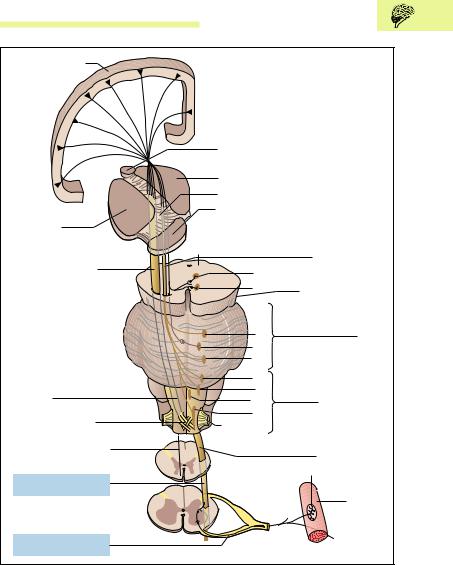
Fig. 13.20 Course of the pyramidal tract. The cranial part of the pyramidal tract (corticobulbar tract) ends at the motor areas of the nuclei of the cranial nerves: III, oculomotor nucleus; IV, trochlear nucleus; V, trigeminal nucleus; VI, abducens nucleus; VII, facial nucleus; IX, glossopharyngeal nucleus; X, vagus nucleus; XI, spinal accessory nucleus; XII, hypoglossal nucleus. The tracts to the nuclei of cranial nerves III, IV, and VI have not been determined with certainty. (After Duus)

566 13 The Central and Peripheral Nervous Systems
Voluntary Motor Tracts of the Head
After leaving the internal capsule, the pyramidal axons run through the cerebral peduncles and enter the medulla oblongata (bulb). While passing through the brainstem, the nerve fibers for the cranial voluntary muscles leave the pyramidal tract (corticobulbar tract, corticonuclear tract), cross the midline, and end in the motor nuclei of the cranial nerves (Fig. 13.20). There they form synapses with the neurons that leave the brainstem with the cranial nerves and supply the cranial striated muscles (e. g., muscles of mastication).
Voluntary Motor Tracts of the Trunk and Extremities
Some 80−90 % of the axons of the pyramidal tract cross to the opposite (contralateral) side of the medulla (pyramidal crossing or decussation), and then descend in the spinal cord. Since the descending fibers run in the lateral funiculus of the spinal cord, the tract is called the lateral corticospinal tract. Those axons that do not cross in the medulla descend in the anterior funiculus of the spinal cord of the same (ipsilateral) side and are therefore called the anterior (or ventral) corticospinal tract (Fig. 13.20).
The axons of the lateral corticospinal tract lose their myelin sheaths at various levels of the spinal cord and enter the gray substance of the anterior horn, where they end in synapses on the motor cells of the anterior horn. At the corresponding sites in the spinal cord, the axons of the anterior corticospinal tract cross to the opposite side, where they also end in synapses with the motor cells of the anterior horn.
It is important to emphasize that both the anterior and lateral corticospinal tracts in their entire course from the precentral gyrus to the anterior horn consist of individual uninterrupted neurons. These neuron are also known as upper motoneurons. They synapse with lower motoneurons, the axons of which run in the anterior roots and supply the peripheral voluntary muscles. These secondary neurons are called lower motoneurons (Fig. 13.20). As we will see, the distinction between upper motoneurons and lower motoneurons is clinically very important (see the sections on lesions of the motoneurons below). In a person 6 feet (184 cm) tall, the axons supplying the toes are almost 36 inches (90 cm) in length. In this case, the upper motoneurons begin in
 (sulcus lateralis)
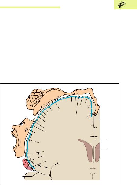
568 13 The Central and Peripheral Nervous Systems
the head at the border of the lateral sulcus of the cerebral cortex. This is called the somatotopic arrangement of the regions of the body, each of which has an area of different size allocated in the precentral gyrus.
Sections of the body where the muscles must execute very finely adjusted movements are represented over especially large areas of the precentral gyrus. For instance, the area of the neurons supplying the hand is exceptionally large in relation to other areas and reflects the large number of neurons necessary to accomplish the complex fine movements of, say, playing the violin, performing surgery, or writing. Another somatotopic arrangement can be found in the internal capsule, the principal path of the ascending and descending fibers. The internal capsule, with its anterior and posterior limbs and their connecting area, the knee, can be seen in a horizontal section through the cerebral hemispheres (Fig. 13.6a, b). The motor fibers supplying the face are located in the genu, and those responsible for the rest of the body in the anterior twothirds of the posterior limb. If the genu is injured, the facial muscles are affected, while if the middle region of the posterior genu is damaged, the muscles of the lower extremity receive no impulses.
Extrapyramidal Motor System
The extrapyramidal motor system includes the basal ganglia (see p. 542), the red nucleus and substantia nigra (see p. 550) in the midbrain, and the vestibular nuclei. In addition, parts of the cerebral cortex (premotor or associative areas), the cerebellum, and the descending extrapyramidal motor pathways to the spinal cord can also be considered components of the extrapyramidal motor system. While the pyramidal tract mainly transmits commands to the voluntary muscles, the extrapyramidal motor system with its motor nuclei (e. g., the basal ganglia) and pathways is responsible for involuntary muscle movements. Among other things, it controls the extent and direction of voluntary movements.
The cerebellum has a special place in the functioning of the extrapyramidal motor system. For instance, it receives a copy of information about planned voluntary muscular movements (corticocerbellar tracts, Fig. 13.14). Since the cerebellum simultaneously receives information from the muscles through the cerebellar tracts of the lateral funiculus, it can coordinate the planning and execution of a movement. It transmits

Central Nervous System 569
corrective commands from the cerebellar nuclei (see p. 553) to the motor centers in the brainstem, the basal ganglia, and the cerebral cortex. For instance, fibers that originate in the premotor area of the cerebral cortex (Fig. 3.10) do not send stimuli to the voluntary muscles, rather they inhibit the lower motoneuron and so prevent overshooting of the muscles during reflex reactions to sensory stimuli.
However, as a result of current understanding, the division into pyramidal and extrapyramidal systems is often put aside, since the functions of the two systems are closely intertwined.
Lesions of the Lower Motoneuron (Flaccid Paralysis)
Flaccid paralysis ensues when, for instance, a peripheral nerve is severed on its path to the muscle or when the cell bodies in the anterior horn are destroyed selectively by the virus of poliomyelitis (infantile paralysis). In both cases, the muscles are deprived of their direct innervation. They cannot contract and show characteristic signs of flaccid paralysis, i.e., they become soft and flaccid and atrophy. Since the efferent limb of the reflex arc (p. 571) is interrupted, the muscles naturally cannot respond to sensory stimuli.
Lesions of the Upper Motoneuron (Spastic Paralysis)
Lesions anywhere in certain parts of the corticospinal tract (e. g., the cell bodies in the precentral gyrus or the descending fibers in the internal capsule, the brainstem, or the spinal cord) cause spastic paralysis. The commonest site of a lesion is within the cerebral hemispheres, before the decussation of the pyramids. Such lesions often occur as a result of arterial occlusions or cerebral hemorrhage, when oxygen deficiency causes destruction of the nerves (cerebral infarct, apoplexy, or stroke). When the lesion is above the decussation of the pyramids, typical paralytic symptoms occur in the muscles of the contralateral side, while after a lesion below the decussation, such as a lesion on the left side of the spinal cord, the resulting paralysis will occur on the same side.
This type of lesion differs from flaccid paralysis in several ways. For one thing, in contrast to a flaccid paralysis, the lower motoneuron is not

570 13 The Central and Peripheral Nervous Systems
damaged, so that the reflex arc (see below) remains intact and reflexes can be elicited. Instead, because the inhibiting extrapyramidal motor fibers run in close association with the upper motoneurons, they are affected by the same lesion, and so can no longer exert their influence over the lower motoneuron. The result is an overshoot of muscular reflex response to sensory stimuli, because the impulses sent out by the lower motoneurons are uncontrolled. This condition is known as hyperreflexia: if, for instance, the wrist joint of the paralyzed arm is grasped and held, a series of muscle contractions following rapidly one on another ensues (clonus). Spastic paralysis is this condition of increased reflex activity resulting from a lesion of the upper motoneuron, combined with increased spastic muscle tone.
Spinal Reflexes
Reflexes are involuntary and unchanging responses of an organism to external or internal stimuli received by the CNS. For instance, a light tap with a reflex hammer on the tendon below the kneecap (patella) leads to a brief contraction of the quadriceps femoris muscle. This is the reflex known as the patellar reflex (knee jerk).
Reflex Arc
The basis of such a spinal reflex is what is called a reflex arc, which is a functional entity with the following components (Fig. 13.22b):
A receptor that registers and transmits the information
An afferent neuron by which an impulses reach the spinal cord
A synapse, in which the impulse is relayed to the motor cell in the anterior horn
An efferent neuron by which the impulses leave the spinal cord
An effector organ
Proprioceptive or Stretch Reflex
In the patellar reflex, only one synapse is interposed between the afferent and efferent neuron and the receptor and effector are common to the


572 13 The Central and Peripheral Nervous Systems
The physiological significance of such a stretch reflex lies in the fact, among others, that it controls the length and tension of a muscle (socalled postural tone) and so balances the effect of gravity. For instance, without postural tone in the quadriceps femoris when standing, our knee joints would constantly give way. The reflex arc therefore ensures that even with slight relaxation of the knee, the corresponding muscle stretch triggers a reflex contraction, thereby restoring the extension of the knee joint.
Sensorimotor or Skin Reflexes
In sensorimotor or skin reflexes, the skin is stimulated and a muscle responds by contracting. When, for instance, the abdominal skin is stroked with a pointed object, the abdominal muscles contract (abdominal reflexes). In contrast to proprioceptor reflexes, in this reflex the receptor and effector are separated and are located in different organs. Moreover, in these reflexes several synapses and intercalated neurons are inserted into the reflex arc. These reflexes are therefore often called polysynaptic reflexes. Intercalated neurons allow the involvement of neighboring spinal segments and contralateral organs.
These reflexes are characterized by prolonged reflex times (e. g., 70− 150 milliseconds for the eyelid closure reflex), rapid fatigability and accommodation, as well as the phenomenon of summation of subthreshold stimuli. The latter term describes the triggering of a reflex by constant repetition of small stimuli each of which by itself would not elicit a reflex response. As an example, prolonged irritation of the nasal mucosa summates to exceed the threshold for a sneeze. Other examples of such protective reflexes include coughing, tearing, and digestive reflexes such as swallowing and sucking.
Pathological Reflexes
A typical example of a pathological reflex is the Babinski reflex, which is the result of a lesion of the pyramidal tract. When the outside edge of the sole of the foot is stroked with a pointed object, all the toes plantarflex reflexly. After a lesion of the pyramidal tract, however, the big toe dorsiflexes, while the other toes open out like a fan and are dorsiflexed.
Central Nervous System 573
Membranes of the Brain and Spinal Cord
The tissues of the brain and spinal cord are the most delicate of all the tissues of the body. These organs are essential to life and so are protected by being encased in closed bony chambers—the skull and the vertebral canal.
Membranes of the Brain
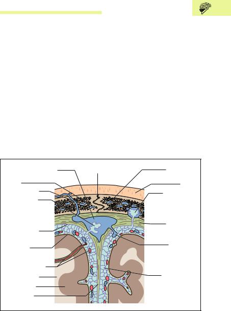
Three membranes, the meninges (Fig. 13.23), surround the brain to protect it from hard bones and blows to the head. The outermost is the densely fibrous and thick dura mater (dura mater cranialis), which is closely applied to the inside of the bones and tightly adherent to the periosteum. Under the dura mater lies the middle layer, the thin and delicate arachnoid (arachnoidea mater cranialis). The third and innermost
Superior sagittal sinus |
Sagittal suture |
Diploic vein |
Bone |
|
Scalp |
Emissary vein |
|
Projection of |
Projection of |
|
arachnoid into an |
arachnoid into |
|
osseous pit |
sinus (arachnoid |
|
(foveola granularis) |
granulation) |
|
|
Arachnoid |
|
Dura mater |
(arachnoidea |
|
|
encephali) |
|
|
Pia mater |
|
Superficial |
|
cerebral vein |
|
|
|
|
|
|
draining into |
Cerebral arteries |
|
sinus |
|
|
|
White matter |
|
Subarachnoid |
Gray matter |
|
space (cavum |
|
subarachnoidale) |
|
Falx cerebri |
|
with arachnoid septa |
|
|
Fig. 13.23 Coronal section through skull, meninges, and brain. The area shown is a section at the level of the falx cerebri

574 13 The Central and Peripheral Nervous Systems
layer is the very thin and sensitive pia mater (pia mater cranialis), rich in capillaries, which is directly applied to the brain and dips into the fissures.
Although the dura is very closely applied to the inside of the skull, it can become detached under certain circumstances (hemorrhage), creating an epidural space (extradural space; e. g., epidural or extradural hemorrhage after skull fractures). The virtual space between the dura and the arachnoid (subdural space) is traversed by veins and can also be the site of clinically significant hemorrhage. The arachnoid and the pia mater are separated by a relatively wide space, the subarachnoid space, which is filled with cerebrospinal fluid (CSF). This clear fluid fills the whole subarachnoid space and so surrounds the brain like a protective cushion. A further layer of protection is added by connective tissue strands, the arachnoid septa, that run between the arachnoid and the pia mater. They attach the brain to the arachnoid and so prevent its excessive movement when the head is exposed to sudden shocks. The cerebral arteries and veins run in the fluid-filled subarachnoid space (Figs. 13.23, 13.27a, b and 13.28). The pia mater is so closely adherent to the underlying brain that there is no space between them, the pia mater tightly holding the brain.
The dura dips into the longitudinal cerebral fissure. This fold between the two hemispheres is known as the falx cerebri (Fig. 13.23). The space between the cerebellum and the occipital lobe of the cerebrum over it is also lined with dura, which thus forms a tentlike cover over the cerebellum, the tentorium cerebelli. Finally, the dura dips between the cerebellar hemispheres to form the falx cerebelli. The membranes of the brain, the subarachnoid space, and the CSF pass through the foramen magnum in the base of the skull.
Membranes of the Spinal Cord
After passing through the foramen magnum, the membranes of the brain continue into the vertebral canal to become the membranes of the spinal cord, surrounding the spinal cord and the roots of the spinal nerves (Fig. 13.24). The dura mater of the spinal cord (dura mater spinalis) forms a strong sac (dural sac) that is anchored around the foramen magnum and the intervertebral foramina and extends to the second sacral vertebra. In contrast to the dura of the brain, which is closely adherent to the
Central Nervous System 575
Spinal arachnoid |
Spinous process |
|
|
Posterior root ganglion |
Periosteum |
(ganglion sensorium |
|
n. spinalis) |
Spinal cord |
|
|
Intervertebral |
(medulla spinalis) |
|
|
foramen |
Epidural space |
|
(spatium epidurale) |
|
with fatty tissue and |
|
venous plexus |
|
Spinal pia mater |
|
Subarachnoid |
|
space (spatium |
|
subarachnoideum) |
|
Spinal dura mater |
Fig. 13.24 Cross-section through the vertebral canal with the spinal cord and spinal meninges
periosteum of the skull, the dura of the spinal cord is not adherent to the bones of the vertebral canal, but separated from it by a space (epidural space, extradural space = spatium epidurale). This space is filled with fatty tissue and contains an extensive venous plexus.
Inside the dura, the arachnoid (arachnoidea mater spinalis) is closely adherent to it, and is connected to the pia mater (pia mater spinalis) by delicate strands of connective tissue. The subarachnoid space, filled with CSF, lies between the arachnoid and the pia mater, which is closely applied to the surface of the spinal cord (Fig. 13.24).
The spinal cord in the vertebral canal ends at the level of the 1st or 2nd lumbar vertebra (Fig. 13.17), but the pia mater of the spinal cord continues as the filum terminale, to be attached to the back of the coccyx

576 13 The Central and Peripheral Nervous Systems
(Fig. 13.16). Additionally, the pia mater over the length of the spinal cord has toothlike projections, the ligamenta denticulata. These are attached to the dura and the arachnoid and stabilize the spinal cord in the coronal plane.
All spinal roots, including those in the cauda equina, are covered by the pia mater, which simply entrains the other two spinal membranes at the exit of the spinal nerves from the vertebral canal. Thus, the spinal nerves on leaving the vertebral canal are also covered by the dura and the arachnoid, which continue as the perineurium and epineurium (see Chapter 3: The Nerves).
Cerebrospinal Fluid (CSF) and the Ventricular System
The cerebrospinal fluid (CSF) is a clear fluid, with a volume of about 130− 150 ml, which fills the whole subarachnoid space. It acts as a protective fluid cushion around the brain and the spinal cord, damping shocks caused by blows or falls. The central canal of the spinal cord is also filled with CSF. The CSF is an important diagnostic tool: using a relatively simple procedure (lumbar puncture), the physician can take a sample of CSF and so obtain a picture of events inside the skull and the spinal cord (CSF diagnosis for disorders of the CNS, see p. 578).
Deep inside the brain lies the ventricular system (Fig. 13.25a, b). This consists of interconnected chambers in which the CSF is secreted and circulates. Each cerebral hemisphere contains a large cavity, the lateral ventricle, which consists of a frontal (anterior) horn in the frontal lobe of the cerebrum, a central part (pars centralis) in the frontal and parietal lobes, an occipital (posterior) horn in the occipital lobe, and a temporal (inferior) horn in the temporal lobe (Fig. 13.25a, b). Each lateral ventricle, as well as the third (diencephalon) and fourth (under the cerebellum) ventricles, contains an arterial network, the choroid plexus. The CSF is produced in the plexus, reaching the ventricle by diffusion and active transport.
From the lateral ventricles, the CSF flows through an opening in each side (interventricular foramen, foramen of Monro) into the single median third ventricle between the walls of the left and right diencephalon. Eventually, the CSF produced in the ventricles flows through a narrow canal (aqueductus mesencephali, cerebral aqueduct, aqueduct of Sylvius)

578 13 The Central and Peripheral Nervous Systems
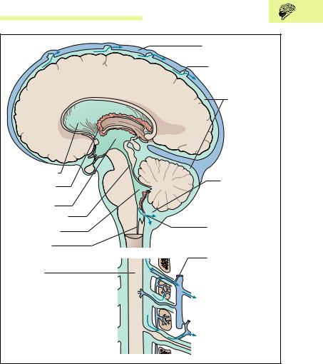
in the midbrain into the single fourth ventricle, from which it flows through three openings (apertures) in the roof of the fourth ventricle into the subarachnoid space (Fig. 13.26). In certain areas the subarachnoid space is distinctly enlarged and forms the so-called cisterns, such as the posterior cerebellomedullary cistern (cisterna magna) (Fig. 13.26).
CSF Drainage
Since CSF is secreted at 30 ml per hour, the question is: where does it drain? At the superior sagittal sinus, the arachnoid projects through small openings in the dura into the venous sinus. The fluid accumulation exerts pressure, which drives the fluid though arachnoid villi into the venous blood. Macroscopically, these arachnoid villi look like grains of salt or sugar, and so are called arachnoid granulations (Fig. 13.23). In the spinal cord, the CSF drains into a dense venous plexus and lymph vessels where the spinal nerves emerge (Fig. 13.26).
Lumbar Puncture
A lumbar puncture (puncture of the subarachnoid space with a needle inserted between two lumbar laminae) can be performed to remove CSF for diagnostic or therapeutic purposes, or to introduce substances into the subarachnoid space (medications, radiological contrast media, anesthetics). When properly performed, lumbar puncture does not endanger the spinal cord, because the cord ends at the L1 or L2 level, while the subarachnoid space continues downward to S2. Lumbar puncture can be dangerous under certain circumstances; a complete discussion of its indications, contraindications, and technique would be beyond the scope of this book.
Blood Supply of the Brain
Nerve cells have high oxygen requirements, so that even 3−4 minutes without oxygen can lead to necrosis of the neurons. The nerve cells of the cerebral cortex are the most sensitive, those of the brainstem the most resistant. The significance of an adequate blood supply to the brain can
Central Nervous System 579
Superior sagittal sinus
Site of CSF drainage through arachnoid villi into venous blood
Subarachnoid space (spatium subarachnoidale)
Lateral ventricle |
Cisterna magna |
|
Choroid plexus |
||
(cisterna |
||
|
||
|
cerebromedullaris |
|
Third ventricle |
posterior) |
|
Cerebral aqueduct |
|
|
Fourth ventricle |
Passage of CSF from |
|
internal to external |
||
|
||
Central canal |
fluid space |
|
|
Drainage of CSF into |
|
Spinal cord |
lymphatics at site of |
|
emergence of spinal |
||
|
nerves |
Fig. 13.26 Diagram of the internal (ventricle) and external (subarachnoid space) fluid-filled spaces. (After Kahle)
be seen by the fact that the brain’s weight is only 2 % of that of the body but it uses 15−20 % of the cardiac output.
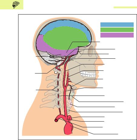
The blood supply to the brain is provided by two sets of bilateral arteries, the internal carotid arteries and the vertebral arteries (Fig. 13.27a,
580 13 The Central and Peripheral Nervous Systems
|
Regions supplied by: |
|
|
Anterior cerebral artery |
|
|
Middle cerebral artery |
|
|
Posterior cerebral artery |
|
|
Posterior communicating |
|
|
artery |
|
|
Basilar artery |
|
Cerebellar |
Ophthalmic artery |
|
Carotid canal |
||
arteries |
||
|
Internal carotid artery |
|
Foramen |
External carotid artery |
|
magnum |
||
|
||
Foramina |
Superior thyroid artery |
|
of transverse |
|
|
processes |
Right common carotid |
|
(foramina |
||
artery |
||
transversaria) |
||
Right vertebral artery |
||
|
||
|
Larynx |
|
|
Thyroid gland |
|
|
Trachea |
|
|
Left subclavian artery |
|
|
Brachiocephalic trunk |
|
|
Right subclavian artery |
|
a |
Aortic arch |
Fig. 13.27 a Arterial blood supply of the brain. Course of the great arteries (vertebral and internal carotid arteries) supplying the brain and areas of the right cerebral hemisphere supplied by the cerebral arteries (seen from the right side)
b). The left and right internal carotid arteries run from the division of the common carotid artery to the base of the skull, and reach the inside of the cranial cavity through a canal (carotid canal). They have no branches. The left and right vertebral arteries arise from the corresponding subclavian artery and run cephalad to the atlas through the foramina in the transverse processes of the upper six cervical vertebrae. By passing
Central Nervous System 581
Circulus arteriosus = (circle of Willis)
A. communicating artery Anterior cerebral artery Internal carotid artery
Middle cerebral artery
Posterior communicating artery
Superior cerebellar artery
Posterior cerebral artery Basilar artery
Vertebral artery Anterior spinal artery
Anterior inferior cerebellar artery
Posterior inferior cerebellar artery b
Fig. 13.27 b Arteries of the base of the brain and circle of Willis (the right cerebellar hemisphere and the right temporal lobe have been removed; the circle of Willis (circulus arteriosus) is marked by the interrupted line)
through the foramen magnum they also reach the inside of the cranial cavity. The four arteries anastomose with each other in an arterial ring (circulus arteriosus, circle of Willis) (Fig. 13.27b). The circle of Willis and all arteries arising from it run in the subarachnoid space.
After passing through the foramen magnum, the two vertebral arteries run on the anterior side of the medulla oblongata, each giving rise to an inferior posterior cerebellar artery to the cerebellum and the spinal arteries (e. g., anterior spinal artery) to supply the spinal cord (Fig. 13.27b). At the lower edge of the pons, the two vertebral arteries join to form the basilar artery, which gives rise to the inferior anterior and the superior cerebellar arteries. At the upper edge of the pons, the basilar artery divides into the left and right posterior cerebral arteries; these run

582 13 The Central and Peripheral Nervous Systems
backward to supply the posterior part of the hemispheres, especially the inferior surface of the temporal lobe and both occipital lobes (Fig. 13.27a).
The two internal carotid arteries give rise to the two ophthalmic arteries (each supplying one eye and part of the nose) immediately after entering the base of the skull, and in their subsequent course divide into the anterior and middle cerebral arteries (aa. cerebri anterior and media). The two anterior cerebral arteries at first run forward in the longitudinal fissure (fissura longitudinalis cerebri), then run backward on the corpus callosum to supply the medial side of the two hemispheres. The two middle cerebral arteries run laterally between the temporal and frontal lobes and fan out in the lateral sulcus to supply the greater part of the lateral side of the cerebral hemispheres (Fig. 13.27a). In their course between the frontal and temporal lobes, the middle cerebral arteries give off important branches to supply the internal capsule (striate arteries, aa. centrales anteromediales). The middle cerebral artery is the largest of the three cerebral arteries and represents the direct continuation of the internal carotid artery.
The two anterior cerebrtal arteries are connected by the anterior communicating artery. The two posterior communicating arteries connect the middle and posterior cerebral arteries (Fig. 13.27b). They form an arterial circle (circle of Willis, circulus arteriosus cerebri) between the vertebral and carotid arteries at the base of the brain. This is of clinical significance, since after occlusion of one of the four contributing vessels the remaining three can supply the affected region. The circle of Willis is often the site of aneurysms, i.e., saccular dilatations of the vessel wall. These weak places in the vascular wall may rupture as a result of a brief rise in blood pressure, with consequent hemorrhage into the subarachnoid space (subarachnoid hemorrhage).
If a blood clot (thrombus) forms along the course of an artery supplying the brain (usually because of atherosclerosis of the arterial wall), or if a thrombus arises elsewhere (e. g., in the heart) and is transported into a cerebral artery by way of the bloodstream (anything transported in this manner is called an embolus), blockage of the artery can result, depriving the tissue normally supplied by the artery of its blood flow. This hypoperfusion (ischemia) results in an inadequate supply of oxygen and nutrients, causing cell death (necrosis). Ischemic necrosis is called infarction, and the area of dead tissue is called an infarct. Cerebral infarction is
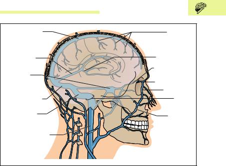
|
Central Nervous System 583 |
Superior sagittal |
Superficial |
sinus |
anastomotic veins |
|
Great cerebral |
|
vein (vena |
Straight sinus |
cerebri magna) |
(sinus rectus) |
Ophthalmic vein |
|
|
Cavernous |
|
sinus |
Nasofrontal vein |
|
Angular vein |
Transverse sinus |
Jugular |
|
foramen |
Sigmoid sinus |
Facial vein |
|
|
Internal jugular vein |
|
Fig. 13.28 Veins of the brain and venous sinuses in lateral view
one of the two most common types of stroke (also called cerebrovascular accident; i.e., any event affecting the blood vessels of the brain that produces a sudden, usually irreversible, or only partially reversible, neurological deficit). Cerebral hemorrhage (bleeding) due to rupture of an artery causes the death of cells adjacent to the hemorrhage; this is the other common type of stroke. (Note: subarachnoid hemorrhage, discussed above, is also a type of stroke according to strict definition, but is nevertheless usually not considered as such.)
Venous drainage occurs essentially through superficial and deep cerebral veins and venous sinuses. The veins from the cerebral capillaries run to the surface and drain into larger veins. These run in the subarachnoid space like the arteries and give off anastomotic veins (Fig. 13.28) that drain into large venous sinuses (e. g., superior sagittal sinus) formed by the dura mater. The blood from the interior of the brain flows through the deep cerebral veins and eventually through the great cerebral vein (vena cerebri magna) into the straight sinus (sinus rectus).

584 13 The Central and Peripheral Nervous Systems
The superior sagittal sinus and the straight sinus join on the internal surface of the occipital bone (confluence of the sinuses), after which they drain into the right and left transverse sinuses; each of these in turn is continued as a sigmoid sinus (Fig. 13.28). Eventually these drain into the internal jugular vein through the jugular foramen, an opening in the base of the skull at the side of the foramen magnum. The cavernous sinus, located around the sella turcica, receives, among others, the drainage of the superficial facial veins (facial vein−angular vein−nasofrontal vein); it also drains through the jugular foramen into the internal jugular vein (Fig. 13.28).
Peripheral Nervous System
The peripheral nervous system includes the pathways (peripheral nerves) running through the spinal nerves and the cranial nerves to the periphery of the body, and in the reverse direction, as well as some collections of nerve cell bodies (ganglia).
Peripheral Nerves
A peripheral nerve is a mixed nerve, since it carries afferent as well as efferent somatic and autonomic nerve fibers (p. 608). Somatic fibers are those that run from a receptor (e. g., skin or pain receptor) to the spinal cord (somatic afferent) or from the motor cells in the anterior horn of the spinal cord to skeletal muscles (somatic efferent). Autonomic fibers are afferent and efferent fibers connected to internal organs, blood vessels, and glands.
Ganglia
Ganglia are knotlike enlargements in nerves and nerve roots, a few millimeters thick, containing collections of nerve cell bodies. Ganglia may be sensory or autonomic. Sensory ganglia (e. g., spinal ganglia or cranial nerve ganglia) contain the cell bodies of the primary afferent nerves of both the somatic and autonomic nervous systems. The cell bodies have a

Peripheral Nervous System 585
peripheral process that may come, for instance, from a pain receptor in the skin or an internal organ, and a central process that connects the ganglion with the spinal cord.
Autonomic ganglia (see Chapter 14: The Autonomic Nervous System), contain cell bodies of the second-order efferent neurons (e. g., cranial parasympathetic ganglia or ganglia of the sympathetic chain). The ganglia relay information transmitted synaptically by autonomic nerve fibers from the spinal cord or the brainstem (preganglionic fibers = firstorder efferent neurons) to the cell bodies of second-order efferent neurons. The latter send nerve fibers (postganglionic fibers) to the effector organ in the periphery (e. g., glands or smooth muscle).
Spinal Nerves
The spinal nerves divide into four branches (rami) after leaving the vertebral canal (Fig. 13.29):
A posterior branch (r. posterior), supplying sensory innervation to the skin of the back and motor branches to a large part of the musculature of the vertebral column (segmental back muscles)
An anterior branch (r. anterior nervi spinalis), supplying sensory and motor innervation to the rest of the trunk and the limbs
A sensory branch to supply the spinal membranes and the ligaments of the vertebral column (r. meningeus)
A branch that connects the sympathetic part of the autonomic nervous system (see Fig. 14.2) with the somatic nervous system (white communicating branch of the spinal nerve, r. communicans albus)
Networks of Nerves (Plexus or Plexuses)
The anterior branches of the spinal nerves form nervous networks in the cervical, lumbar, and sacral regions: the cervical plexus (C1−C4), the brachial plexus (C5−T1), the lumbar plexus (T12−L4), and the sacral plexus
(L5−S4).
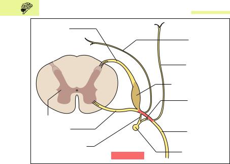
586 13 The Central and Peripheral Nervous Systems
Posterior root (radix posterior)
|
Sensory branch |
|
|
to the meninges |
|
|
(r. meningeus) |
|
|
Posterior branch |
|
|
of spinal nerve |
|
|
(r. posterior) |
|
|
Posterior root ganglion |
|
|
(ganglion sensorium) |
|
|
Gray communi- |
|
|
cating branch |
|
|
to the spinal nerve |
|
Spinal cord |
(r. communicans griseus) |
|
|
||
Anterior root |
Anterior branch |
|
(radix anterior) |
||
of spinal nerve |
||
|
||
White communicating |
(r. anterior) |
|
|
||
branch to sympathetic chain |
Sympathetic chain |
|
(r. communicans albicans) |
Spinal nerve |
Fig. 13.29 Spinal nerve roots, a spinal nerve, and its branches. (After Faller)
Cervical Plexus
The nerves of the cervical plexus supply sensory innervation to the regions of the neck and shoulder and provide motor innervation to the hyoid muscles and the diaphragm (phrenic nerve).
Brachial Plexus
From the brachial plexus arise motor nerves to the muscles of the shoulder girdle and the upper limb, and sensory nerves to the skin in the region of the shoulder and upper limb. The most important nerves supplying the upper limb are (Figs. 13.30a, b and 13.31):
The ulnar nerve (n. ulnaris)
The musculocutaneous nerve (n. musculocutaneus)
The median nerve (n. medianus)
The radial nerve (n. radialis)
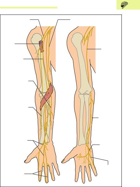

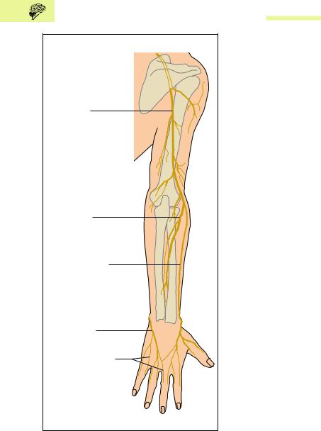

Peripheral Nervous System 589
Lumbar Plexus
The sensory branches of the lumbar plexus supply the skin of the abdomen, the genital region, and the anterior thigh (Fig. 13.32a, b). The motor branches supply chiefly the muscles of the hip, and through the femoral nerve the quadriceps femoris muscle.
Sacral Plexus
Finally, the sacral plexus supplies sensory and motor innervation for the muscles of the pelvic floor, the muscles of the lower extremity, as well as the skin of the posterior genital region (perineum) and the lower limbs. It also gives rise to the largest nerve of the whole body (Fig. 13.32a, b), the sciatic nerve (n. ischiadicus).
Cranial Nerves
The 12 pairs of cranial nerves (Fig. 13.33) can be grouped in various ways, e. g., by their central location: cranial nerves I and II, the olfactory nerve and the optic nerve are not true nerves, but extensions of the cerebrum and diencephalon respectively. The oculomotor and trochlear nerves, cranial nerves III and IV, are related to the midbrain. The trigeminal nerve (V), the abducens (VI), and the facial nerve (VII) are located at the level of the pons. The remaining cranial nerves, the auditory nerve (VIII, vestibulocochlear nerve), the glossopharyngeal nerve (IX), the vagus nerve (X), the spinal accessory nerve (XI), and the hypoglossal nerve (XII) arise from the medulla.
Another way of classifying the cranial nerves is by the function of their components. A few nerves are composed exclusively of sensory neurons. These include:
Olfactory nerve (I), for the sense of smell
Optic nerve (II), for vision
Vestibulocochlear nerve (VIII), which conducts the sensations of hearing and equilibrium
Other cranial nerves contain exclusively motoneurons supplying voluntary muscles. They include:
Trochlear nerve (IV), which supplies one of the external muscles of the eye, the superior oblique muscle
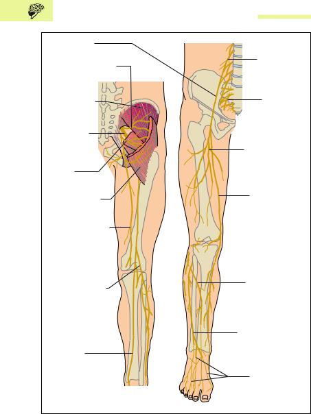
590 13 The Central and Peripheral Nervous Systems
Femoral nerve (n. femoralis)
Superior gluteal nerve |
Lumbar plexus |
|
(plexus lumbalis) |
||
(n. gluteus superior) |
||
|
||
Gluteus |
Sacral plexus |
|
medius muscle |
||
(plexus sacralis) |
||
|
||
Sciatic nerve |
|
|
(n. ischiadicus) |
|
|
Inferior gluteal |
Muscular branches |
|
of femoral nerve |
||
nerve (n. gluteus |
(r. musculares |
|
inferior) |
n. femoralis) |
|
Gluteus |
Saphenous nerve |
|
maximus muscle |
||
|
||
Posterior cutaneous |
|
|
nerve of thigh |
|
|
(n. cutaneus |
|
|
femoris posterior) |
|
|
Common peroneal |
Deep fibular nerve |
|
(n. peroneus |
||
nerve (n. fibularis |
profundus) |
|
communis) |
|
|
|
Superficial pernoneal |
|
|
nerve (n. fibularis |
|
Tibial nerve |
superficialis) |
|
|
||
(n. tibialis) |
|
|
|
Dorsal cutaneous |
|
|
nerves of the foot |
|
|
(n. cutanei dorsales pedis) |
|
a |
b |
Fig. 13.32 a, b Course of the most important nerves of the leg from the lumbar and sacral plexus, on the back (a) and the front (b) of the lower extremity. (After Feneis)
Peripheral Nervous System 591
Cranial n. I |
Crossing of optic nerves |
Frontal lobe |
(olfactory n.) |
(optic chiasma) |
|
Cranial n. II |
|
|
(optic n.) |
|
|
Cranial n. III |
|
Temporal lobe |
|
|
|
(oculomotor |
|
|
Cranial n. IV |
|
|
(trochlear n. |
|
|
Cranial n. V |
|
Pituitary |
(trigeminal |
|
|
|
gland |
|
|
|
|
Cranial n. VI |
|
(hypophysis) |
|
|
|
(abducens n |
|
|
Cranial n. VII |
|
|
(facial n.) |
|
Pons |
Cranial |
|
|
|
|
|
n. VIII |
|
|
(vestibulo- |
|
Bulb |
cochlear n.) |
|
(medulla |
Cranial n. |
|
oblongata) |
|
|
|
IX (glosso- |
|
|
pharyngeal |
|
Spinal cord |
Cranial n. X |
|
|
|
(medulla spinalis) |
|
(vagus n.) |
|
|
Cranial n. XI |
|
|
(spinal accessory n.) |
|
|
Cranial n. XII |
|
Cerebellum |
(hypoglossal n.) |
|
|
Fig. 13.33 Base of the brain with emerging cranial nerves
Abducens (VI), which also supplies one external eye muscle, namely, the lateral rectus muscle
Spinal accessory nerve (XI), which supplies two important muscles outside the head, namely, the trapezius and the sternocleidomastoid
Hypoglossus (XII), which supplies all the muscles of the tongue The remaining cranial nerves have mixed functions. They are:
Oculomotor nerve (III), which includes motor and parasympathetic
components (see Chapter 14: The Autonomic Nervous System, p. 605):

592 13 The Central and Peripheral Nervous Systems
1.Voluntary motor fibers to four of the external ocular muscles (superior, inferior, and medial rectus muscles and the inferior oblique) and the levator palpebrae superioris muscle
2.Parasympathetic nerve fibers to the pupillary sphincter (m. sphincter pupillaris, constricts the pupil) and the ciliary muscle (accommodation)
Trigeminal nerve (V), with its three terminal branches, the ophthalmic, maxillary, and mandibular nerves, which have sensory as well as motor components.
1.Sensory fibers transmit pain, temperature, touch, pressure, and proprioceptive sensation from the face, the cornea, the mouth, the nasal sinuses, the tongue, the teeth, the meninges, the external surface of the ear drum, and the jaw joint.
2.Motor components to the muscles of mastication (masseter, temporalis, lateral and medial pterygoid muscles) and the muscles of the floor of the mouth (anterior belly of the digastric muscle, mylohyoid muscle), the soft palate (m. tensor veli palati), and the eustachian tube (m. tensor tympani).
Facial nerve (VII), a complex nerve with three main components:
1.Special fibers for taste sensation in the anterior two-thirds of the tongue (chorda tympani)
2.Parasympathetic fibers to the salivary glands (lingual and submandibular glands), the lacrimal gland (tear gland), and the glands of the mouth and nose (palatine and nasal glands)
3.Motor fibers to the voluntary muscles of expression, as well as the muscles of the hyoid bone (stylohoid muscle), the floor of the mouth (posterior belly of the digastric muscle), and the middle ear (m. stapedius)
Glossopharyngeal nerve (IX), which also has three main components:
1.Special fibers for taste sensation in the posterior third of the tongue
2.Parasympathetic fibers to the parotid gland through the autonomic otic ganglion
3.Sensory neurons (pain, temperature, pressure, and touch sensation) to the eustachian tube, the palatine end of the tongue, the internal surface of the ear drum, the throat, and the carotid sinus (changes in blood pressure)
Vagus nerve (X), a nerve essential to life, with three main components:
Summary 593
1.Parasympathetic neurons to all the autonomic structures of the thoracic and abdominal cavities as far as the left flexure of the transverse colon (e. g., heart, coronary arteries, bronchi, stomach, arteries and arterioles of the small bowel, and glands)
2.Motor fibers to those muscles of the larynx and the throat that are involved in swallowing and speech
3.Sensory nerves from the larynx, the internal organs, the carotid body (a chemoreceptor), the carotid sinus, the meninges of the posterior cranial fossa, and the lower part of the throat
Summary The Central and Peripheral Nervous Systems
Classification and Role
According to its spatial arrangement the nervous system is divided into a central and a peripheral nervous system:
Central nervous system (CNS): brain and spinal cord
Peripheral nervous system: peripheral nerves (spinal and cranial nerves, collections of nerve cells = ganglia)
Functionally the CNS and peripheral nervous systems can be divided into a somatic and an autonomic (= vegetative) nervous system:
Somatic nervous system (organism—environment): responsible for
(1) conscious perception, (2) voluntary movements, and (3) rapid processing of information
Autonomic (vegetative) nervous system (organism—internal organs): responsible for (1) maintaining a constant internal milieu (homeostasis) and (2) regulation of organic functioning in response to environmental demands
The somatic and autonomic nervous systems both include afferent and efferent impulses:
Afferents: all impulses transmitted from the periphery (e. g., skin, internal organs) centrally (to brain, spinal cord)
Efferents: all impulses reaching the periphery (e. g., striated skeletal muscle, smooth muscle of the internal organs and blood vessels, glandular cells) from the center (brain, spinal cord)

59413 The Central and Peripheral Nervous Systems
Development
The central and peripheral nervous systems develop from the outer germ layer (ectoderm) of the three-layered embryonic disk. The rudiment appears at the end of the third week as an ectodermal thickening (neural plate = neural ectoderm) above the notochord (chorda dorsalis). Lateral folds in the neural plate (neural folds) lead to the formation of the neural groove, which eventually closes to form the neural tube. Parts of the neural folds become the neural crests. The neural tube develops into the CNS, the neural crests into the peripheral nervous system.
Central Nervous System (CNS)
The central nervous system consists of the brain (encephalon) and the spinal cord (medulla spinalis); both parts develop from the neural tube. The wall of the neural tube develops into the gray and white substance of the brain and spinal cord, while the anterior part of the lumen develops into the ventricles of the brain and the posterior part into the central canal of the spinal cord. The cranial part of the neural tube (primary brain vesicles) becomes the brain, the caudal part forms the spinal cord.
Derivatives of the three primary brain vesicles (prosencephalon, mesencephalon, and rhombencephalon):
−Prosencephalon (anterior brain vesicles): telencephalon (cerebrum) and diencephalon
−Mesencephalon (middle brain vesicle): mesencephalon (midbrain)
−Rhombencephalon (posterior brain vesicle): pons, cerebellum, and medulla oblongata
The Brain
Average weight: in women 1245 g, in men 1375 g, in the newborn 400 g; definitive weight is reached at age 5−7 years Weight in relation to body weight (b.w.): women 22 g/kg b.w., and men 20 g/kg b.w.
Cerebrum (Telencephalon)
This most highly developed part of the brain consists of the two cerebral hemispheres, the basal ganglia, the two lateral ventricles, and the limbic system. The surface of the cerebrum is extended by convolu-

Summary 595
tions (gyri) and fissures (sulci). The central fissure (sulcus centralis) and the lateral fissure (sylvian fissure, sulcus lateralis) divide each hemisphere into four lobes: the frontal, parietal, temporal, and occipital lobes. The cerebral cortex (cortex cerebri) contains primarily cell bodies (gray matter, gray substance); the inner white matter (white substance) contains nerve fibers or axons. Nerve fibers may connect the two hemispheres (commissural fibers), begin and end within a hemisphere (association fibers), or run from the cerebral cortex to other parts of the CNS (projection fibers). The largest commissure, with about 200 million axons, is the corpus callosum; the most important projection tract is the pyramidal tract (corticospinal tract). Almost all projection fibers are closely bundled with the tracts ascending to the cortex as they pass through the internal capsule, which is bounded by the basal ganglia and parts of the diencephalon (thalamus).
Within the cerebral cortex are located highly developed functional cortical areas with specific functions: primary areas (where the projection tracts begin and end) and secondary areas (association areas, which process information and make up about 80 % of the cortical surface).
The most important primary areas are:
−The precentral gyrus (primary motor cortex): origin of large parts of the pyramidal tract (execution of voluntary movements)
−The postcentral gyrus (primary sensory cortex): terminal area of somatic sensory tracts for conscious perception
−The calcarine fissure (primary visual cortex): terminal area of the optic pathways
−The transverse temporal gyrus (primary auditory cortex): terminal area of the acoustic pathways.
The most important association areas are:
−The inferior frontal gyrus (motor speech center = Broca’s area): coordination of speech (80−90 % located in the left, dominant hemisphere; lesions cause motor aphasia.
−The superior temporal gyrus (secondary auditory center/sensory language center = Wernicke’s area: auditory association area; lesions cause sensory aphasia.

596 13 The Central and Peripheral Nervous Systems
Essentially the left hemisphere is responsible for the right half of the body and the right hemisphere for the left half of the body (crossover of the afferent and efferent pathways). Certain functions are located preferentially in one hemisphere (hemispheric dominance); left hemisphere: reading, speech, writing; right hemisphere: memory, language comprehension, visual/spatial imagination and musical comprehension.
The basal ganglia are collections of nerve cell bodies in the white matter. They have an important role in the extrapyramidal motor system (control of extent and direction of voluntary movements). They include the globus pallidus, the putamen, and the caudate nucleus: The globus pallidus and the putamen together are known as the lentiform nucleus, while the caudate nucleus and the lentiform nucleus together form the corpus striatum (striate body).
The limbic system runs along the corpus callosum like a hem. It takes part in creating memories and in triggering and processing emotional reactions. It is formed from phylogenetically older parts of the telencephalon: paleocortex and archicortex; those parts of the brain that are phylogenetically younger (essentially the cerebral hemispheres) are called the neocortex (isocortex, neopallium). The limbic system includes: (1) derivatives of the paleocortex—olfactory brain; (2) derivatives of the archicortex—amygdala, hippocampus, dentate gyrus (gyrus dentatus), cingulate gyrus (gyrus cinguli), indusium griseum (supercallosal gyrus), and the most important fiber tract to the hypothalamus, the fornix.
Diencephalon
The diencephalon lies between the cerebral hemispheres under the corpus callosum and surrounds the third ventricle. It includes:
The thalamus: central relay station of the sensory tracts (pain, temperature, pressure, touch)
The metathalamus (medial and lateral geniculate bodies): central relay station of the visual and auditory pathways
The hypothalamus: center controlling the autonomic nervous system (e. g., body temperature, water balance, nutrition)
The hypophysis (pituitary gland): principal organ controlling the endocrine system

Summary 597
The epithalamus (pineal body): biological clock controlling the circadian rhythm
Brainstem
The brainstem includes the midbrain, the pons, and the medulla oblongata and contains the reticular formation and the nuclei of the cranial nerves.
Midbrain (Mesencephalon)
The midbrain is the smallest part of the brain and lies between the diencephalon and the pons. It contains the tectum, the tegmentum, the cerebral aqueduct (conduit between the third and fourth ventricles), and the two crura cerebri. The tectum is formed by the corpora quadrigemina (superior and inferior colliculi = relay stations of the visual and auditory reflex pathways). The tegmentum contains two basal ganglia (red nucleus and the substantia nigra) and the nuclei of cranial nerves III and IV (oculumotor and trochlear nerves). The ascending tracts to the cortex pass through the tegmentum, the descending tracts through the crura cerebri.
Pons
The pons lies between the midbrain and the medulla and is separated from the cerebellum by the superior part of the fourth ventricle. Besides the ascending and descending tracts the pons contains the nuclei of cranial nerves V, VI and VII (trigeminal, abducens and facial nerves).
Medulla Oblongata
The medula oblongata forms the transition from the brain to the spinal cord and extends to the foramen magnum. The medulla contains the respiratory and circulatory centers and the nuclei of cranial nerves VIII−XII (vestibulocochlear, glossopharyngeal, vagus, spinal accessory, and hypoglossal nerves). Anteriorly, the pyramids lie on either side of the median fissure, and a little below them is the decussation of the pyramidal tract. The posterior side of the medulla is partly covered by the cerebellum, and together with the rhomboid fossa forms the floor of the fourth ventricle (the upper part of the rhomboid fossa is considered part of the pons). Below the rhomboid fossa lie two raised areas which are relays for the tracts of the posterior funiculus (fasciculus cuneatus and fasciculus gracilis).

598 13 The Central and Peripheral Nervous Systems
Cerebellum
The cerebellum lies in the posterior cranial fossa under the tentorium (tentorium cerebelli). It consists of the vermis and two cerebellar hemispheres, the surface of which is greatly extended by parallel folia (gyri). The cerebellum weighs 130−140 g and is connected to the brainstem by the peduncles. The parts of the cerebellum can be classified by their phylogenetic age: an oldest part (archicerebellum) a second oldest part (paleocerebellum), and a new part (neocerebellum).
The archicerebellum: includes the nodulus and the bilateral flocculus (flocculonodular lobe). It is responsible for maintaining the equilibrium.
The paleocerebellum: formed by the anterior lobes of the cerebellar hemispheres and part of the vermis. It regulates muscle tone.
The neocerebellum: includes the posterior lobe of the cerebellar hemispheres and the rest of the vermis. It is responsible for the coordination of voluntary muscles.
Spinal Cord (Medulla Spinalis)
The spinal cord represents an extracranial part of the central nervous system and lies in the vertebral canal, extending from the foramen magnum to the 1st−2nd lumbar vertebra. The spinal cord contains ascending and descending bundles of nerve fibers through which the brain communicates with the peripheral nervous system.
Spinal Nerves
The 31 pairs of spinal nerves each are formed from an anterior and a posterior root (radix anterior and radix posterior). They leave the vertebral canal through the intervertebral foramina. The spinal nerves arise in pairs, of which there are eight cervical (from the cervical cord C1−C8), 12 thoracic (from the thoracic cord T1−T12), five lumbar (from the lumbar cord L1−L5), five sacral (from the sacral cord S1−S5) and 1−2 coccygeal (from the coccygeal cord Co1−Co2).
Gray Matter of the Spinal Cord
The gray matter (substantia grisea) of the spinal cord, in cross-section shaped like a butterfly, lies in the middle of the spinal cord. It contains the bodies of the nerve cells and contains:

Summary 599
The anterior horn (anterior column): contains motoneuron cells, the axons of which leave the spinal cord by the anterior root and supply the striated skeletal muscles.
The posterior horn (dorsal column): contains the sensory nerve cells, where some of the peripheral afferent nerve cells entering through the posterior root end in synapses.
The lateral horn (intermediate column): contains motor nerve cells of the autonomic system, the efferent axons of which leave the spinal cord by the anterior roots to innervate among others the smooth muscle of the internal organs.
White Matter of the Spinal cord
The gray matter is surrounded by the white matter (substantia alba), which contains the ascending and descending myelinated fibers. It is divided into dorsal, lateral (or anterolateral), and ventral funiculi. The intersegmental (intercalated) fibers of the spinal cord itself (fasciculi proprii) run next to the gray substance.
Ascending tracts. tracts of the ventral funiculus (afferent tracts to the thalamus for pressure, coarse touch and tactile discrimination, as well as pain and temperature sensation from the extremities and the trunk). tracts of the dorsal funiculus (afferent pathways to the thalamus for deep sensation, vibration, light touch, and tactile discrimination on the limbs and trunk); spinocerebellar tracts of the lateral funiculus (afferent tracts to the cerebellum for unconscious deep sensations from muscles, tendons, and joints).
Descending tracts: pyramidal tract (efferent tract from the motor cortex to the anterior horn cells carrying voluntary motor fibers to the extremities and the trunk); extrapyramidal tracts (efferent tracts for involuntary movements, such as position, posture and automatic movements, from the brainstem, the basal ganglia, and the premotor areas to the anterior horn cells).
Spinal Reflexes
The basis for all spinal reflexes (e. g., the patellar reflex) is the reflex arc, consisting of the following components: (1) receptor, (2) afferent neuron, (3) synapse, (4) efferent neuron, and (5) effector organ. Reflexes include proprioceptive or stretch reflexes, sensorimotor or skin reflexes, and pathological reflexes.

600 13 The Central and Peripheral Nervous Systems
Tracts for the Voluntary Innervation of Muscles (Pyramidal Tracts)
Pyramidal Tracts
Principal pathway for all voluntary muscular activity. It begins in the precentral gyrus of the frontal lobe and runs through the internal capsule in the brainstem, ending either in the nuclei of the cranial nerves (corticobulbar tract) or in motor cells of the anterior horn of the spinal cord (corticospinal tract). Hence these tracts include:
The corticobulbar tract (corticonuclear tract) for the innervation of the cranial voluntary muscles: the efferent axons of the corticobulbar tract leave the pyramidal tract in the brainstem, cross to the opposite side and end in synapses in the motor nuclei of the cranial nerves.
The corticospinal tract for the innervation of the muscles of the trunk and extremities: 80−90 % of the axons cross in the medulla in the decussation of the pyramidal tracts to the opposite side. They run as the lateral corticospinal tract to the corresponding cells in the spinal cord; the uncrossed axons descend through the anterior corticospinal tract of the same side and do not cross until they reach the level of the anterior horn cells.
The axons of the pyramidal tract are called upper motoneurons regardless of their target area; axons to the striated skeletal muscles arising after relay in the nuclei of the cranial nerves or the anterior horn cells are called lower motoneurons. Lesions of the upper motoneuron cause spastic paralysis; lesions of the lower motoneuron flaccid paralysis.
Extrapyramidal Motor System
The extrapyramidal motor system includes among others the basal ganglia of the cerebrum (globus pallidus, putamen, caudate nucleus) and the midbrain (red nucleus, substantia nigra), the vestibular nuclei, the premotor areas of the cerebral cortex, the cerebellum, and the extrapyramidal descending motor tracts of the spinal cord. It is responsible for involuntary muscular activity and controls the extent and direction of voluntary movements.
Membranes of the Brain and Spinal Cord
To protect them from external influences, the brain and spinal cord are surrounded by connective tissue membranes (meninges) as
Summary 601
well as bony structures (skull and vertebral canal). The membranes include a dense fibrous outer dura mater of the brain and spinal cord (dura mater cranialis and spinalis) a thin and delicate middle arachnoid (arachnoidea cranialis and spinalis) and a soft inner membrane (pia mater cranialis and spinalis) directly applied to the surface of the brain and spinal cord. Between the arachnoid and the pia mater lies the subarachnoid space, which is filled with CSF and contains the cerebral and spinal vessels as well as the cranial nerves and the roots of the spinal nerves. While the dura mater is directly adherent to the bony skull, an epidural space filled with fatty tissue and veins lies between the vertebrae and the spinal dura.
Cerebrospinal Fluid (CSF) and the Ventricular System
Cerebrospinal fluid: the CSF is contained in the internal (ventricles of the brain) and external (subarachnoid space) fluid-filled spaces (total amount: 130−150 ml). About 500−600 ml of CSF is secreted daily from the choroid plexus of the ventricles; it reaches the subarachnoid space through three openings in the fourth ventricle and is reabsorbed through the arachnoid villi (projections from the arachnoid into the superior sagittal sinus) and where the spinal nerves emerge (fluid is replaced 3−4 times a day).
The ventricular system: includes one lateral ventricle for each cerebral hemisphere (first and second ventricles), each connected to the third ventricle through an interventricular foramen (foramen of Monro) (in the diencephalon). A fourth ventricle between the brainstem and the cerebellum is connected to the third ventricle by the cerebral aqueduct (of Silvius), which runs through the midbrain. The apertures to the subarachnoid space lie at the level of the fourth ventricle, which also contains an aperture to the central canal of the spinal cord.
Blood Supply of the Brain
Arterial supply is provided by the vertebral arteries (through the foramen magnum) and the internal carotid arteries (through the carotid canal), the branches of which form the circulus arteriosus (of Willis) at the base of the brain, which supplies blood to the brain. The larger vessels run at first in the subarachnoid space, and

602 13 The Central and Peripheral Nervous Systems
from there they pass into the brain. The most important are the anterior, posterior, and middle cerebral arteries.
Venous drainage is by the deep (great cerebral vein) and superficial (e. g., superior cerebral) veins that give off anastomotic branches to the venous sinuses: superior sagittal, straight, transverse, sigmoid, and cavernous sinus. Each sinus collects venous blood and channels it through the sigmoid sinus to the jugular foramen, where it drains into the two internal jugular veins.
Peripheral Nervous System
The peripheral nervous system includes the nerves that originate in the spinal cord (spinal nerves) and those that originate in the brainstem (cranial nerves III through XII) to run to the peripheral body, those running in the reverse direction (peripheral nerves), and collections of nerve cells bodies (ganglia).
Peripheral nerves are mixed nerves, that is, they contain efferent and afferent somatic as well as autonomic axons.
Ganglia are knotlike enlargements, a few millimeters thick, in peripheral nerves or nerve roots. They include:
−Sensory ganglia: e. g., spinal root ganglia, containing the cell bodies of primary afferent neurons (somatic and autonomic)
−Autonomic ganglia: e. g., parasympathetic ganglia, containing the cell bodies of secondary efferent neurons (autonomic)
Spinal nerves are formed by the junction of the posterior and anterior roots to continue as mixed nerves (afferent and efferent nerve fibers):
−The posterior root contains exclusively afferent nerve fibers, the cell bodies of which lie in a knot (spinal ganglion, dorsal root ganglion) in the root.
−The anterior root contains exclusively efferent nerve fibers, the cell bodies of which are located in the anterior and lateral horns.
Spinal nerves divide into four branches:
−A posterior branch (r. posterior): sensory and motor supply to the skin of the back and the segmental muscles of the back

Summary 603
−An anterior branch (r. anterior): sensory and motor supply for the rest of the trunk and the extremities
−A recurrent branch (r. meningeus): sensory supply to the spinal meninges and the ligaments of the vertebral column
−A communicating branch (r. comunicans albus): a communication from the sympathetic part of the autonomic nervous system to the somatic nervous system
Nervous networks (plexuses) are formed by the anterior branches of the spinal nerves (except T2−T11); cervical plexus (C1−C4), brachial plexus (C5−T1), lumbar plexus (T12−L4), and sacral plexus (L5−S4).
Cranial nerves: there are 12 pairs of cranial nerves:
(I) |
olfactory n. |
(VII) facial n. |
(II)optic n. (VIII) vestibulocochlear n.
(III) |
oculomotor n. |
(IX) |
glossopharyngeal n. |
|
|
|
(IV) |
trochlear n. |
(X) |
vagus n. |
|
|
|
(V) |
trigeminal n. |
(XI) |
spinal accessory n. |
|
|
|
(VI) |
abducens n. |
(XII) |
hypoglossal n. |
|
|
|
|
|
|
|
|
|
|
|
|
|
|
|
|
|
|
|
|
|
|
|
|
|
|
|
|
|
|
|

604