


624 15 Sense Organs
Receptors and Sensory Cells
Sensation results from the excitation of sensors (receptors) by internal or external stimuli and the ensuing processing of nerve impulses in the peripheral and central nervous systems. Superfical, deep, and visceral sensation are respectively subserved by receptors in the skin and mucous membranes (e. g., of the oral and nasal cavities), in the muscles, tendons, and joints, and in the internal organs. These types of sensation are known collectively as somatovisceral sensation, in distinction to the special senses: sight, hearing, smell, and taste.
The function of a sensory receptor is to convert (transduce) a physical or chemical stimulus into an electrical excitation (receptor potential). Receptors can be classified according to the type of stimulus that excites them as mechanoreceptors, thermoreceptors, and chemoreceptors, and according to their sensitivity as low-threshold or high-threshold receptors (i.e., relatively sensitive or relatively insensitive receptors). Nociceptors (receptors that subserve pain), for example, are high-threshold receptors responding only to stimuli that cause actual, or threatened, tissue damage.
The neural output of a receptor cell is a measure of the intensity of the corresponding type of stimulus in the area of tissue for which the cell is responsible (its receptive field). The graded receptor potential is translated into nerve impulses (action potentials) of higher or lower frequency, which travel, either directly or after synaptic transmission, through an afferent nerve fiber toward the central nervous system.
Receptors can be classified according to their structure as follows:
Primary sensory receptors
Secondary sensory receptors
The axon terminals of afferent fibers (as a “free nerve terminal” or as a specialized receptor organ with an encapsulated nerve terminal)
Primary sensory receptors are specialized nerve cells possessing both a receptor process and a neural process (neurite, axon) that transmits impulses toward the central nervous system. Olfactory receptor cells and the rod and cone photoreceptors of the retina are of this type.
Secondary sensory receptors make synaptic contact with the terminal of an afferent nerve fiber. When stimulated, the receptor releases a neu-

The Eye 625
rotransmitter that excites an action potential in the afferent fiber. The receptors found in taste buds and in the organs of hearing and equilibrium are of this type.
Sometimes the axon terminal of the afferent fiber is itself the site at which the receptor potential originates. This may be a free nerve terminal (by far the commonest type of receptor for somatovisceral sensation) or a specialized receptor organ (e. g., muscle spindles, Golgi tendon organs, Vater−Pacini corpuscles).
The Eye
The actual organ of seeing includes the eyeball with the optic nerve and accessories including eyelids, the lacrimal apparatus, and the extraocular (outside the eye) muscles.
The Eyeball (Globe, Bulbus Oculi)
Location of the Globe and Structure of its Wall
The approximately spherical eyeball lies in the bony eye socket (orbit), embedded in fatty tissue. The wall of the eyeball consists of three layers, which have different tasks in the anterior and posterior halves of the globe. They are, starting from the from outside (Fig. 15.1):
The fibrous tunic (tunica fibrosa bulbi), which forms the sclera posteriorly and the cornea and conjunctiva anteriorly.
The vascular tunic (tunica vasculosa bulbi) or uvea, which forms the choroid in the posterior half of the eyeball and the iris as well as the ciliary body in the anterior half.
The internal tunic of the eye (tunica interna bulbi), whose posterior half is the retina, composed of a photoreceptor layer (stratum nervosum) and a layer of pigment epithelium (stratum pigmentosum), and whose anterior half is the pigment epithelium of the ciliary body and iris.

626 15 Sense Organs
Limbus |
Anterior |
Cornea |
Iris |
of cornea |
chamber |
|
|
|
|
|
Pigment |
Schlemm’s |
|
|
epithelium |
|
|
of iris |
|
canal |
|
|
|
|
|
Conjunctiva |
|
Posterior |
|
|
|
|
|
Ciliary body |
|
chamber |
|
|
|
Zonular fibers |
|
|
Pigment |
(suspension |
|
|
epithelium |
apparatus |
|
|
of ciliary body |
of lens) |
|
|
|
Lens |
Vitreous body |
Retina |
|
|
|||
|
|
||
|
|
|
Choroid |
|
|
|
Sclera |
Blind spot |
|
|
|
(papilla of |
|
|
Macula |
optic nerve) |
|
|
and fovea |
Lamina |
|
|
|
cribrosa |
|
Optical axis |
|
Optic nerve |
Meninges |
|
|
Fig. 15.1 Horizontal section through the eyeball. Different colors indicate the three tunics of the eyeball: internal (yellow), middle (red), and external (green). The egress of fluid from the anterior chamber is marked with red arrows
Anterior Part of the Globe
The anterior part of the globe contains the optical (refractory) apparatus, which projects an image onto the retina and is composed of the following structures:
The anterior and posterior chambers of the eye
The lens and ciliary body
The iris with a central opening (pupil)
The cornea
The vitreous body

|
|
|
The Eye |
627 |
Schlemm’s canal |
Anterior chamber of eye |
Stratified nonkeratinous |
||
Corneoscleral |
(camera anterior bulbi) |
|
epithelium |
|
|
|
|
|
|
junction |
|
|
|
Connective |
(limbus corneae) |
|
|
|
tissue stroma |
Conjunctiva |
|
|
|
of cornea |
|
|
|
|
|
Ciliary muscle |
|
|
|
Simple |
|
|
|
epithelium |
|
|
|
|
|
|
Sclera |
|
|
|
Sphincter |
Pigmented |
|
|
|
pupillae |
|
|
|
muscle of iris |
|
epithelium of |
|
|
|
|
ciliary body |
|
|
|
|
Choroid |
|
|
|
Anterior |
|
|
|
|
|
|
|
|
|
epithelium |
Optic part |
|
|
|
of lens |
of retina |
|
|
|
|
Transition |
|
|
|
Lens capsule |
|
|
(capsula lentis) |
||
optic to ciliary |
|
|
||
|
|
|
|
|
part of retina |
|
|
|
Vitreous body |
Posterior chamber of |
|
|
(corpus vitreum) |
|
|
Fibers of lens |
Dilator pupillae muscle |
||
eye (camera posterior bulbi) |
||||
with zonular fibers |
|
(fibrae lentis) |
|
|
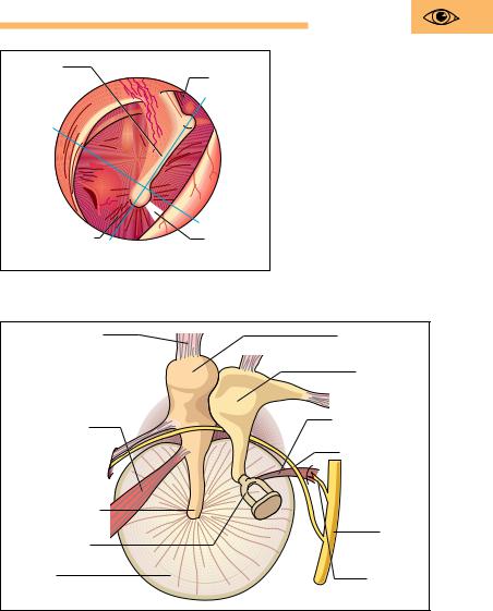
Fig. 15.2 Horizontal section through the anterior part of the eyeball. (After Faller)
Anterior and Posterior Chamber of the Eye
The eye can be divided into three spaces (Figs. 15.1 and 15.2): the anterior chamber, the posterior chamber, and the cavity of the eye containing the vitreous body (corpus vitreum). The anterior chamber lies behind the cornea. It is filled with aqueous humor, and extends posteriorly to the pupil and the iris. At the iridocorneal angle lies a meshwork of connective tissue trabeculae. The aqueous humor flows through its spaces to reach a circular vein, the Schlemm canal (sinus venosus sclerae). At the pupil the anterior chamber is connected to the posterior chamber, where the aqueous humor is secreted. The posterior chamber is bounded by the posterior surface of the iris and the ciliary body (ciliary muscle and

628 15 Sense Organs
zonular fibers) together with the anterior part of the vitreous body (vitreous humor). The vitreous body contains neither vessels nor nerves and is composed of a clear glassy gelatinous substance (98 % water) that is essentially composed of collagen and hyaluronic acid (binds water). It stabilizes the bulb and takes up about two-thirds of the total volume of the eyeball.
Intraocular (inside the eye) Pressure
The external shape of the globe is maintained by its connective tissue coat, the sclera, and especially by an internal pressure that is elevated above atmospheric pressure by about 15−20 mmHg (2−3 kPa). The internal pressure of the eye is created by the aqueous humor, and the equilibrium between secretion and drainage of the aqueous humor plays an important role in its maintenance. If, for instance, drainage from Schlemm’s canal (see above) is impeded, the internal pressure of the eyeball can rise dangerously (glaucoma). The rise in pressure must be treated with medication, since an elevated pressure can reduce perfusion, thereby damaging the retina and leading to blindness.
Lens and the Process of Accommodation
Light falling on the lens is focused because the posterior surface of the lens is more curved than the anterior surface. The lens consists of a nucleus, epithelium, and fibers, and it is completely transparent. It is suspended from the circular ciliary muscle (m. ciliaris) by a similarly circular arrangement of fibers (zonular fibers) (Figs. 15.1 and 15.2). By changing its shape, the lens can vary its refraction (see below). By this mechanism the eye can bring objects at varying distances into sharp focus (accommodation).
When the ciliary muscle contracts, the zonular fibers relax, because the distance between the muscle and the lens becomes smaller (muscle contraction shortens the muscle and thickens its belly). Hence the zonular fibers no longer pull on the lens, which now becomes increasingly spherical as a result of its own elasticity, i.e., its radius of curvature decreases. Consequently, the incident light rays are more strongly refracted and the eye accommodates to near vision. When the parasympathetically innervated ciliary muscle relaxes, the distance between lens


630 15 Sense Organs
width of the pupil is reflexly controlled and depends on the intensity of the light falling on it, among other factors. It varies between 1.5 mm (miosis) and 8 mm (mydriasis). The pupillary light reflex can also be triggered when light does not fall on the eye. If, for instance, the light from a flashlight enters one eye, the pupils of both eyes constrict, the so-called consensual light reflex. Failure to trigger this reflex suggests a serious condition in the CNS. Fixing a nearby object also leads to constriction of the pupil (increasing depth of focus) as well as convergence of the axes of the two eyes.
The color of the iris depends on the quantity and location of pigments in the connective tissue and the pigment epithelium behind the iris. If the iris is completely devoid of pigment, it appears red, because blood vessels can be seen through it (as in albinos).
Cornea
The cornea, the most anterior portion of the outer coat of the eye, is more strongly curved than the sclera, like a flattened dome inserted into the anterior aspect of the globe (Fig. 15.2). It is devoid of blood vessels and receives nutrients by diffusion through the aqueous humor. The outer surface of its connective tissue stroma is covered by a stratified noncornified (i.e., the name cornea is misleading) squamous epithelium, while its inner surface is covered by a single epithelial layer. Its transparency rests on a certain fluid content and the degree of swelling of the lamellar collagen fibers of the stroma. If this specific degree of swelling changes, the cornea becomes cloudy.
Posterior Part of the Globe
The posterior part of the globe consists of the sclera, the uvea, and the perceptive parts in the form of the sensory cells of the retina, the processes of which leave the posterior wall as the optic nerve.
Sclera
The opaque sclera is made up of tough bundles of collagen fibers, and its inelastic connective tissue capsule maintains the shape of the eyeball, supported by the internal pressure of the globe and the pull of the ex-
The Eye 631
traocular muscles. At the corneoscleral junction or corneal limbus
(Fig. 15.2) anteriorly, the sclera blends into the cornea, which represents about one-sixth of the surface of the eyeball. Where the optic nerve leaves the eyeball, the sclera is pierced like a sieve (lamina cribrosa) and continues as the dura mater and arachnoid covering the optic nerve (Fig. 15.1).
Vascular Tunic (Uvea)
The vascular tunic is 0.2 mm thick and consists of the iris, the ciliary body, and the choroid. It is applied to the inside of the sclera. In front of the corneal limbus it forms the ciliary body which, in contrast to the smooth choroid, is covered with ridges, folds, and processes. Its connective tissue stroma is continued into the iris. The choroid consists of delicate pigmented connective tissue and contains numerous blood vessels (Fig. 15.5), which supply nutrition to the adjacent layers, especially the avascular outer layers of the retina.
Retina
The retina is composed of a posterior light-sensitive segment (pars optica retinae) and an anterior light-insensitive part (pars ciliaris and pars iridica). The boundary between the two parts of the retina is a serrated line (ora serrata) at the posterior border of the ciliary body. The pars ciliaris and the pars iridica are simple epithelia covering the ciliary body and the posterior surface of the iris respectively. Where it covers the iris, the epithelium is densely pigmented.
The optical part of the retina coats the entire posterior area of the globe and consists of two layers, an outer pigmented epithelium (pars pigmentosa) and an inner light-sensitive layer (pars nervosa). The simple pigmented epithelium directly adjoins the choroid and contains elongated brown pigment granules. With cellular processes of varying shapes, the pigmented layer extends to the photoreceptors of the nervous layer. Its main function is the nutrition of the photoreceptors.
The light-sensitive part of the retina contains three layers of neurons of the visual pathway. These layers, from outside inward, contain (Figs. 15.4 and 15.5):

632 |
15 Sense Organs |
|
|
|
|
|
Fig. 15.4 Neuronal con- |
|
|
|
nections in the retina. In |
Optic nerve |
|
|
the region of the fovea |
3rd neuron: |
|
light |
centralis of the macula |
|
lutea, the only sensory |
||
Ganglion cells |
of |
cells are cones; outside the |
|
= stratum |
|
||
|
Direction |
||
|
direction of the light. |
||
ganglionare |
|
|
fovea centralis (periphery), |
nervi optici |
|
|
they include rods. Note the |
2nd neuron: |
|
(After Duus) |
|
Bipolar cells |
|
|
|
= stratum |
|
|
|
ganglionare |
|
|
|
retinae |
|
Excitation |
|
1st neuron: |
|
|
|
|
|
|
|
Rods and cones |
|
|
|
= stratum |
|
|
|
neuroepitheliae |
|
|
|
Pigment |
|
|
|
epithelium |
Fovea |
Periphery |
|
|
centralis |
|
|
Fovea centralis |
Ganglion cells |
|
|
|
Bipolar cells |
|
Rods and |
|
cones |
|
Pigmented |
|
epithelium |
|
Choroid |
|
Sclera |
Fig. 15.5 Longitudinal section through the macula lutea (yellow spot) of a human eye. In the fovea centralis the light impinges directly on the cones

The Eye 633
The first neurons, i.e., photoreceptors (rods and cones; stratum neuroepitheliale retinae)
The second neurons, i.e., bipolar retinal ganglion cells (ganglionic layer of retina, stratum ganglionicum retinae)
The third neurons, i.e., the ganglion cells of the optic nerve (stratum ganglionare nervi optici)
The retina is divided by anatomists into 10 layers, though the borders between adjacent layers are admittedly somewhat indistinct. The pattern of layers is produced by the various components of the types of neurons listed above lying at characteristic depths within the retina.
Rods and Cones
The light-sensitive photoreceptors, the rods and cones, lie in the outermost layer overlying the pigmented layer, and are covered by the nerve cells of the two inner layers. Hence, the light-sensitive cells are turned away from the incident light, i.e., the light must first penetrate the inner layers of the retina before reaching the rods and cones. Hence the eye is sometimes called “inverted.”
The light-sensitive sensory cells consist of about 120 million rods (light and dark sensitive, as well as vision in dim light) and about 6 million cones (color vision). They synapse with relay cells (bipolar retinal ganglion cells, 2nd neuron), the axons of which end in synapses on the ganglion cells of the optic nerve (3rd neuron) (Fig. 15.5). There are considerably fewer retinal and optic nerve ganglion cells than sensory cells. The result is that the impulses from several sensory cells are conducted to only one retinal or optic ganglion cell (convergence of impulse conduction).
Optic Disk (Discus Nervi Optici)
Finally, the central axons of the optic ganglion cells reach a point in the posterior pole of the eyeball where they collect (optic disk, papilla nervi optici, discus nervi optici) and leave the eye through a sievelike interruption in the sclera (lamina cribrosa). They then continue as the optic nerve (n. opticus) to the diencephalon. The disk is devoid of sensory cells (blind spot). The vessels of the optic nerve (central artery of the retina) enter here (Fig. 15.6).

634 15 Sense Organs
Optic disk |
Macula lutea |
(papilla nervi optici) |
(yellow spot) |
|
Fovea centralis |
Branches of central artery |
Branches of central vein |
of retina (a. centralis retinae) |
of retina (v. centralis retinae) |
Fig. 15.6 Normal fundus of a left eye. At the optic disk (papilla) the nerve fibers collect to become the optic nerve and the central artery of the retina enters
Macula Lutea (Yellow Spot)
The retina is almost devoid of blood vessels in the macula lutea, a depression lying about 4 mm lateral to the optic disk (Figs. 15.5 and 15.6). It contains a depression (fovea centralis) in which only cones are found. The remaining retinal layers are pushed aside at this point, so that light falling on it reaches the sensory cells directly. The macula and fovea are therefore the points where vision is most acute.

The Eye 635
Eye Ground (Fundus)
The redto orange-colored fundus can be viewed directly with an ophthalmoscope (Fig. 15.6). The optic disk, where all the nerve fibers converge to leave the eye, lies in the nasal half. The central retinal artery enters in the middle of the disk and divides into several branches, of which some run toward the macula. The somewhat darker and wider veins join in a central retinal vein and also leave the retina through the papilla. Ophthalmoscopic examination allows an examiner to view the vessels and to determine retinal changes.
The Optic System
The optical (dioptric) apparatus projects an inverted, markedly reduced image of the world on the retina. Visible light entering the eye is composed of wavelengths between 400 and 700 nm (1 nm = one billionth of a meter = 10−9 m). These waves induce a photochemical excitation in the sensory cells, which the visual pathways conduct to the visual cortex of the cerebrum (see The Visual Pathway below).
Power of Refraction
The image on the retina is formed by the refraction of light rays by the curved surfaces (e. g., cornea, lens). For sharp vision, all the rays coming from a specific point on an object must reunite in a point on the retina. Thus the optic system of the eye functions as a focusing lens. The more curved the lens (i.e., the more convex) the more strongly the rays are refracted (refraction increases) and the shorter their focal length. When looking at close objects (positive accommodation), refraction must be increased; when viewing distant objects (negative accommodation), it must be reduced, and the lens flattens.
The measure of the refractive power of the optic system of the eye is the diopter (D). Refractive power is calculated as follows:
Refractive power (D) = 1/focal length (m)
For a human eye with maximal negative accommodation (lens flattened), the whole optical system has an anterior focal length of 0.017 m

636 15 Sense Organs
(17 mm); the total power of refraction is therefore 1/0.017 = 59 D. For an eye with maximal positive accommodation (lens curved), the power of refraction increases by about 10 D. This increase in power of refraction is also known as the power of accommodation. Because the lens becomes less and less elastic with increasing age (the ability to relax is lost), the power of accommodation decreases, leading to presbyopia (old sightedness). Here the ability to see distant objects is unimpeded, but for close viewing (e. g., reading) glasses with convex lenses are necessary.
Cloudiness of the lens (cataract), a condition most often encountered in the elderly but which may occur at any age, can be treated by surgical removal of the lens if necessary. The loss in refractory power of the lens must be replaced either by the use of strong convex glasses (cataract lens) or by replacing the lens that has been removed with a plastic lens.
Errors of Refraction
Apart from the refractive error of old age (presbyopia), congenital malformations of the globe can cause visual defects. The anterior surface of the cornea is normally exactly 24.4 mm distant from the retina. A distance that is too short or too long results in an error of refraction. If the eyeball is too long the result is nearsightedness (myopia); when the distance is too short the result is farsightedness (hyperopia) (Fig. 15.7a, b).
Nearsightedness
In cases of nearsightedness, light rays originating at infinity come together before reaching the retina and then diverge again. Thus the refractive power of the eye is too great relative to the length of the eye, and the image projected on the retina will be blurred. Hence myopic people, having eyeballs that are too long, can only focus on nearby objects. In order to be able to focus on distant objects, they must wear glasses with concave lenses (diverging lenses) (Fig. 15.7b).
Farsightedness
Because the eyeball is too short in cases of farsightedness, light rays originating at infinity would converge behind the retina. In order to see

|
The Eye |
637 |
a |
b |
|
Fig. 15.7 a, b Farsightedness (hyperopia) and nearsightedness (myopia).
a When the globe is too short (farsightedness): correction by convex lens (+ diopters) b When the globe is too long (nearsightedness) correction by concave lens (− diopters)
distant objects clearly in spite of this, hyperopic persons must constantly accommodate, i.e., increase the power of refraction. In this way, farsighted people can bring distant objects into focus even though their eyeball is too short. However, their power of refraction is not sufficient to see close objects, and they need correction with a convex lens (converging lens) (Fig. 15.7a). This is also required for distant vision, in order not to tire the eye by constant contraction of the ciliary muscle (producing headaches).
Astigmatism
In astigmatism an irregular curvature of the cornea results in a point being projected not as a point but as a line. This error is corrected by cylindrical glasses that are curved in the appropriate direction.

638 15 Sense Organs
50 |
50 |
35 |
35 |
25 |
25 |
20 |
20 |
15 |
15 |
10 |
10 |
7,5 |
7,5 |
5 |
5 |
4 |
4 |
Fig. 15.8 Eye charts. If the chart is placed at a distance of 20 feet , the person tested should be able to read the lowest line prescribed for that distance. Vision is then said to be 20/20. If the line read is that which would normally be legible at 40 feet, vision would be 20/40
Visual Acuity
The ability of the eye to distinguish between two points at a certain distance is called resolution or visual acuity. Visual acuity is tested with special test charts that are usually read at a distance of 20 feet (Fig. 15.8). In good lighting conditions, the normal eye should be able to see two points separated by 1.5 mm as distinct points.

The Eye 639
The Visual Pathway
Each eye has a lateral (temporal) and medial (nasal) visual field. The incident light from the temporal field falls on the nasal part of the retina, and the nasal visual field is projected on the temporal part of the retina (Fig. 15.9).
The visual pathway begins at the retina and ends in the visual cortex in the calcarine fissure of the occipital lobe. It comprises four successive neurons, the cell bodies of the first three of these being located in the retina (Fig. 15.4).
The photoreceptors (1st neuron)
The retinal ganglion cells (2nd neuron)
The ganglion cells of the optic nerve (3rd neuron), the axons of which run posteriorly in the optic nerve
At the crossing of the optic nerves (optic chiasma) below the diencephalon, the axons of the nasal halves of the retina cross to join the uncrossed axons of the temporal halves of the retina. Together they continue backward as the optic tract and end in the lateral geniculate body of the diencephalon. There they synapse with the 4th neurons, the axons of which form the optic radiations, ending in the visual cortex (Fig. 15.9).
Thus the left visual field of each eye is represented in the cortex of the right hemisphere, while the right visual fields are projected to the visual cortex of the left hemisphere. The area with greatest visual acuity, the macula lutea with the fovea centralis, is projected to by far the largest area in the visual cortex.
Visual Field Defects
During the examination of the eye, the visual fields of both eyes are tested and recorded. If, for instance, the left optic nerve is injured (example a in Fig. 15.9), both visual fields in that eye are affected, resulting in blindness (amaurosis) in the left eye. If, on the other hand, a pituitary tumor presses on the nasal axons of the two optic nerves where they cross in the chiasma (example b in Fig. 15.9), this leads to blindness in the temporal visual fields of both eyes (bitemporal hemianopsia). An injury to the left optic tract (example c in Fig. 15.9) leads to a defect of the right visual field, known as right homonymous hemianopia (blindness in

640 |
15 Sense Organs |
|
|
Medial (nasal) |
Lateral (temporal) |
|
visual field of right eye |
visual field of |
|
|
right eye |
Lateral |
Medial (nasal) |
|
(temporal) |
retina |
|
retina |
|
|
Optic nerve
ab
Crossing of optic nerves (optic chiasma)
Pituitary gland (hypophysis) c
Optic tract (tractus opticus)
Thalamus
Pineal gland (glandula pinealis)
Lateral geniculate body  (corpus geniculatum d laterale)
4th neuron of
the visual pathway (optic radiation)
Visual cortex (calcarine fissure)
Occipital pole
Fig. 15.9 The visual pathway. The medial (nasal) visual field is projected to the lateral (temporal) half of the retina. The lateral (temporal) visual field is projected to the medial (nasal) half of the retina. Fibers of the optic nerve from the temporal retina (green) do not cross. Fibers from the medial half of the retina (pink) cross to the opposite side. For description of defects following lesions of the optic nerve (a), inside the crossing of the optic fibers (b), the optic tract (c), and the 4th neuron in the optic radiation (d), see Visual Field Defects in the text
The Eye 641
the temporal visual field of the right eye and the nasal visual field of the left eye). A similar visual disturbance can be due to injury to e. g., the left optic radiation (example d in Fig. 15.9).
Accessory Structures
Eyelids (Palpebrae)
In front, the cover of the eyelids protects the eyeball (Fig. 15.10). The upper and lower lids bound the palpebral fissure, and each is made more rigid by a connective tissue plate (tarsal plate, tarsus). Sebaceous glands embedded in the margin of the lid grease this margin, from which grow several rows of eyelashes. The outer part of the lid is covered by keratinous stratified squamous epithelium, while the inner side is lined by the conjunctiva. This consists of a stratified squamous epithelium, which is reflected from the sclera at the upper and lower conjunctival fornix (fold).
The most important muscles of the eyelids are the m. levator palpebrae superioris, supplied by the oculomotor nerve (cranial III), and the m. orbicularis oculi, supplied by the facial nerve (cranial VII), which closes the palpebral fissure. Both muscles are voluntary muscles.
Lacrimal Apparatus
The lacrimal apparatus includes the lacrimal gland (glandula lacrimalis), and the pathways by which the tears drain. The lacrimal glands lies laterally and above the eyeball (Fig. 15.10), and drain through several ducts into the outer part of the fornix. The lacrimal fluid constantly moistens the anterior surface of the globe and cleans and nourishes the cornea. It is distributed evenly by blinking and accumulates in the inner canthus (angle) of the eye.
The pathways by which the tears drain begin with the lacrimal punctum (punctum lacrimale), from which the lacrimal fluid drains through the lacrimal canal (canaliculus lacrimalis) into the lacrimal sac (Fig. 5.10). From there the tears drain through the lacrimonasal duct (ductus nasolacrimalis) into the inferior nasal meatus of the nasal cavity.


 retinae)
644 15 Sense Organs
Superior oblique muscle |
Medial rectus muscle |
|
|
Trochlea |
Superior rectus |
|
|
muscle |
|
Superior rectus |
|
|
|
|
|
|
|
|
muscle |
|
|
Levator |
Trochlea |
|
|
palpebrae |
|
|
|
muscle |
|
|
a |
|
b |
Levator |
Inferior rectus |
Lateral rectus |
Inferior oblique |
palpebrae |
|||
superior |
muscle |
muscle |
muscle |
muscle |
|
|
|
Fig. 15.12 a, b Extraocular muscles of the right eye seen from above (a) and from the side (b). In a the anterior portion of the levator palpebrae superioris muscle has been removed. (After Kahle)
The Ear
The ear contains two sensory organs with different functions (organs of hearing and of equilibrium) that nevertheless form an anatomical unit, the inner ear. It lies in the petrous pyramid of the temporal bone and consists of the cochlea and the vestibular apparatus (labyrinth), the latter including two fluid-filled pouches (utricle and saccule) and three semicircular canals, also fluid-filled. The organ of hearing, in contrast to the vestibular apparatus, has accessory structures to conduct sound waves: the external ear and the middle ear.

The Ear 645
The Organ of Hearing
External Ear
The external ear includes the auricle or pinna (auricula), the external auditory meatus (meatus acusticus externus), about 3 cm long, and the eardrum (membrana tympani). The pinna consists mainly of elastic cartilage, which continues into the outer end of the external auditory meatus. This then continues as the bony part of the external auditory canal, which is somewhat S-shaped. The cartilaginous part contains numerous ceruminous glands that secrete the “earwax” (cerumen). The eardrum stretches across the inner end of the bony canal and forms the boundary of the middle ear.
Middle Ear
The middle ear (Fig. 15.13a, b) includes the tympanic cavity, lined with mucous membrane and containing the auditory ossicles (ossicula auditus) (malleus, incus and stapes); its continuation anteriorly into the pharynx, the eustachian tube (tuba auditiva); as well as numerous mu- cosa-lined cavities in the mastoid process. The eardrum is nearly circular with a diameter of about 1 cm and forms the outer wall of the tympanic cavity. It is made up of three layers. The mainly tense connective tissue framework of the eardrum (membrana tensa, pars tensa) is weaker in only a small section at its upper end (membrana flaccida, pars flaccida) (Fig. 15.14). Its inner surface is lined with a mucous membrane, its outer surface with skin. The long handle of the malleus is fastened to the tympanum, bulging it inward like a funnel (Fig. 15.15).
The auditory ossicles, together with the tympanum, form the soundconducting apparatus. Malleus (hammer), incus (anvil), and stapes (stirrup) form a jointed chain between the eardrum and the oval window (fenestra vestibuli), into which the footplate of the stapes is inserted. The auditory ossicles conduct vibrations elicited by sound waves in the eardrum through the oval window to the inner ear. The latter, together with the first turn of the cochlea, forms the inner bony boundary of the tympanic cavity (promontory). The plate of the stapes in the oval window transmits the vibrations to the fluid of the inner ear. The malleus and stapes are also stabilized by two muscles, the m. tensor tympani, and the m. stapedius. Both can influence sensitivity to the transmission.

646 15 Sense Organs
External auditory |
Stirrup |
Perilym- |
Membranous |
|
meatus (meatus |
(stapes) |
semicircular canal |
||
phatic space |
||||
acusticus externus) |
in oval |
(ductus semicircularis) |
||
|
||||
Pinna |
window |
Endolym- |
Utricle |
|
(fenestra |
||||
(auricula) |
||||
vestibuli) |
phatic space |
|
||
Hammer |
|
|||
|
|
Endolymphatic canal |
||
(malleus) |
|
|
||
Anvil |
(ductus endolymphaticus) |
|||
|
||||
|
(incus) |
|
|
|
|
|
|
Saccule |
|
|
|
|
Tympanic |
|
|
|
|
cavity |
|
a |
|
|
|
|
Eardrum |
|
|
|
|
(membrana tympani) |
|
|
|
|
Round window |
|
Cochlea |
Tympanic canal |
|
(fenestra cochleae) |
|
(scala tympani) |
||
|
|
|||
Vestibular canal |
|
|
Internal |
|
(scala vestibuli) |
|
|
carotid artery |
|
Foramen |
|
|
Cochlear canal |
|
magnum |
|
|
(ductus cochlearis) |
|
Petrous bone |
|
|
|
|
|
|
Semicircular canals |
Eustachian tube |
|
b |
|
|
(tuba auditiva) |
|
Fig. 15.13 a, b Diagrammatic view of the external ear, the middle ear, and the inner ear (coronal section). (After Kahle)
aExternal ear (to the eardrum), middle ear (auditory ossicles and eustachian tube), and inner ear (labyrinth and cochlea)
bPosition of the inner ear in the skull (base of skull seen from above), with a cast of the organs of hearing and equilibrium
 (secretory and taste nerve for anterior twothirds of tongue)
648 15 Sense Organs
Inner Ear
The inner ear (Fig. 15.13) is surrounded by a hard bony capsule, and consists of a fluid-filled (perilymph) labyrinthine system of passages and cavities (bony labyrinth). The membranous labyrinth, also fluid-filled (endolymph), lies inside the bony labyrinth. Perilymph and endolymph differ chiefly in their sodium and potassium contents. The membranous labyrinth contains the organs of hearing and equilibrium.
The bony spiral (cochlea) of the inner ear, about 3 cm long, makes up a passage that in humans winds about 21/2 times around a bony axis, the central pillar or columella (modiolus). A cross-section of the cochlea (Fig. 15.16 and 15.17) shows three separate cavities: the cochlear canal
(ductus cochlearis) in the middle, the vestibular canal (scala vestibuli) above it, and the tympanic canal (scala tympani) below. The scala tympani and the scala vestibuli join at the apex of the cochlea (helicotrema). They are filled with perilymph and end at the round window (fenestra cochleae) and the oval window (fenestra vestibuli), respectively (Fig. 15.13a).
The cochlear duct is filled with endolymph and abuts the scala tympani by the basilar lamina (lamina basilaris), and the scala vestibuli by
Reissner’s membrane (paries vestibularis). The organ of Corti (spiral organ) (Fig. 15.17) rests on the basilar membrane. It contains some 15 000 auditory sensory cells arranged in rows (inner and outer hair cells) as well as numerous support cells. The sensory hairs of the hair cells are connected to a gelatinous layer (membrana tectoria) overlying it.
Auditory Pathway
The hair cells synapse with neurons the cell bodies of which lie in the spiral ganglion of the cochlea at the modiolus (Fig. 15.17). From here the central axons run with the cochlear and the vestibular nerves in cranial nerve VIII (vestibulocochlear nerve) to the brainstem. There the axons of the cochlear nerve end in the nuclei of the cochlear nerve (nucleus cochlearis), while those of the vestibular nerve terminate in the vestibular nuclei. On the way to the auditory area in the anterior transverse gyrus of the temporal lobe, the auditory pathway synapses several times, including synapses in the medial geniculate body of the diencephalon.


650 15 Sense Organs
Mechanism of Hearing
Vibrations (sound waves) impinging on the oval window, after transmission by the eardrum and the auditory ossicles, create pressure waves in the perilymph of the vestibular canal. These pressure waves continue to the apex of the cochlea and then return through the tympanic canal (Fig. 15.13a). The opposing flows of the perilymph in the vestibular and tympanic canals create vibrations in the endolymph of the cochlear duct, and this excites the sensory cells by moving the sensory hairs in the membrana tectoria (Fig. 15.17). Since the basilar membrane is narrow at the base of the cochlea, and broader at its apex, the frequency of the pressure waves decreases toward the apex. By this mechanism, highpitched tones are heard at the base of the cochlea, low-pitched tones at the apex.
Pure tones or musical notes are produced by regularly recurring (periodic) vibratory processes of a defined frequency and intensity, while noises are characterized by nonperiodic (irregular) auditory events. In general it can be said that sound waves with frequencies between 20 and 16 000 hertz (1 Hz = one vibration per second) excite the hair cells in the inner ear and so elicit sound sensations (tones, notes, or noises). The higher the frequency of the sound waves, the higher pitched the tone that is experienced (see above). Frequencies below 20 Hz (infrasonic frequencies) and above 16 000 Hz (ultrasound) do not elicit excitations in the human inner ear. On the other hand, auditory events must exceed a certain intensity (minimal pressure) before they can be heard. The auditory threshold, that is the sound pressure that just elicits sound perception, for a tone with a frequency of 1000 Hz would be about (2 × 10⁻⁵ Newton/m2 (N/m2 = Pascal). The ear is most sensitive in a range between 2000 and 5000 Hz, where even very low sound pressures are sufficient to exceed the auditory threshold.
The reference power level has been introduced to obtain an objective measure of sound pressure. It is usually expressed in decibels (dB). By convention, the sound pressure that just elicits a perceived sound has been pegged at 0 dB. Every tenfold increase in sound pressure corresponds to an increase in the reference power level of 20 decibels.
The Ear 651
Hearing Problems
Hearing problems are a widely distributed phenomenon affecting millions of people. In general they may be divided into two kinds. The first is middle ear or conduction deafness, in which there is a mechanical impediment to the tone reaching the cochlea. This impediment may, for example, be a torn drum or blockage of the auditory meatus by earwax. The commonest cause of conduction deafness, however, is otosclerosis, in which the stapes in the middle ear is ankylosed (fixed) and so cannot transmit vibrations. The second type of hearing problem is inner ear deafness, caused, for example, by lesions of the cochlea or the auditory nerve. An infection with German measles during the first four months of pregnancy can often lead to the birth of a completely deaf infant.
The Organ of Equilibrium
The organ of equilibrium (vestibular organ) includes three semicircular ducts with their dilatations (ampullae), in which lie the acoustic crests (cristae ampullares) with their sensory cells, and the utricle and saccule, each with a sensory area (macula utriculi and macula sacculi). They are filled with endolymph and form the membranous labyrinth (Figs. 15.13 and 15.18). They register acceleration and changes in position, and so determine spatial orientation. Specialized sensory cells project into the endolymph, and these react to fluid movements, which they sense. The endolymph moves with shifts in posture or changes in position of the head. The sensory cells so stimulated send information to the cerebellum, which responds reflexly to the changes.
Macula of Utricle and Saccule
The two sensory areas of the larger and smaller vestibular pouches register primarily straight-line accelerations, especially when they are horizontal (e. g., the braking of an automobile) or vertical (e. g., traveling in an elevator).
The epithelium lining the saccule and utricle is thickened in certain areas, where it is differentiated into special sensory and support cells. These sensory areas (maculae) are covered with a gelatinous substance

652 15 Sense Organs
Posterior semicircular |
Anterior semicircular duct |
|
duct |
|
|
|
Dilatations of semicircular ducts |
|
|
(ampullae) |
|
|
Endolymphatic duct |
|
Lateral |
|
|
semicircular |
Vestibular nerve |
|
duct |
||
|
||
Sensory area |
|
|
(macula) of utricle |
|
|
Sensory area |
Auditory |
|
(macula) of saccule |
nerve |
|
|
(n. cochlearis) |
|
Cochlear canal |
Cochlea |
|
|
Fig. 15.18 Right membranous labyrinth
(membrana statoconiorum macularum), which is weighted down by tiny calciferousgranules(otoliths,statoconia,otoconia).Frombelow,processes of the sensory cells, the sensory hairs or cilia, project into the gelatinous membrane (Fig. 15.19). When the sensory epithelium and the statoconic membrane move against each other, the shear force displaces the cilia. Since the macula in the floor of the utricle is oriented almost horizontally, its sensory cells are stimulated mainly by horizontal acceleration. On the other hand, the macula of the saccule is oriented nearly vertically on the anterior wall of the saccule and so responds mainly to vertical acceleration.
Acoustic Crest (Crista Ampularis)
The acoustic crests (cristae ampullares) in the dilatations (ampullae) of the semicircular ducts register especially rotary acceleration moving the endolymph. They lie in the three principal spatial planes and are essentially connected to reflex eye movements.

 Support cells
654 15 Sense Organs
Gustatory papilla |
Pit |
(papilla vallata) |
Wall of |
|
|
|
vallate papilla |
|
Taste buds |
|
Mucoserous |
|
glands |
|
(of von Ebner) |
|
Nerve fibers |
Fig. 15.21 Longitudinal section through a taste papilla (papilla vallata). For the indicated section see Fig. 15.22
The Sense of Taste
The sensory cells for the perception of various tastes lie together with support cells in the taste buds, which are arranged in the taste papillae of the tongue (see Chapter 9: The Oral Cavity, The Tongue). The taste buds are shaped like tulip buds and lie in the stratified squamous epithelium of the tongue (Figs. 15.21 and 15.22). On the epithelial surface they are provided with a depressed pit with an opening, the taste pore, into which the sensory cells project with taste hairs. Each taste bud is supplied with several nerve fibers, which connect to fibers supplying other taste buds. The anterior two-thirds of the tongue are supplied by neurons of the facial nerve (cranial nerve VII) whose fibers travel in the chorda tympani), the posterior part by sensory neurons from the glossopharyngeal nerve (cranial nerve IX). The four taste sensations (sweet, sour, salty, and bitter) are perceived in different areas of the tongue and probably registered by different receptors in individual taste buds. Sour is perceived especially at the lateral border of the tongue, salty at the borders and the tip of the tongue, bitter at the base of the tongue, and sweet especially at the tip of the tongue (see Fig. 9.5). The grooves adjacent to the taste papillae contain mucoserous glands (of von Ebner) (Fig. 15.21) that secrete a watery fluid thought to have a “rinsing” function.

The Sense of Smell 655
Taste pore with |
Fig. 15.22 |
Longitudinal |
|
section through a taste |
|||
taste rods of the |
bud. Section from |
||
sensory cells |
|||
Fig. 15.21 |
|
||
|
|
||
Epithelium of |
|
|
|
surface of tongue |
|
|
|
Sensory cell |
|
|
|
Support cell |
|
|
|
The Sense of Smell
The human olfactory mucous membrane encompasses an area of about 500 mm2 in the superior nasal turbinate and on each side of the upper nasal septum (regio olfactoria, see Fig. 8.1). It can be distinguished from the remaining nasal mucosa (regio respiratoria) by an epithelium that is distinctly higher, containing mostly basal cells (proliferative cells), supportive (sustentacular) cells, and sensory cells (Fig. 15.23). In humans there are about 10 million sensory cells (olfactory cells). These bipolar nerve cells (fila olfactoria) are the 1st neurons of the olfactory pathway. At their upper end (dendrite) they project a small knob, the olfactory rod, with numerous olfactory hairs. Their axons pass into the anterior cranial fossa through a bony plate, which is perforated like a sieve (cribroid plate of the ethmoid bone, lamina cribrosa) (Fig. 4.55, p. 192). They synapse with the nerve cells (2nd neurons) of the olfactory bulb, an enlargement of the olfactory nerve. The olfactory nerve runs to the olfactory cortex, where the corresponding perceptions are generated.
Under the olfactory mucosa lie numerous mucous glands (Bowman’s glands), the ducts of which end on the surface of the epithelium and the secretions of which dissolve and remove olfactory substances.


Summary 657
Outer coat: sclera (posteriorly) and cornea (anteriorly)
Middle coat: choroid (posteriorly) and iris and ciliary body (anteriorly)
Inner coat: retina (posteriorly) and pigmented epithelium of the ciliary body (anteriorly).
Anterior Part of the Globe
The anterior part of the globe contains the optical apparatus and is made up of the following structures:
Anterior and posterior chambers of the eye: contain the aqueous humor, which is secreted in the posterior chamber and reaches the anterior chamber by the opening in the iris (pupil). It drains through Schlemm’s canal in the iridocorneal angle. The aqueous
humor generates the intraocular pressure (15−20 mmHg |
= 2− |
3 kPa). A rising intraocular pressure leads to glaucoma, |
with |
danger of retinal damage. |
|
Cornea: bounds the anterior chamber of the eye. Consists of an avascular, transparent, stratified nonkeratinous (!) epithelium.
Vitreous body: Consists of a clear, glassy, gelatinous substance (collagen and hyaluronic acid).
Lens: consists of nucleus, epithelium, and fibers and is completely transparent (clouding of the lens = cataract). The perfectly round lens is attached along its entire circumference to the ciliary muscle by the zonular (zonula = belt) fibers (zonular fibers + ciliary muscle = ciliary body). The curvature of the lens, and hence its refractive power, is changed by contraction and relaxation of the ciliary muscle (accommodation).
Iris: forms a variable aperture (pupil) in front of the lens, controlling the amount of light entering the eye (color of the iris depends on the density of its pigments). The sphincter of the pupil (m. sphincter pupillae) is innervated by the parasympathetic, and constricts the pupil (miosis), while the dilator of the pupil (m. dilator pupillae) dilates it (mydriasis). Pupillary reflexes: consensual light reflex (when the incident light hits only one eye, both eyes are reflexly constricted); convergence (internal rotation of the axes of both eyes and constriction of the pupils when fixating close objects).

658 15 Sense Organs
The optical apparatus acts like a convex lens and projects an inverted and markedly reduced image of the world on the retina. The more curved the lens, the greater its power of refraction (measured in diopters) and the shorter the focal length: refractive power (D) = 1/focal length (m). When the eye is maximally accommodated for distance (flattened lens with a focal length of 0.017 m) the power of refraction 1/0.017 m = 59 D. When accommodation is maximal for close vision (curved lens), the power of refraction increases by about 10 D (increase in refractive power = range of accommodation). Because the lens becomes less elastic, the range of accommodation decreases with aging, resulting in presbyopia (convex glasses needed for close vision).
Posterior Part of the Globe
The posterior part of the globe includes the sclera, the choroid and the retina.
Sclera: forms an inelastic connective tissue capsule, into which the extraocular muscles are inserted. It is pierced like a sieve where the optic nerve exits.
Choroid: consists of delicate pigmented connective tissue containing numerous blood vessels.
Retina: separates the choroid and the vitreous and consists of two layers, the outer pigmented layer (stratum pigmentosum) and the inner light-sensitive layer (stratum nervosum), which consists of three layers (starting from the outside):
1.Layer of photoreceptors (1st neuron) with rods and cones: light-sensitive photoreceptors (about 120 million rods for dark and light vision, and 6 million cones for color vision)
2.Layer of bipolar retinal ganglion cells (2nd neuron)
3.Ganglionic layer of optic nerve (3rd neuron), the axons of which form the optic nerve
Optic disk (papilla nervi optici): area where the central axons of the ganglion cells of the optic nerve collect and pierce the sclera, origin of the optic nerve, and point of entry of the central artery of the retina.
Macula lutea (yellow spot): with the fovea centralis, the site of maximal visual acuity, 4 mm lateral to the optic disk. Contains only cones, and the overlying retinal layers are pushed aside (light reaches sensory cells directly).

Summary 659
Eye ground (fundus): examination of the fundus with an ophthalmoscope allows direct examination of vessels (central artery and vein of the retina).
The Visual Pathway
The visual pathway starts at the retina and ends in the primary visual cortex at the calcarine fissure (occipital lobe); it consists of four successive neurons, the first three neurons being located in the retina. The axons of the 3rd neurons (ganglion cells) of the optic nerve form the optic nerve, which crosses at the optic chiasma to continue as the optic tract to the diencephalon, where it synapses with the 4th neuron in the lateral geniculate body. The axon of the 4th neuron form the optic radiation, which ends in the visual cortex of the cerebrum.
Visual fields: the visual field for each eye can be divided into an inner (nasal) and outer (temporal) field. The image from the inner field projects on the temporal, that from the outer field on the nasal half of the retina. The optic nerve fibers from the nasal halves of the retina then cross to the opposite side at the optic chiasma, while the fibers from the temporal side remain uncrossed. By this mechanism, the left visual field of each eye is represented in the right hemisphere and the right visual field in the left hemisphere.
Visual field defects: lesions of the neurons at various points may create partial or total visual field defects.
Accessory Structure of the Eye
These include the eyelids, the lacrimal apparatus, and the extraocular muscles.
Eyelids: the upper and lower lids protect the anterior surface of the eyeball and form the edges of the palpebral fissure. They are reinforced by connective tissue plates, and their outside is covered with stratified keratinous epithelium, their inside with conjunctiva. The levator palpebrae superior muscle is innervated by cranial nerve III, the circular muscle (orbicularis orbis muscle) that closes the palpebral fissure by cranial nerve VII.
Lacrimal apparatus: this includes the lacrimal gland and the pathways by which the tears drain. The tears secreted by the lacrimal gland clean and nourish the cornea. They flow through the lacrimal

660 15 Sense Organs
puncta and the lacrimal canal into the lacrimal sac, and from there through the nasolacrimal duct into the nasal cavity.
Extraocular muscles: striated muscles that move the two bulbs. There are four rectus muscles on each side (superior rectus, inferior rectus, medial rectus, and lateral rectus) and two oblique muscles (superior oblique and inferior oblique), innervated by cranial nerves III, IV, and VI.
The Ear
The ear contains two sensory organs, the organ of hearing and the organ of equilibrium. Both lie in the petrous pyramid of the temporal bone. They form the inner ear, consisting of the cochlea and the vestibular apparatus (labyrinth consisting of two vestibular pouches and three semicircular canals). Their afferent nerves run to the brainstem by way of the vestibulocochlear nerve (cranial nerve VIII ) and continue to the auditory cortex in the temporal lobe or to the cerebellum.
The Organ of Hearing
The organ of hearing includes the inner ear and its accessory structures, the external ear and the middle ear:
External ear: this includes the pinna, the external auditory meatus, which is about 3 cm long, and the eardrum, which separates it from the middle ear.
Middle ear: this includes the tympanic cavity, the eustachian tube (connects it to the pharyngeal cavity), and the auditory ossicles (hammer, anvil, and stirrup). Together with the tympanum, the latter form the sound-conducting apparatus. Vibrations elicited by sound waves in the tympanum are conducted by the auditory ossicles to the oval window of the inner ear and so transmitted to the perilymph. Two muscles, the stapedius muscle and the tensor tympani muscle can influence the sensitivity of the transmission.
Inner ear: this is a fluid-filled (perilymph) labyrinthine system of cavities and spaces (bony labyrinth), which contains the membranous labyrinth (contains the actual organs of hearing and equilibrium) filled with endolymph. Endolymph and perilymph differ principally in their sodium and potassium contents. The cochlea, a spiral about 3 cm long, contains a passage winding about 21/2 turns around its bony axis (modiolus); in cross-section it contains three
Summary 661
separate cavities: the middle cochlear canal (ductus cochlearis), the vestibular canal (scala vestibuli) above it, and the tympanic canal (scala tympani) below it. The scala tympani begins at the oval window, the scala vestibuli ends at the round window. They are connected at the apex of the cochlea (helicotrema) and are filled with perilymph. The cochlear duct is filled with endolymph and contains the organ of Corti, which is fitted with the sensory auditory cells (hair cells).
The Mechanism of Hearing and the Auditory Pathway
Sound waves traveling along the sound-conducting apparatus impinge on the oval window and create continuous pressure waves in the perilymph of the scala vestibuli; these waves then run in the opposite direction in the scala tympani. They initiate vibrations in the endolymph of the cochlear duct, creating frequency-dependent excitations in the hair cells of the organ of Corti (high-pitched tones are heard at the base of the cochlea, low-pitched at the apex). The action potentials are transmitted to the cochlear nucleus of the brainstem through the cochlear part of the vestibulocochlear nerve (cranial nerve VIII), and eventually through the auditory pathway to the primary acoustic cortex (anterior transverse temporal gyrus).
The Organ of Equilibrium
This includes the three semicircular ducts with their dilatations (ampullae) and the two vestibular pouches, the utricle (larger) and the saccule (smaller), all filled with endolymph. The ampullae and the utricle and saccule contain the sensory areas and their hair cells (acoustic crests, macula utriculi, and macula sacculi) that register acceleration and changes in position:
Acoustic crests: rotational acceleration in the three principal spatial axes
Macula utriculi: horizontal accelerations
Macula sacculi: vertical accelerations
The action potentials reach the vestibular nuclei in the brainstem by the vestibular part of the vestibulocochlear nerve (cranial nerve VIII). From there they run to the cerebellum and to the nuclei of the extraocular muscles.

662 15 Sense Organs
The Sense of Taste
The secondary sensory cells (altered epithelial cells for the sense of taste lie with supportive cells in the intraepithelial taste buds of the tongue. These taste buds lie in depressed pits in the epithelium at the edge of taste papillae, into the bottom of which drain mucoserous glands. The innervation of the sensory cells is by the facial nerve (anterior two-thirds of tongue) and the glossopharyngeal nerve (posterior two-thirds of the tongue). The four taste sensations (sweet, sour, salty, and bitter) are sensed by different receptors in taste buds in different parts of the tongue.
The Sense of Smell
The olfactory mucosa (about 500 mm2) in the superior nasal turbinates and the upper portion of the nasal septum contains about 10 million olfactory cells (bipolar nerve cells), which form the 1st neuron of the olfactory pathway in the form of the olfactory nerve. They pass through the lamina cribrosa of the ethmoid bone to the olfactory bulb, where they synapse with the 2nd neuron, the afferent axons of which run in the olfactory nerve (cranial nerve I) to the olfactory cortex.