


506 12 Reproduction, Development, and Birth
Human sexual reproduction is characterized by the union of a male sperm cell with a female ovum (fertilization), the ensuing transport of the germ cell through the uterine tube, its nidation (implantation) into the uterine mucosa, and the subsequent development into a viable newborn infant (embryonic and fetal development). At the end of the 8th week of gestation, embryonic development (embryogenesis), during which the primordial organs are laid down, becomes fetal development (fetation), characterized by the growth and differentiation of the organ systems.
Germ Cells
The development of germ cells (oogenesis and spermatogenesis) begins when diploid primitive germ cells (oogonia and spermatogonia) first form more diploid primary oocytes and spermatocytes (primary oocytes and primary spermatocytes) by mitosis. These develop into haploid cells (spermatozoa and ova) during the first and second maturation division (see Chapter 1). During fertilization, the sperm cell and the ovum fuse and form a zygote (see below).
A female diploid germ cell (primary oocyte) with 44 autosomes and 2 sex chromosomes (44, XX) eventually forms four haploid daughter cells, each with 22 autosomes and one X chromosome (22, X) (Fig. 12.1a). Only one, however, develops into an ovum, the other three form polar bodies that contain hardly any cytoplasm and degenerate during further development.. The male diploid germ cell (primary spermatocyte) with 44 autosomes and 2 sex chromosomes (44, XY) also forms four daughter cells (spermatids), of which two each possess 22 autosomes and either an X or a Y chromosome (22, X and 22, Y) (Fig. 12.1b). In contrast to oogenesis, however, all develop into mature sperm cells.
Fertilization
The spermatozoa must seek the ovum actively to fertilize it. The arduous passage through the uterus to the ampulla of the uterine tube at a migrating speed of 3 mm per minute requires about 1−3 hours. Of the 200− 300 million spermatozoa deposited in the posterior vaginal fornix

|
Fertilization 507 |
Oogenesis |
Spermatogenesis |
Oogonium |
Spermatogonium |
(44, XX) |
(44, XY) |
|
Mitoses |
Primary |
Primary |
oocyte |
spermatocyte |
(44, XX) |
(44, XY) |
1st maturation division
Secondary |
|
|
Secondary |
|
|
spermatocyte |
|
oocyte |
|
|
|
|
|
|
|
|
2nd maturation |
|
|
|
division |
(22 + X) |
(22 + Y) |
|
|
||
Mature |
|
|
Sperma- |
ovum |
|
|
|
|
|
tids |
|
(22+X) |
|
|
|
Polar bodies |
|
|
|
|
|
|
|
|
(22+X) |
|
|
|
|
|
Sperma- |
|
|
|
tozoa |
a |
b |
|
|
Fig. 12.1 a, b Formation of ovum and spermatozoa. (After Sadler.) a Oogenesis: the oogonia first form primary oocytes with a diploid set of chromosomes (44, XX) by mitosis. The primary oocytes then pass through the first and second maturation divisions, which lead to four haploid cells (one mature ovum and three polar bodies). b Spermatogenesis: diploid spermatogonia divide by mitosis and form primary spermatocytes (44, XY). These divide by meiosis and form four haploid spermatids, each with one X (22, X) or one Y (22, Y) chromosome. The spermatids develop into the motile spermatozoa
during intercourse only about 300 reach the ovum. In contrast to the sperm cells, which can survive up to 4 days in the female genital apparatus, the ova must be fertilized within 6−12 hours after ovulation, otherwise they perish.

508 12 Reproduction, Development, and Birth
Cells of the corona radiata |
1st polar body in |
|
process of |
|
division |
|
|
Phase 2 |
|
Phase 1 |
|
|
|
Dissolution |
Nucleus of |
Acro- |
of the acro- |
spermato- |
some |
some membrane |
zoon |
|
Oocyte in |
|
|
|
|
Cell |
process of 2nd |
|
maturation |
|
|
membrane |
division |
|
|
Zona pellucida |
|
Fusion of cell membranes |
Phase 3 |
of ovum and spermatozoon
Fig. 12.2 Schematic representation of the process of fertilization. (After Sadler.) During the first stage, the spermatozoon penetrates the cells of the corona radiata; during the second stage, the acrosome dissolves and the zona pellucida is digested by enzymes. In the third stage, the cell membranes of ovum and spermatozoon fuse and the sperm cell enters the ovum
During fertilization, the spermatozoon penetrates the attached follicular epithelial cells (corona radiata) and the zona pellucida by the action of the enzymes in the acrosome. This process of fertilization can be divided into three phases (Fig. 12.2):
Phase 1: Penetration of the corona radiata
Phase 2: Dissolution of the zona pellucida
Phase 3: Fusion of the cell membranes of ovum and sperm
As a spermatozoon passes through the corona radiata and the zona pellucida, the so-called acrosome reaction takes place. Initially this involves the liberation of acrosomal enzymes (hyaluronidase and proteases) to disperse the follicular epithelial cells. Once the sperm cell has made contact with the glycoproteins of the zona pellucida, which have specific binding sites for spermatozoa, the acrosomal membrane dis-

Fertilization 509
solves and the enzymes so released facilitate the passage of the spermatozoon to the ovum. The cell membranes of the ovum and sperm cell then fuse and the spermatozoon penetrates the ovum. When the sperm cell touches the ovum, it elicits the so-called cortical reaction. Specific membrane receptors on the surface of the ovum are activated, triggering an action potential throughout the cell membrane. This leads to a sort of chain reaction that leads to exocytosis (outpouring) of intracytoplasmic vesicles from the ovum (cortical granules), the content of which empties into the space (perivitelline space) between the cell membrane of the ovum and the zona pellucida. This reaction (known as polyspermy block) prevents more than one sperm cell from entering the ovum. The head of the spermatozoon that has penetrated the ovum swells and forms the male pronucleus (Fig. 12.3a−f). Almost simultaneously, the ovum completes its second maturation division and forms the female pronucleus and the third polar body (Fig. 12.3). The first polar body also completes the second maturation division. Thus, at the end of oogenesis, a female pronucleus and three polar bodies have been formed, each with a haploid set of chromosomes. The two haploid pronuclei with 23 maternal or paternal chromosomes fuse into one diploid cell, called a zygote. The first mitotic division, in which two daughter cells, each with 46 chromosomes, is formed follows after about 30 hours. The number of cells then continues to be doubled by mitotic cell divisions.
Sex Determination
The genetic sex is determined during fusion by the sex chromosome (X or Y) of the male pronucleus. While all haploid ova always contain an X chromosome, sperm cells possess either an X or a Y chromosome (Fig. 12.4). Depending on whether the sperm cell meeting an ovum contains an X or a Y chromosome, the zygote that is formed will have XX (female) or XY (male) sex chromosomes.

Transport through the Uterine Tube and Segmentation 511
Transport through the Uterine Tube and Segmentation
After fertilization in the ampulla, the ovum divides in the course of its 4− 5-day migration through the fallopian tube to the uterus (Figs. 12.5 and 12.6) and arrives in the uterine lumen at approximately the 16-cell stage. The fertilized ovum is transported in the direction of the uterus by a fluid stream and by the cilia of the epithelial cells beating toward the uterus. Should this tubal transport be impeded, the germ cell can become implanted in the tubal mucosa, leading to a tubal pregnancy. In such a case, the growth of the embryo can lead to rupture of the tube after a short time (6−9 weeks), followed by massive, mostly life-threatening hemor-
Fallopian tube |
Corpus luteum |
|
|
(tuba uterina) |
|
|
|
4 |
30 hours |
|
|
5 |
|
|
|
|
|
|
|
3 |
6 |
3 days |
|
|
|
7 |
4 days |
|
|
|
|
|
|
|
412 –5 days |
2 |
|
|
8 |
|
|
|
|
1 |
Uterine muscle |
512 –6 days |
12–24 hours |
|
||
|
|
||
|
(myometrium) |
9 |
|
|
|
||
|
|
|
Fimbria Graafian follicle Uterine mucosa (endometrium)
Fig. 12.5 Schematic representation of human developmental processes during the first week. (After Sadler)
1 Ovum immediately after ovulation
2 Fertilization within 12 hours
3 Stage of male and female pronucleus
4 First cleavage division
5 2-cell stage
6 Morula stage
7 Entry into the uterine cavity
8 Blastocyst
9 Early nidation (implantation) stage

512 12 Reproduction, Development, and Birth
rhage. A fertilized ovum reaching the abdominal cavity leads to an abdominal pregnancy. In this case the ovum usually implants in the pouch of Douglas (see Chapter 11, Figs. 11.8 and 11.14).
The segmented cells (blastomeres) formed during continued cell divisions form a morula, which is hardly larger than the fertilized ovum (Fig. 12.6). The morula enters the lumen of the uterus, where it develops, over the ensuing two days, into a blastocyst consisting of an outer cellular envelope (trophoblast) and an inner cell mass (embryoblast). Development up to the morula stage takes place within the zona pellucida; the blatocyst usually exits from the zona pellucida on the fifth day. At this point, the blastocyst becomes embedded in the endometrium with the aid of enzymes, a process called nidation or implantation. (Premature implan-
2-cell stage |
4-cell stage |
Morula |
||
|
|
|
Trophoblast |
|
|
|
|
cells |
|
Inner cell cluster (embryoblast) |
Mucosal epithelium |
Endometrium |
||
|
|
|
|
|
Cavity of blastocyst |
|
Outer layer |
|
of cells |
|
(trophoblast) |
Embryoblast |
Fig. 12.6 Schematic representation of the development of a zygote (zygote = fertilized ovum) from the two-cell stage to implantation of the blastocyst in the endometrium on day 5−6 (After Sadler)

 Implantation and Development of the Placenta (Afterbirth) 513
tation into the wall of the fallopian tube is prevented by the zona pellucida.) While the embryoblast goes on to develop into the embryo, the trophoblast forms the fetal part of the placenta.
Implantation and Development of the Placenta (Afterbirth)
Normally, the germ cell is implanted into the part of the uterine cavity near the fundus (fundus uteri, see Fig. 11.14). At this time, about 5−6 days after ovulation, the endometrium is at the height of its secretory phase. For the rest of the period of gestation it is called decidua (membrane that falls off), because after delivery it is expelled as part of the afterbirth.
When it is implanted into the uterine mucosa, the trophoblast forms villi (chorionic villi or chorion) (Figs. 12.7 and 12.9) that first contain a connective tissue core and later fetal blood vessels (fetal part of the placenta). These, together with parts of the uterine mucosa (maternal
|
Smooth |
|
chorion |
Villous chorion |
(chorion laeve) |
|
|
(chorion |
|
frondosum) |
|
|
Embryo |
Umbilical cord |
|
(funiculus |
|
umbilicalis) |
|
Amnion |
|
Fig. 12.7 Human embryo in the 2nd month of gestation. The gestational membranes have been opened

514 12 Reproduction, Development, and Birth
|
Placental disk |
|
|
with amniotic |
|
|
covering |
|
Maternal part |
|
|
of placenta |
|
|
Maternal |
Plane of section |
|
blood |
in Fig. 12.9a |
|
Fetal |
Uterine muscle |
|
chorionic villi |
||
(myometrium) |
||
|
||
Umbilical cord |
|
|
(funiculus |
Internal os |
|
umbilicalis) |
||
|
(ostium uteri internum) |
|
|
Cervix |
|
|
(cervix uteri) |
|
|
External os |
|
|
(ostium uteri externum) |
|
|
Vaginal part of uterus |
Fig 12.8 Uterus, fetus, afterbirth (placenta), umbilical cord, and fetal membranes in the 6th month of gestation (plane of cross-section shown in Fig. 12.9a)
part of the placenta), form the discoid placenta (afterbirth). The umbilical cord connects the embryo to this organ, which nourishes the growing germ and takes over the gas and substance exchange between the maternal and fetal bloodstreams.
Structure of the Placenta
The mature, fully formed placenta has a discoid shape with a diameter of about 18 cm (Figs. 12.8, 12.9a, b, and 12.10a, b). It weighs between 450 and 500 g and has the shape of a flat pot. The bottom of the pot is formed

 Implantation and Development of the Placenta (Afterbirth) 515
Maternal |
Umbilical |
Chorionic |
Maternal blood |
Placental |
Amnion |
||
blood |
vessels |
villi |
entering the |
disk |
|
||
|
|
|
|
intervillous space |
|
|
|
|
|
|
|
|
|
Myometrium |
|
|
|
|
|
|
Maternal |
|
|
a |
Blood |
Placental |
Maternal |
Decidua |
spiral arteriole |
|
|
basalis |
|
|
|
||||
|
|
|
|
||||
|
vessels |
septa |
veins |
|
|
|
|
|
of villus |
|
|
|
|
|
|
|
|
|
|
|
Villous epithelium |
||
|
|
|
|
|
Villous connective |
||
|
|
|
|
|
tissue |
|
|
|
|
|
|
|
Maternal blood |
|
|
|
|
= Placental |
|
|
|
|
|
|
|
barrier |
|
|
Fetal vessels |
|
|
|
|
|
|
|
|
||
|
|
b |
|
|
|
|
|
Fig. 12.9 a, b Cross-section of a placenta (a) and a placental (chorionic) villus (b) a See Fig. 12.8 for the plane of cross section
bThe chorionic villi are bathed in maternal blood and contain fetal vessels. The placental barrier (arrow) consists of the villous epithelium, the villous connective tissue, and the wall of the fetal vessels
by the uterine mucosa (decidua basalis), the lid by parts of the fetal trophoblast (chorionic disk). From the chorionic disk project about 15−20 cotyledons, distinct structures formed by villi branching like trees from that part of the placenta that is filled with maternal blood. The maternal blood flows through spiral arterioles into the intervillous spaces, that are divided incompletely by placental septa. The intervillous spaces of a mature placenta contain about 150 ml of blood, which is exchanged about 3 to 4 times a minute. The placental villus is lined by an epithelium, and

516 12 Reproduction, Development, and Birth
carries fetal vessels in the villous connective tissue inside it (Fig. 12.9). The total surface of the villi of a mature placenta is between 8 and 14 m2. The gas and substance transport between maternal and fetal blood must pass through the epithelium and connective tissue of the villi and the wall of the fetal vessels (placental barrier) (Fig. 12.9b). As a rule, maternal and fetal blood do not mix. The placenta becomes detached from the uterine wall after the birth of the infant and is itself “born” (afterbirth). On examination of the maternal side of the placenta, 15−20 slightly elevated areas can be seen, the cotyledons, covered by a thin layer of decidua basalis. The grooves between the cotyledons are created by the placental septa (Fig. 12.10a and b).
The placenta has numerous tasks beside gas and substance transport. It ensures the stability of the pregnancy by taking over the tasks of the hypophysis and the ovary, forming important hormones such as estrogens, progesterone, and chorionic gonadotropins (placental hormones).
Umbilical Cord (Funiculus Umbilicalis)
After being oxygenated in the placenta, fetal blood reaches the fetus through a single umbilical vein. Deoxygenated blood returns to the placenta through two umbilical arteries (see Chapter 5: The Fetal Circulation). The umbilical arteries and vein run in a gelatinous connective tissue envelope, the umbilical cord, about 1.5 cm thick and up to 1 m long (Figs. 12.8, 12.9,and 12.10).
Development of the Embryo
After completed implantation, the cells of the embryoblast form a twolayered embryonic disk, consisting of an inner (endoderm) and an outer (ectoderm) germ layer, from which the embryo develops. The endoderm and ectoderm are each covered by a fluid-filled sac, the endoderm by the yolk sac and the ectoderm by the amnion, which forms the amniotic sac (Fig. 12.11). While the yolk sac regresses slowly, the embryo grows into the amniotic cavity that forms in the amnion. By the end of pregnancy, the latter contains about 1 liter of amniotic fluid (“waters”), the function of which is to protect and nourish.

Development of the Embryo 517
Amnion |
Umbilical cord |
Decidua basalis |
|
|
removed |
a |
Placental disk |
Chorionic vessels |
b |
Placental septa |
Cotyledon |
Fig. 12.10 a, b Placenta after birth. a View from the fetal side (the amniotic epithelium has been partially removed). b View from the maternal side (the decidua basalis has been partly removed. The cotyledons bulge out and are separated from each other by the placental septa
Endometrium |
Maternal |
blood vessels |
Amniotic sac |
Embryonic disk with |
two germ layers |
Ectoderm |
Endoderm |
Yolk sac |
Trophoblast |
Endometrium |
Fig. 12.11 Diagrammatic representation of a 12-day-old human blastocyst. (After Sadler)

518 12 |
Reproduction, Development, and Birth |
|
|
Cut end of |
Secondary |
Primitive streak |
Amnion |
amnion |
yolk sac |
|
|
Plane of |
|
Ectoderm |
|
cross- |
|
|
|
|
|
|
|
section |
|
|
|
shown |
|
|
|
in b |
|
|
|
Primitive |
|
|
|
node |
|
|
|
Primitive streak |
b |
|
|
|
|
|
|
a |
Invagination of mesoderm Endoderm |
Yolk sac |
|
Fig. 12.12 a, b Diagrammatic representation of a 16-day-old human embryonic disk
(After Sadler)
a View of the embryonic disk from above
bCross-section of the embryonic disk; in the region of the primitive streak cells migrate inward and form the middle germ layer, the mesoderm, between ectoderm and endoderm
The primitive streak (Fig. 12.12a, b), a narrow groove that deepens and becomes the primitive groove, appears on the ectoderm of an embryo when it is about 16 days old. The cranial end of the primitive streak (primitive node) develops into the primitive pit, from which later the rudimentary notochord (chorda dorsalis) slides under the ectoderm in the direction of the eventual head. In this area occur extensive cell migrations and shifts (gastrulation). In the area of the primitive streak cells migrate inward and form the middle germ layer, the mesoderm between the endoderm and the ectoderm (forming the three germ layers).
Derivatives of the Germ Layers
The rudimentary organs develop from the three germ layers laid down at the beginning of embryonic development (3rd week of gestation). The outermost germ layer (ectoderm) essentially forms the rudiments of the central nervous system (brain and spinal cord, as well as the otic vesicle, nasal pit, and optic lenses) and the surface epithelium (epidermis). The skeleton, the skeletal musculature, the circulatory organs, and the genitourinary organs develop from the middle germ layer (mesoderm). Finally

Development of the Embryo 519
Head bend (cephalic flexure)
Neck bend (cervical flexure)
Rudimentary ear
Eye rudiment
Heart and liver bulge
Upper and lower limb buds
Primitive segments |
Umbilicus |
Rump
Fig. 12.13 Human embryo at 6 weeks. Natural size (crown−rump) about 13 mm
the endoderm (innermost germ layer), as it develops, forms chiefly the epithelial rudiments of the digestive and respiratory organs.
Evolution of the Body
The embryo is at first flat and shaped like a shield; it then begins to constrict at the anterior and posterior ends. At the end of the 4th week, the basic form of the trunk has been elaborated. The head grows very rapidly and occupies about one third of the total length of the embryo. Its shape is determined by the three primary brain vesicles and the ocular vesicles. At the beginning of the 5th week, the upper and lower limbs (limb buds) appear as plump buds on the lateral walls of the trunk. The trunk bulges owing to the rudiments of the cardiac tube and the liver. Caudad, it tapers to the rump (Fig. 12.13).
In the course of the 2nd month, the embryo bends markedly, with emphasis on bends in the neck and head (cervical and cephalic flexures). The head by this time takes up about half of the total length; the rudimentary forebrain is especially prominent. The eyelids are laid down as folds, and nose, lips, and chin can be identified. The pinnae of the exter-

520 12 Reproduction, Development, and Birth
nal ear can be seen where the neck begins. Fingers and toes are seen as rays in the limb buds.
Development of the Fetus
The germ is called a fetus from the beginning of the 3rd month of gestation. Fetal development involves growth and the differentiation of the organ systems. Growth occurs in spurts. While the rate of growth is small up to the 26th week of gestation, the rate of physical growth then accelerates until the 27th week. This is followed by a period of maximal growth that continues until the 37th week. During this time the amniotic cavity (Fig 12.14a, b) also reaches its final size.
At the beginning of the 3rd month, the head takes up about one-half the size of the whole body, in the 5th month one-third, and at birth onequarter (see Fig. 12.17).
The relationship between total length of the embryo and its age is given by Haase’s rule, which can indicate the age of a fetus from the measurement of its length. According to this rule the total length (crown− heel length) of the fetus in centimeters (Arey’s rule gives an equivalent calculation in inches) in the 4th and 5th months of gestation equals the square of the age in months, while from the 6th month on it equals 5 times the number of months. Table 12.1 additionally lists the body weights in grams. The data for length in the 1st, 2nd, and 3rd months of gestation deviate from Haase’s rule and refer to the crown−rump length, since the lower extremity is at first present only as a short bud.
Body length increases about 50-fold from the first month of gestation to birth, while weight increases about a thousandfold. After birth, until growth is complete, body length increases only about 3.5 times, while body weight increases 20 times.
Fetal growth can be monitored very precisely by the use of ultrasound. One measure used for this purpose is the distance between the two parietal bones (biparietal diameter), since the head is easily visualized by ultrasound. An approximate determination of fetal length can be obtained by the formula:
Biparietal diameter in cm × 5.5 = body length in cm
At birth, the biparietal diameter measures about 9 cm.
|
Development of the Fetus |
521 |
Decidua basalis |
Myometrium |
Placenta |
Villous chorion
Chorionic sac
Smooth chorion (chorion laeve)
Aminotic cavity |
|
|
Yolk sac |
|
|
a |
Amniotic cavity |
b |
with amniotic fluid |
Fig. 12.14 a, b Diagrammatic representation of the relation between fetal membranes and uterus. (After Sadler)
aAt the end of the 2nd month of gestation. Chorion frondosum (= villous chorion) becomes the fetal part of the placenta; chorion laeve (= smooth chorion) does not take part in the formation of the placenta. The chorionic sac disappears at the end of the
2nd month. Decidua basalis = maternal part of the placenta b End of the 3rd month. The placenta is fully developed
Table 12.1 Body length and weight of the human fetus
Month of gestation |
Body length (cm) |
Body weight (g) |
|
|
|
|
|
At the end of: 1st month |
0.4 |
1 |
|
2nd month |
3 |
|
3 |
3rd month |
6 |
|
20 |
4th month |
16 (4 × 4) |
130 |
|
5th month |
25 |
(5 × 5) |
400 |
6th month |
30 |
(6 × 5) |
700 |
7th month |
35 |
(7 × 5) |
1100 |
8th month |
40 |
(8 × 5) |
1800 |
9th month |
45 |
(9 × 5) |
2750 |
10th month |
50 |
(10 × 5) |
3300 |
|
|
|
|

522 12 Reproduction, Development, and Birth
Signs of Maturity
A full-term infant has a crown−heel length of about 49−51 cm with a sitting length (crown−rump length) of about 33 cm. Average weight is 3200 g for girls and 3400 g. for boys. (A neonate weighing less than 2500 g, regardless of gestational age, is said to be of low birth weight, by definition.) Lanugo hair is barely present and head hair is about 2 cm long. The fingernails and toenails project beyond the ends of the digits. In the male infant the testes have reached the scrotum and in the female infant the labia minora are just covered by the labia majora.
Other criteria used to determine the maturity of the newborn are the condition of the skin (color, turgidity, development of subcutaneous fat), stiffness of the nasal and auricular cartilages, the presence of certain neuromuscular reflexes, and the respiratory state, heart rate, and muscle tone. Radiologically, an epiphyseal center can be seen at the lower end of the femur.
Duration of Gestation and Calculation of the Date
of Delivery
The date of reference for determining the duration of gestation is the date of last menstruation. Using the first day of the last menstrual period for this calculation, the average duration of pregnancy is 280 days, while the mean duration of gestation calculated from the time of ovulation is 266 days. For the sake of simplicity, the normal duration of pregnancy is given as 10 lunar months of 28 days = 280 days = 40 weeks.
Naegele’s rule is used to calculate the precise expected date of delivery on the basis of a 28-day menstrual cycle:
Subtract 3 calendar months from the first day of the last menstrual period, then add 7 days and 1 year = expected date of delivery.
If the menstrual cycle is shorter (e. g., 25 days) or longer (e. g., 30 days) the difference above or below the normal must be subtracted (3 days) or added (2 days).
Example: first day of the last menstrual period: 22/10/1999. Calculated on the basis of a 28 day cycle:

Birth 523
−Subtract 3 months from 22/10/1999 (= 22/07/1999)
−Add 7 days (= 29/07/1999)
−Add 1 year (expected date of delivery = 29/07/2000)
For a 25-day cycle the expected date would be 26/07/2000; for a 30-day cycle it would be 31/07/2000.
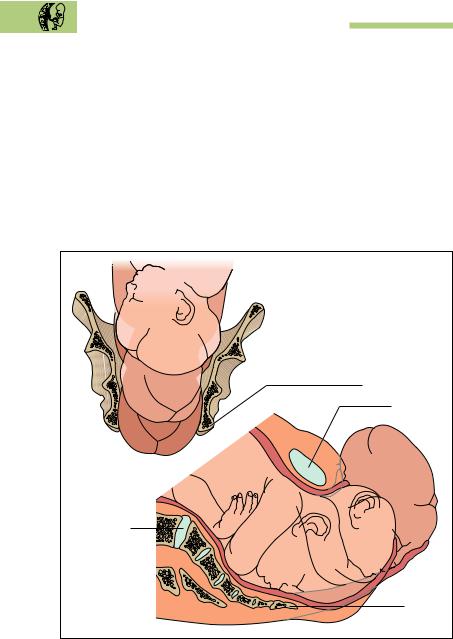
Birth
At the end of the period of gestation, the infant lies in the uterus ready for delivery with its body bent and arms and legs crossed (Fig. 12.15); the head (cephalic presentation) or the breech (breech presentation) may be the presenting part that is directed toward the cervix. Labor is usually divided into three stages, of which the first is the stage of dilatation, the second the stage of expulsion, and the third the placental stage.
The descent of the fetus, i.e., the entry of the head of the fetus into the pelvic inlet, occurs toward the end of pregnancy in a woman who is giving
Afterbirth |
|
|
(placenta) |
|
|
Uterus |
|
|
Internal os |
|
|
(ostium uteri internum) |
Rectum |
|
|
|
|
External os |
|
|
(ostium uteri externum) |
Bladder (vesica) |
|
Vagina |
|
|
Fig. 12.15 |
Section through the uterus with full-term infant. (After Leonhardt) |
|
524 12 Reproduction, Development, and Birth
birth for the first time (primipara), or at the beginning of labor in one who has given birth before (multipara). During the first stage, the “waters,” a sac filled with amniotic fluid, forms and precedes the head, widening the soft tissues of the birth canal. For this stage, the cervix, vagina, and perineum form a canal of constant diameter. The amnion usually ruptures (“breaking of the waters”) toward the end of the first stage of labor, though it may do so earlier; indeed, amniotic rupture sometimes triggers the first stage. The amniotic fluid drains through the vagina.
The expulsion stage begins as soon as the external os of the cervix is fully dilated (fully dilated cervix). While the abdominal muscles bear down in support, rhythmic contractions of the uterus shorten the uterine musculature (Fig. 12.16a, b) and the child is pushed into, and through,
Ischial tuberosity |
Symphysis |
a |
Lumbosacral |
disk |
Coccyx |
b |

Postnatal Development 525
the birth canal. Because the pelvic inlet is oval with the longer diameter set transversely and the pelvic outlet is oval with the longer diameter set anteroposteriorly, the infant’s head must rotate 90° during passage through the canal. Following the head, the shoulders align themselves first with the transverse diameter, then the anteroposterior diameter. The head, which has already been born, is forced into a renewed 90° rotation, which is assisted and supported by the obstetrician or midwife. The passage of the baby’s head through the vulva is known in obstetrics as “crowning.”
The third stage of labor begins with the completed expulsion of the child and ends with the expulsion of the placenta and fetal membranes. The cord is clamped and cut when delivery of the infant is complete. This procedure leads to carbon dioxide enrichment in the baby’s blood and this helps to activate the respiratory center. The newborn begins to breathe with the “first cry.” At the same time, the fetal circulation changes to the baby’s own systemic and pulmonary circulation. After delivery, the uterus contracts, so that the placenta separates from the uterine wall. It is usually “born” about 1/4−1/2 hour later (afterbirth) (Fig. 12.10a, b).
Postnatal Development
At birth, prenatal development ends and postnatal development begins. It is divided for practical purposes into various stages of development and age:
Infancy: from birth to the end of the 1st year of life
Early childhood: from the beginning of the 2nd year of life through the 6th year
School years: from the 7th year of life to the beginning of puberty
Fig. 12.16 a, b Childbirth (parturition). (After Leonhardt) a Bony pelvis and the baby’s head seen from in front
b “Crowning” of the baby’s head during childbirth, seen from the side. Because the pelvic inlet is oval with the longer diameter set transversely and the pelvic outlet is oval with the longer diameter set anteroposteriorly, the infant’s head must rotate 90° during crowning

526 12 Reproduction, Development, and Birth
Puberty: the period from the appearance of the first secondary sexual characteristics up to complete sexual maturity
Youth or adolescence: continues to the completion of physical growth
The rate of growth differs among these various developmental stages. Normal physical development is best defined by correlating age with body length or height and body weight. As growth is followed from birth to its completion, it may be seen at different times to occur at slower or more rapid rates.
Body Length
The most rapid rate of growth is noted during the first year of life. For instance, growth during the first half-year is about 16 cm, while in the second half-year it is about half that (8 cm). About 50 % of definitive body length is attained as early as the end of the 2nd year of life. To the beginning of puberty, annual growth is about 5−6 cm. After that, both sexes show a noticeable growth spurt (pubertal growth spurt), more marked in boys than in girls (9−10 cm as opposed to 8−9 cm). At 12 years of age, girls are in general taller than boys of the same age, as their pubertal growth spurt begins about two years earlier. Growth ends at different ages in different populations. In central Europe, for example, growth usually ends at age 17 or 18 years in women and at about age 19 in men The mean height of an adult woman is about 167 ± 11 cm, of an adult man 177 ± 13 cm.
Body Weight
Body weight, like body length, shows characteristic changes during the developmental stages. Thus birth weight (3200−3400 g) has about doubled by 5 months and has tripled by the end of the first year of life. It is fivefold by age 4−5 years. By the end of growth, body weight is about 20 times birth weight.


528 12 Reproduction, Development, and Birth
Growth of the Skeleton
The development of the skeleton, especially the part played by bone growth, is closely related to the total development of the organism and is considered when evaluating disorders of growth and in estimating eventual body height. In this estimation, radiological examination plays a major diagnostic role, for example, to determine at what point in time the secondary ossification centers appear. From these it is often possible to draw conclusions about general development by referring to the socalled “skeletal age.” Apart from the secondary ossification centers, an especially important criterion used for this evaluation is the fusion of the epiphyseal and apophyseal disks (growth disks). For this, by convention, a radiograph is performed on the left hand of the infant or small child. The rate of development may be retarded or accelerated. Growth and development depend on many factors. For instance, body weight and especially growth in height depend on genetic influences, but external influences, such as the quality and quantity of the nutrients ingested with food, can also be demonstrated.
Summary Reproduction, Development, and Birth
Human sexual reproduction takes place in several stages:
Union of a male sperm cell with a female ovum (fertilization)
Subsequent transport of the germ cell through the uterine tube (tubal transport)
Implantation (nidation) in the uterine mucosa (endometrium)
Development into a viable infant (development of the embryo and fetus)
Germ Cells
Oogenesis: oogonia (primitive germ cells, 44, XX)—primary oocytes (44, XX)—secondary oocytes (22, X)—ovum (mature egg cell, 22, X) plus 3 polar bodies (22, X).
Spermatogenesis: spermatogonia (primitive germ cells, 44, XY)— primitive spermatocytes (44, XY)—secondary spermatocytes (22, X

Summary 529
or 22, Y)—spermatids (22, X or 22, Y)—sperm cells (spermatozoa, 22, X or 22, Y)
Fertilization
The spermatozoa must find the ovum after ovulation in the uterine tube (only 200−300 of 200−300 million reach the ovum, speed of migration: 3 mm/min). While spermatozoa can survive in the uterine tube up to 4 days, ova must be fertilized within 6−12 hours.
Process of fertilization (3 phases): Acrosomal reaction (phase 1 and phase 2): penetration of the corona radiata and enzymatic dissolution of zona pellucida. Phase 3: fusion of the cell membranes of ovum and sperm cells, followed immediately by the cortical reaction (prevents penetration of the ovum by other spermatozoa = polyspermy block).
At the time of fertilization the ovum ends its second maturation division and forms the female pronucleus, which then fuses with the male pronucleus to form the diploid zygote. A female (XX) or male (XY) germ cell then forms, depending on the sex chromosome of the male pronucleus (X or Y).
Transport through the Uterine Tube and Cleavage
The zygote begins to divide (cleavage) during the 5 days of transport through the uterine tube into the uterus: zygote—2-cell stage—4-cell stage, etc. After the 16-cell stage, the germ is called a morula (mulberry), which is followed by a blastocyst with an outer shell of cells (trophoblast) and an inner cluster of cells (embryoblast).
When transport through the tube is impeded, a tubal pregnancy occurs.
Implantation and Placenta Formation
Implantation: Nidation of the blastocyst into the endometrium 5−6 days after ovulation or fertilization (endometrium at the peak of the secretory phase):
Trophoblast: forms the fetal part of the placenta
Embryoblast: forms the embryo, later fetus
Placenta:
Function: provides the growing germ with nutrition, takes over substance and gas exchange, secures maintenance of pregnancy,

530 12 Reproduction, Development, and Birth
and secretes hormones (e. g., estrogens, progesteone, chorionic gonadotropins)
Structure: consists of a fetal part with chorionic disk and chorionic villi, containing the fetal vessels, and a maternal part with decidua basalis (endometrium), spiral arterioles, and placental septa, between which the intervillous spaces are filled with maternal blood. Total surface of the villi: 8−14 m2; diameter of a mature placenta: ca. 18 cm; weight: 450−500 g.
Placental barrier: separates maternal and fetal blood; formed by the villous epithelium, villous connective tissue, and the walls of the fetal vessels; selectively permeable to various substances.
Umbilical cord (funiculus umbilicalis): connects the placenta with the fetal organism; contains an umbilical vein (v. umbilicalis; oxygenated blood flows from the placenta to the fetus) and two umbilical arteries (aa. umbilicales: deoxygenated blood flows from the fetus to the placenta).
Development of the Embryo
The period of embryonic development (formation of the embryonic disk, formation of the germ layers and organogenesis) begins following early development (transport through the uterine tube and implantation, 1st and 2nd week).
Formation of the embryonic disk: at first two germ layers (inner endoderm, outer ectoderm), later three germ layers including the middle germ layer (mesoderm). The mesoderm develops in the primitive streak on the ectodermal surface of the embryonic disk and migrates inward. The rudimentary head (notochord, chorda dorsalis) forms from its anterior end (primitive node).
Derivatives of the germ layers: ectoderm (central nervous system and surface epithelium); mesoderm (skeleton, skeletal muscle, circulatory organs, genitourinary apparatus), endoderm (epithelial lining of the digestive and respiratory passages).
Development of the body: folds form in the embryonic disk at the end of the 4th week; rudimentary extremities form at the beginning of the 5th week; embryonic curvature and appearance of the cephalic and cervical flexures between weeks 5 and 7. At the end of the embryonic period, the head makes up about 50 % of the total length.
Summary 531
At the end of embryonic development, the embryo lies protected by amniotic fluid in the amniotic cavity, which continues to grow and contains about 1 liter of amniotic fluid at the end of gestation.
Development of the Fetus
At the beginning of the 9th week of gestation, the germ becomes a fetus. During the fetal period the organ systems grow and differentiate.
Growth in length (crown−heel length or crown−rump length depending on the month of gestation). Haase’s rule: total length of the fetus in centimeters during the 4th and 5th month is the square of the number of months; from the 6th month onward it is 5 times the number of months.
Monitoring of fetal growth by ultrasonography: biparietal diameter in cm × 5.5 = body length in cm.
Anatomical signs of maturity at term: crown−heel length (49−51 cm); crown−rump length (33 cm); weight (3200−3400 g); fingernails and toenails project beyond the ends of the digits. In boys the testes are in the scrotum; in girls the labia majora cover the labia minora.
Functional signs of maturity at term: evaluation of skin color, respiration, heart rate, muscle tone, and neuromuscular reflexes.
Duration of pregnancy: 280 days (= 10 lunar months of 28 days), calculated from the first day of the last menstrual period; 266 days, calculated form the time of ovulation or fertilization.
Naegele’s rule to calculate the expected date of delivery: subtract 3 calendar months from the first day of the last menstruation, add 7 days, add 1 year (valid for a 28 day cycle).
Labor
Divided into three stages. first stage (stage of dilatation), second stage (stage of expulsion), third stage (placental stage). During the phase of dilatation, the fetal head enters the pelvic inlet; the amnion, filled with amniotic fluid, dilates the soft tissues of the birth canal (soft tissue canal: cervix, vagina, and perineum) and the external os of the cervix dilates. The expulsion phase begins with rhythmic uterine contractions (supported by bearing down of abdominal muscles), rota-

532 12 Reproduction, Development, and Birth
tion of the baby’s head in the birth canal followed by the passage (“crowning”) of the head through the vulva. When parturition is complete, the umbilical cord is divided. The placenta separates and appears as afterbirth about 30 minutes later.
Postnatal Development
Divided into stages of development and age: infancy (1 year); early childhood (2nd to 6th years); school age (7th year to puberty); puberty (to full sexual maturity); youth or adolescence (to the completion of physical growth).
Physical development is described by correlating rapidity of growth, body length or height, body weight, and physical proportions with age.
Skeletal age (timely appearance of centers of ossification and epiphyseal fusion); used to evaluate disorders of growth and estimate eventual body height. Left hand is examined radiologically.