


378 9 The Digestive System
By taking up nourishment or nutrients (proteins, fats, carbohydrates, vitamins, minerals, and trace elements) the body can create energy by the “combustion” of nutrients to maintain its organization (structure, catabolic, or energy metabolism) and the functions derived from it (growth, renewal of cells, body temperature, mechanical and chemical work). The nutrients are degraded by the enzymes of the digestive glands in the various divisions of the digestive system, broken down into chemical combinations that can be absorbed, and taken up (resorbed) by the gastrointestinal mucosa. These energy-rich combinations (e. g., fatty acids, amino acids, or glucose) enter the bloodstream and next reach the liver by way of the portal vein (see Liver, Function below). Eventually they reach the cells of the body, where they are broken down oxidatively in the mitochondria to energy-poor combinations (CO2 and H2O) (biological oxidation). The energy thus liberated is stored in energy-rich ATP following a chain reaction (mitochondrial respiratory chain). ATP in its turn is made available to processes requiring energy (e. g., protein synthesis or muscular work). ATP splits off phosphate molecules to liberate energy (see Chapter 1: Mitochondria).
Metabolism, Energy Requirements and Nutrients
Metabolism
Metabolism, which includes all the biochemical processes needed to maintain life by building, transforming, and breaking down the organism, can be divided into constructive (productive, anabolic) and energy transforming (catabolic) metabolism. Anabolism (constructive metabolism) includes the production of cellular substances, i.e., the synthesis of endogenous (the body’s own) substances (.e. g., proteins, carbohydrates, fats) involved in the growth of the organism.
In general we call processes in which substances foreign to the body are transformed into the body’s own substances assimilative energy-con- suming (anabolic) processes. In plants assimilation (photosynthesis) involves capturing energy from sunlight, by which energy-poor inorganic substances are transformed into energy-rich organic substances (autotrophic organisms). Heterotrophic organisms, such as humans and ani-

Metabolism, Energy Requirements and Nutrients 379
mals, acquire energy by taking up nourishment that already contains energy-rich substances formed by other organisms.
Energy-transforming metabolism (catabolism) involves the breakdown of energy-rich substances (e. g., fats, carbohydrates, proteins) into those containing less energy with the liberation of energy (= dissimilation). Dissimilative processes are breaking down or catabolic processes. One of the most important catabolic processes is the biological oxidation described above. The energy stored in ATP is used for anabolic as well as energy-transforming metabolism, that is, it serves the maintenance of processes needed to maintain life, such as the maintenance of body temperature, muscular work, absorption and transport at cell membranes, the transmission of nerve impulses, etc. In the course of these processes, a considerable amount of energy is lost, e. g., as heat. Since, unlike plants, humans and animals cannot build organic substances by photosynthesis, they must regularly take in food to make energy available.
Energy Requirements
The energy requirements and energy transformation of the human body depend on many factors such as age, sex, weight, ambient temperature, and physical activity and vary considerably even at complete rest (basal metabolism). Hence, metabolism may be divided into basal metabolism, measured under standard conditions (in the morning, fasting, resting quietly, normal body temperature, comfortable ambient temperature) and the excess metabolism of exercise measured under varying conditions.
Energy Content of Nutrients
The energy gained from metabolic processes depends among other things on the nature of the metabolized substances (proteins, fats, and carbohydrates). If, for instance, we want to measure the energy content of individual nutrient materials outside the body, we can burn them in a combustion chamber (calorimeter) and measure the heat or energy liberated (physical caloric value). The results can be given in calories (cal) or joules (J), though the unit most often used in practice is the calorie: 1 cal

380 9 The Digestive System
corresponds to 4.185 J. In nutrition, the old distinction between a “small calorie” and a “large calorie” has largely been abandoned in favor of the larger Calorie (1 Cal = 1000 small calories or 1 kcal), which is equivalent to the old large calorie. Such a measurement gives the following results for the three most important energy-providing nutrients: carbohydrates 4.1 kcal/g (17 kJ/g), fats 9.3 kcal/g (39 kJ/g) and proteins 5.3 kcal/g (22 kJ/g).
If the nutrients are “burnt” completely in the organism, i.e., broken down oxidatively to CO2 and water, the energy available to the body (physiological caloric value) from fats and digestible carbohydrates approximates the physical caloric value (fuel value). During the breakdown of protein, however, urea is formed in addition to CO2 and water. Since this would generate more energy if it underwent complete combustion, the fuel value of proteins is higher than the physiological caloric value. Hence, under physiological conditions, energy generated by the same amounts of different substances is:
1 g carbohydrate (e. g., starch): |
4.2 kcal (17.6 kJ) |
1 g fat (e. g., triglyceride): |
9.3 kcal (38.9 kJ) |
1 g protein (e. g., animal protein): |
4.1 kcal (17.2 kJ) |
1 g alcohol (e. g., ethyl alcohol): |
7.1 kcal (30.0 kJ) |
Energy Requirements at Rest
(Basal Metabolism, Basal Metabolic Rate, BMR)
As already noted, the basal metabolic rate depends on a variety of factors. As a guide, the following facts may help: the value of the BMR in a grown man per kilogram body weight is about 1 kcal (4.2 kJ) per hour. For a weight of 70 kg this correspond to round 1700 kcal (7000 kJ) a day. The corresponding value for a woman of equal weight is lower by about 10−20 %. However, the body’s energy metabolism depends on factors other than height, weight, age, and sex. For instance, intellectual activity, emotional reactions (joy, fear), fever, or thyroid overactivity (hyperthyroidism) increase metabolism, while sleep, anesthesia, or thyroid deficiency (hypothyroidism) lower it.
Metabolism, Energy Requirements and Nutrients 381
Energy Requirements during Physical Activity (Excess Metabolism of Exercise)
Physical activity increases metabolism (excess metabolism of exercise). As a result the metabolism developed during working hours exceeds that developed during leisure hours:1
About 2760 kcal (12 000 kJ) during light work (e. g., desk work)
About 3680 kcal (16 000 kJ) during moderately heavy work (e. g., mowing the lawn)
About 4600 kcal (20 000 kJ) during very heavy work (e. g., moving furniture)
Exceptionally top-ranked athletes may attain 16 100 kcal (70 000 kJ) for brief periods (e. g., marathon runners, triathletes), but of course their daily metabolism is much lower.
Determination of Energy Metabolism
In order to determine energy metabolism, the total heat production of the body would have to be measured. It is, however, simpler to calculate the energy consumption of the body from the oxygen consumption (O2 taken up in the lung). For instance 134 liters of oxygen are used in the complete combustion (oxidation) of 1 mole (=180 g) of a simple sugar (glucose), involving the generation of about 664 kcal (2,780 kJ) of energy, i.e., 4.95 kcal (20.7 kJ) energy is transformed for each liter of oxygen. This value is called the “combustion equivalent”.
Since in each case the quantities of CO2 produced and of O2 consumed depend on the type of nutrient (carbohydrate, protein, or fat) being oxidized, the quotient of the two values (CO2 production/O2 consumption), called the “respiratory quotient” (RQ), gives information on the nature of the substance undergoing combustion. With approximately equal amounts of protein present, the RQ of a preponderantly carbohydrate diet is 1.0, while a preponderantly fatty diet has an RQ of 0.7, as can be determined from the following equations:
Carbohydrate (glucose): C6H12O6 + 6 O2 = 6 CO2 + 6 H2O (RQ = 6/6 = 1)
Fat (triglyceride): 2 C51H98O6 + 145 O2 = 102 CO2 + 98 H2O (RQ = 102/ 145 = 0.7
1 Energy metabolism developed during today’s typical average leisure activity, i.e., without much movement.

382 9 The Digestive System
Nutrients
In addition to proteins, carbohydrates, and fats, a balanced human diet should include vitamins, minerals, trace elements, and adequate water. Also desirable is a daily intake of fiber, i.e., substances derived from plants (e. g., cellulose) that are not digested and stimulate intestinal activity by their ability to swell.
The daily protein intake of a normal adult should account for 10−15 % of the total daily caloric intake (0.8 g per kg body weight, or about 56 g for a 70 kg man). Fat intake should account for a further 25−30 % (about 78 g of fat), and carbohydrate intake for the remaining 55−60 %. Carbohydrates are thus the body’s main source of energy.
Proteins
The principal task of proteins is to provide the body with amino acids, which the body requires for the biosynthesis of its endogenous proteins. The human body cannot synthesize eight of the 20 naturally occurring amino acids that it needs to manufacture its own proteins. These amino acids are called “essential amino acids” and include leucine, lysine, methionine, phenylalanine, isoleucine, valine, threonine, and tryptophan. Other amino acids can be essential under special circumstances, e. g., tyrosine and cysteine in premature infants and in persons suffering from hepatic cirrhosis; histidine in uremic patients; and arginine in immunocompromised patients. Apart from lysine, which is absent or present only in insufficient quantities in many vegetable nutrients, both vegetable and animal proteins contain the essential amino acids.
Fats (Lipids)
Fats are dietary substances that are rich in energy (see Energy Content of Nutrients above) and they function primarily as energy providers, but they also store energy. They also include the essential fatty acids (polyunsaturated fatty acids occurring in vegetable oils, especially linoleic and linolenic acid). A diet lacking these substances leads to manifestations of deficiency. Lipids are present in high concentrations in the sex organs, for example, and form a large part of the lipids in the cell membrane. They also include cholesterol, which forms part of the structure of the

Metabolism, Energy Requirements and Nutrients 383
cell (cell membrane) and is a hormone precursor. Finally, fats are solvents that allow the complete absorption of fat-soluble vitamins (e. g., vitamin A, vitamin E) from the gastrointestinal tract.
Triglycerides constitute the major portion of the fats included in the diet. These are neutral fats, each consisting of three fatty acids attached to the trivalent alcohol glycerol. Among the more frequently encountered fatty acids are palmitic, stearic, oleic, and linoleic acids. While animal fats are predominantly saturated fatty acids (exception: salt water fish), vegetable oils have a higher content of unsaturated fatty acids (exception: coconut oil).
Carbohydrates
Carbohydrates are the preferred sources of energy of many organisms. The combinations most important in the human diet are the monosaccharides (e. g., glucose = dextrose), disaccharides (e. g., lactose = milk sugar), and polysacccharides (e. g., starch). Our diet contains chiefly monosaccharides contained especially in honey and fruit, disaccharides in milk and in all foods sweetened with the common household sugar saccharose (sucrose, cane sugar), and polysaccharides in vegetable (starch) and animal (glycogen) products. Carbohydrates can only be stored in small quantities in the body. For instance, during starvation the total store of glycogen in the liver and the skeletal muscles (about 300−400 g) is used up in a day and a half. Because of their sweet taste, simple sugars (monosaccharides and disaccharides) are very popular and have multiple uses. They are also suitable for protecting food from deterioration and therefore for preserving. On the other hand, simple sugars put a considerable burden on the pancreas. The small molecules reach the bloodstream rapidly and the blood sugar level rises rapidly. The pancreas must secrete large quantities of insulin in order to lower the blood sugar level, which then sinks to such low levels that it evokes a strong sensation of hunger with fatigue. With renewed sugar intake, the level rapidly rises again. A diet rich in sugar entails strong swings in blood sugar level, with consequent variability in performance. Moreover, simple sugars have practically no nutritional value, they are “empty calories.” Whole-grain products (starch + vitamins + fiber; see Fiber below) are more filling in the long run and avoid blood sugar peaks.

384 9 The Digestive System
Vitamins
Vitamins are organic compounds that are indispensable for humans and animals and are synthesized insufficiently or not all in the body. Hence they must be ingested regularly in food or as supplements. In our diet they are present in quite variable quantities in the form of vitamins or vitamin precursors known as provitamins, that can be transformed into vitamins in the body. The best-known example of the latter is -carotene, also known as provitamin A. On the other hand, vitamin D3 can be synthesized from the provitamin 7-dehydrocholesterol, an intermediate metabolic product, by the effect of sunlight on the skin.
Vitamins do not play a role in the production of energy or in the structure of the body, but fulfill chiefly a catalytic or regulatory function. According to their solubility vitamins are divided into:
Fat-soluble vitamins: vitamin A (retinol and others), vitamin D (er-
gocalciferol, vitamin D2 or cholecalciferol, or vitamin D3) vitamin E (α- tocopherol and others) and vitamin K (phytonadione or vitamin K1, and menaquinone or vitamin K2)
Water-soluble vitamins: vitamin C (ascorbic acid), vitamin B1 (thiamin), vitamin B2 (riboflavin), niacin (antipellagra factor), pantothenic acid, vitamin B6 (pyridoxine and others), folic acid, vitamin B12 (cyanocobalamin and others), inositol, and biotin. Inositol (a component of lecithin) is sometimes included among the B vitamins, even though it is synthesized in the body in quantities much greater than its dietary intake.
For practical purposes, water-soluble vitamins cannot be stored in the body. They circulate in the blood until they are needed for cellular reactions. Fat-soluble vitamins are chiefly contained in fatty foodstuffs and can only be absorbed in adequate quantities if fat digestion and absorption are intact. They can be stored in the liver and in fatty tissues.
Chemically, vitamins belong to several different types of substance and are defined according to their actions. By their function they can be divided into two major groups: vitamins of the B complex and vitamin K are components of coenzymes that catalyze carbohydrate, fat, and protein metabolism. Thus, by taking part in fundamental intermediary metabolic processes, they are indispensable for every living cell. Vitamins A, D, E, and C, on the other hand, can only be demonstrated at a more ad-

Metabolism, Energy Requirements and Nutrients 385
vanced stage of evolution, where the maintenance of specific organ functions is essential. These vitamins are highly specialized active substances coupled to certain cell and organ systems. Apart from vitamin A they are not components of coenzymes. In evolutionary history (phylogenesis), dependence on these vitamins can be found only in the more highly developed invertebrates, while the need for vitamin D is only found in vertebrates.
Age, sex, and physiological conditions such as pregnancy, physical stress, and nutrition influence vitamin requirements in humans. Vitamin deficiency (hypovitaminosis) can be due to such conditions as malnutrition, faulty nutrition (undiversified diet, e. g., in the elderly or alcoholics, a fast-food diet), or malabsorption in the gastrointestinal tract. Treatment with medications, e. g., those that damage the intestinal flora, may lead to vitamin deficiencies by eliminating bacterial vitamin synthesis (especially of vitamins B12 and K).
The following well-known severe illnesses are due to vitamin deficiencies: scurvy (vitamin C deficiency), beri-beri (vitamin B1 deficiency), pellagra (niacin deficiency), and rickets (vitamin D deficiency). Moreover, vitamin A deficiency leads to night blindness, vitamin B12 deficiency to pernicious anemia, and vitamin K deficiency to clotting disorders.
Toxicity due to vitamin overdose (hypervitaminosis) occurs only with the fat-soluble vitamins A and D. This does not apply to the watersoluble vitamin precursor -carotene. Normally, excess water-soluble vitamins are rapidly excreted in the urine.
Minerals (Macrominerals and Trace Minerals)
Minerals are classified according to their concentration in the body as macrominerals ( 50 mg/kg body weight) or trace minerals (trace elements) ( 50 mg/kg body weight). The recommended daily intake of the trace minerals is less than 100 mg/day. Iron is exceptional, in that it is present in high concentration in the body, and thus counts as a macromineral, but its recommended daily intake lies in the range of the trace minerals, because it is effectively recycled in the body, rather than being excreted. Trace elements are minerals the requirement of which is less than 100 mg/day. The total weight of all trace elements in the body is 8−9 g. They include chromium, iron, fluorine, germanium, iodine, cobalt, cop-

386 9 The Digestive System
per, manganese, molybdenum, nickel, selenium, silicone, vanadium, zinc, and tin. The significance of some others also present in the blood, such as aluminum, arsenic, barium, gold, and rubidium, is not entitrely clear.
Adequate intake must be ensured especially for iron and iodine. The most important function of iron, which at 4−5 g is quantitatively the most important of the trace elements in the body, is as component of hemoglobin and myoglobin (daily requirement 10 mg for men, 15 mg for women). Iodine is part of the structure of the thyroid hormone thyroxin, and is contained especially in salt-water fish, iodized table salt, and drinking water (daily requirement 0.18−0.2 mg). Although fluorine is not necessary for life, daily use promotes remineralization of the teeth. The remaining trace elements are primarily components of important enzymes.
Calcium, magnesium, phosphorus, sodium, potassium, and sulfur belong to the macroelements. The most abundant of these is calcium with 1.5 kg (99 % skeletal, 1 % in bodily fluids and tissues). Minerals do not have a uniform biological function in the body. They take part in the structure, maintenance, and constant renewal of bones and teeth and in the activation of enzymes. They are responsible for the conduction of impulses in the nervous system, the functioning of muscles, the constant ion content of bodily fluids, and the regulation of water balance. They also take part in the maintenance of a constant osmotic pressure and pH in the blood and the other parts of the body.
Antioxidants (Free Radical Scavengers)
Vitamins A ( -carotene), E, and C, like the trace elements selenium, manganese, zinc, and molybdenum as well as several active plant ingredients (see below), possess anitoxidative activity, which gives them special significance. They inactivate free radicals that are metabolic by-products (e. g., superoxide anions, hydroxyl radicals) and so supplement the body’s own antioxidative enzymes (e. g., glutathione peroxidase). Free radicals are molecules marked by high chemical reactivity, because they possess unpaired electrons (i.e., they are chemically unstable). Even oxygen, which is required for the oxidation of nutrients, is a radical in its basic configuration (a dioxygen radical, with unpaired electrons). The energy-creating (ATP) reaction in the cells (oxygen + hydrogen water)

Metabolism, Energy Requirements and Nutrients 387
is only possible because of the high reactivity of oxygen. The same process may create other reactive forms of oxygen that are not radicals (e. g., hydrogen peroxide), some of which are even more reactive and so aggressive that they can extract electrons from other molecules. The result is new radicals that oxidize other molecules as they try to become stabilized, producing damage to cellular structures:
Damage to DNA in the chromosomes may entail mutations in the genes or uncontrolled cell division (cellular degeneration or cancer production).
Oxidized LDL (see Chapter 6: Plasma Proteins) in the blood are deposited preferentially in damaged vascular walls (arteriosclerosis); antioxidants prevent the oxidation.
Cell membranes are damaged when fatty acids in phospholipids are oxidized to peroxides (peroxidation of lipids). Premature senility may ensue (increased cell death, deposition of the products of oxidation). Antioxidants protect the cell membrane from oxidation and so prevent death of the cell or impairment of cellular function by the products of oxidation.
Extreme physical exertion and acute inflammation, as well as products that damage the environment (e. g., ozone, nitric oxide) and ultraviolet and radioactive radiation, lead to increased formation of free radicals during cell metabolism (oxidative stress).
Active Substances in Plants
As well as vitamins, minerals, trace elements, and fiber, fruit and vegetables contain a number of substances that protect them from dangerous components of sunlight, from pests, and from adverse environmental influences. Such substances also protect people from certain diseases. From a scientific point of view, the most interesting chemical compounds among the many thousand contained in every kind of fruit and vegetable are those that act as antioxidants or play a role in cancer prevention. Many of the active substances found in plants that have been studied are free radical scavengers; others show configurations that may deactivate carcinogenic (cancer-causing) substances or prevent their formation. These include:

388 9 The Digestive System
p-Coumarins and quinic acid compounds (found in tomatoes, pepperoni, strawberries, pineapple): inhibit the formation of carcinogenic nitrosamines.
Indoles (found especially in broccoli, cauliflower, Brussels sprouts, kale, and cabbage): reduce estrogen synthesis and so reduce the risk of diseases due to hormone-dependent tumors. Moreover, they increase the activity of certain detoxifying enzymes in the liver that also break down carcinogens.
Allicins (in alliums such as garlic, leeks, onions, chives): these have antibacterial activity. Alliums also contain organic sulfur compounds that activate enzymatic detoxifying systems in the liver.
Isocyanates (all crucifers): activate detoxifying enzymes; inhibit reactions that alter DNA (phenethyl isocyanate). Sulforaphan is one of the very few active substances of which the activity has been studied in isolation, and which can be produced synthetically. The substance renders carcinogens harmless by activating certain detoxifying enzymes.
Flavonoids and isoflavonoids (almost every fruit and vegetable contains its characteristic flavonoids): of the over 800 flavonoids known so far, many have antioxidative, antifungal (effective against fungi), anti-in- flammatory (fighting inflammation), antiviral (fighting viruses), antiallergenic or anticarcinogenic effects or promote blood perfusion. For instance, substances such as catechin, nobiletin, hesperidin, quercetin, quercitrin, morin, robinin, myrecitin, rutin, kaemferol, and neoponcerin have antioxidative or anticarcinogenic properties. The isoflavonoid genistein (found in soy products) can inhibit tumor growth and prevent the development of metastases. Other flavonoids can guard against arteriosclerosis or reduce cholesterol synthesis. For instance, morin and sylimarin block oxidation of LDL and so prevent its deposition in vascular walls (antioxidative action).
Saponins (found in soy products): prevent the synthesis of DNA in tumor cells.
Terpenes: these include many aromatic vegetable oils, e. g., the limonene of citrus oil found in citrus fruit. All have marked antioxidative activity.
In contrast to vitamins, most active plant substances survive all processes used in preparation, industrial processing, and prolonged storage. Tomatoes alone contain 10 000 different active substances, of

The Digestive Organs 389
which only a fraction have been studied. Whether these substance continue to be active when they have been isolated from the other active substances in the plant (e. g., there might be interactions between the individual compounds), or if their action might then reverse has, with few exceptions, not yet been explored. Fruit and vegetables must continue to be included in the diet, especially in view of their fiber content.
Fiber
Fiber plays a special role in our diet. It includes indigestible plant carbohydrates such as cellulose and hemicellulose. Cellulose is a component of the cell wall of plants. Hence a diet rich in fiber includes fruit, vegetables, and whole-grain products. Substances rich in fiber not only promote intestinal activity, they also slow gastric emptying and are therefore more filling. This effect prevents blood sugar peaks. They have a high capacity for binding water and so stimulate intestinal activity, shortening the time of passage of materials through the intestines and so leading to regular emptying of the bowels.
The Digestive Organs
The digestive organs may be divided according to their function into the cephalic (foregut) and those of the trunk (midgut and hindgut). The cephalic digestive organs include the oral cavity with its salivary glands, the oropharynx, and the hypopharynx. The digestive organs of the trunk (Fig. 9.1) include the gullet (esophagus), stomach, small bowel (duodenum, jejunum, and ileum), large bowel (caecum, vermiform appendix, ascending, transverse, descending and sigmoid colon, and rectum) and the digestive glands (liver, pancreas).
The Oral Cavity
The oral cavity is bounded by the lips in front, the cheeks laterally, the muscles of the floor of the mouth below, and the hard and soft palate above. The back of the oral cavity (fauces) is formed by the oropharyn-


392 9 The Digestive System
Intrinsic |
Septum of tongue |
Hard palate |
Cheek |
muscles of |
|
|
muscle |
the tongue |
|
|
(buccinator |
|
|
|
muscle) |
Vestibulum |
|
|
Premolar |
|
|
tooth |
|
|
|
|
|
Sublingual |
|
|
Muscle of |
|
|
the floor |
|
gland |
|
|
|
|
|
of the |
|
|
|
|
|
|
|
|
mouth |
|
|
|
(mylohyoi |
|
|
|
d muscle) |
Genioglossus |
|
|
Lower jaw |
muscle |
|
|
|
|
|
|
(mandible) |
Platysma |
|
|
Anterior belly of |
|
|
|
digastric muscle |
Fig. 9.3 Coronal section through the oral cavity. (After Faller)
The muscles of the tongue are divided into intrinsic and extrinsic muscles (Fig. 9.3). The most important and strongest extrinsic tongue muscle is the genioglossus muscle, which takes its origin in the middle of the mandible and radiates fanwise from the tip of the tongue to its palatine end. It pulls the whole tongue forward and at the same time flattens the dorsum of the tongue. The intrinsic tongue muscles traverse the organ in all three directions. Their function is chiefly to deform the body of tongue.
Tactile and Taste Papillae
The dorsum of the tongue contains numerous papillae of various kinds, subserving the sensations of touch and taste (Fig. 9.4). The filiform (threadlike) papillae are distributed over the whole dorsum of the tongue and serve especially the perception of touch, pressure, temperature, and pain. The taste papillae include fungiform (mushroom-shaped) papillae, vallate (walled) papillae and foliate (leaf-shaped) papillae. They contain taste buds and can be found at specific locations on the dorsum of the


394 9 The Digestive System
tonsil. In the midline, immediately behind the apex of the V, lies the foramen caecum, where the thyroid gland has developed.
The Teeth (Dentes)
The human dentition is arranged in two rows of teeth, the upper and lower dental arches, anchored in the upper and lower jaws. The first teeth formed are the milk (deciduous) teeth, replaced later by the permanent teeth. Human teeth have various shapes and have different functions. The incisors are the cutting teeth, the canines next to them help with tearing and fixing. Beyond these, the chewing surfaces of the premolars and molars grind and perform the major part of the work of chewing. Incisors and canines are also called the anterior (labial) teeth and the premolars and molars the posterior (buccal) teeth.
Structure of the Teeth
The tooth is divided into crown, neck, and root. The crown extends above the gum and is covered by enamel (Fig. 9.6). The root sits in an alveolus in the upper or lower jaw and is covered with cementum (cement). It is anchored in the bone by the desmodontium (periodontal ligament, periodontal membrane). The part of the tooth where cementum and enamel meet is called the neck of the tooth. The apex of the root is pierced by the root canal, through which nerves and vessels reach the dental cavity (pulp cavity). The dental cavity contains the dental pulp, a connective tissue structure containing blood vessels and nerves through which the tooth obtains its nutrition. The odontoblasts, dentin-forming cells, spread out like an epithelium, lie at the free border where pulp and dentin meet. They form dentin when needed and send processes into the dentin. These processes are accompanied by blood vessels and nerves that run in dental canaliculi (dental tubules) that give dentin a slightly wavy radial striation.
Each tooth consists of three hard substances that resemble bone: dentin (dentine), enamel, and cementum (cement). Dentin forms the largest portion of the tooth and surrounds the dental cavity (Fig. 9.6). At the crown, dentin is covered by enamel, at the root by cementum. Dentin is sensitive to pain. Enamel is the hardest substance in the human body

|
The Digestive Organs 395 |
Enamel |
Gum |
|
(gingiva) |
Dentine |
|
Dental canaliculi |
Crown (dental |
corona) |
|
Tooth cavity with |
|
dental pulp |
|
Odontoblasts |
|
Artery |
Neck (cervix |
Vein |
dentis) |
|
|
Collagen fibers |
|
Periodontal |
|
ligament |
|
Cementum |
Root (radix |
|
|
Alveolar bone |
dentis) |
|
Root canal
Apex
Fig. 9.6 Schematic view of a longitudinal section through a lower incisor
and consists of 97 % inorganic salts (chiefly hydroxyapatite). Dentin includes 70 % inorganic substances, cementum 65 %.
Support Structures of the Teeth (Parodontium)
The collagen fibers of the periodontal ligament (desmodontium) anchor the tooth elastically in its bony alveolus (Fig. 9.6). The collagen fibers (Sharpey’s fibers) run between the alveolar wall and the cementum, and are mainly oriented toward the apex of the root. Because of this orientation, chewing puts pressure on them. The periodontal membrane, which covers and protects the gum (gingiva) to the neck of the tooth, contains a prominent vascular network and sensory nerves (sensitive to pressure). The support structures of the tooth include the alveolus, the edge of the gum, the periodontal ligament, and the cementum.

396 9 The Digestive System
The Dental Formula
The human permanent dentition consists of 32 teeth (8 incisors, 4 canines, 8 premolars, and 12 molars). On each side of either jaw they have the following sequence from front to back (Fig. 9.7a, b)
2 incisors (dentes incisivi = I)
1 canine (dens caninus = C)
2 premolars (dentes premolares = P)
3 molars (dentes molares = M)
The number and sequence of the teeth can be expressed in brief by the dental formula. Where the structure is symmetrical, this is written for only one half of the mouth, with the maxillary teeth above the line and the mandibular teeth below, thus: I2/2 C1/1 P2/2 M3/3.
In dental practice the teeth are numbered in a particular order. The upper right third molar is given the number 1, and the teeth are then numbered in order along the upper alveolar margin from 1 to 16, 16 being the upper left third molar. The numbers then continue along the lower dental arch from left to right, the third left lower molar being 17, and the third right lower molar 32. The medial upper left incisor is 9, the medial lower left incisor is 24.
Left upper jaw |
|
|
|
Right upper jaw |
|
|
|
||||||||||
|
|
|
|
|
|
||||||||||||
16 |
15 |
14 |
13 |
12 |
11 |
10 |
9 |
8 |
7 |
6 |
5 |
4 |
3 |
2 |
1 |
|
|
|
|
|
|
|
|
|
|
||||||||||
Left lower jaw |
|
|
|
Right lower jaw |
|
|
|
||||||||||
17 |
18 |
19 |
20 |
21 |
22 |
23 |
24 |
25 |
|
26 |
27 |
28 |
29 |
30 |
31 |
32 |
|
Shape of the Permanent Teeth
The crown of the incisors is shaped like a chisel, with a sharp horizontal cutting edge. The canines are the longest teeth and their crowns each have two cutting edges that come together in a point. The premolar teeth have a chewing surface and their crown has two cusps (bicuspids). Their roots are often bifurcated, particularly in the upper premolars. The molars are oriented in the direction of the masticatory muscles. Their chewing surfaces mostly form four cusps, and when the teeth are apposed the cusps of the upper molars are placed between those of the lower molars and vice versa. While the upper molars have three roots,

The Digestive Organs 397
a |
b |
Fig. 9.7 a, b |
Deciduous (milk) and permanent dentitions (both upper jaw) |
a Deciduous dentition: incisors (blue), canines (yellow), deciduous molars (violet)
bPermanent dentition: incisors (blue), canines (yellow), premolars (green), molars (violet); the 3rd molars (wisdom teeth) not yet erupted
the lower mostly only form two. The third molars (wisdom teeth) vary in shape and sometimes do not develop.
Milk Dentition and the Eruption of Teeth
The permanent dentition must be distinguished from the first (milk, deciduous) dentition. The latter consists of 20 teeth (Figs. 9.7a, b and 9.8). With the exception of the premolar teeth, it is the same as the permanent dentition (8 incisors, 4 canines, 8 milk molars (deciduous molars). Between the 6th to 12th month of life the incisors are the first deciduous teeth to erupt. At 2 years the deciduous dentition is usually complete.
The first permanent teeth to erupt are the first molars. Since they appear in the 6th year of life they are sometimes called the six-year-old teeth. The last molar (wisdom tooth) often erupts late and may be malformed. The following are the times (years of life) of eruption of the permanent teeth:
|
Tooth: |
I1 |
I2 |
C |
P1 |
P2 |
M1 |
M2 |
M3 |
|
Eruption (year of life) |
7 |
8 |
11 |
9 |
10 |
6 |
12 |
? |


The Digestive Organs 399
Parotid gland
Parotid duct
Masseter muscle
Cheek muscle (buccinator muscle)
Sublingual ducts
Sublingual caruncle (sublingual papilla)
Sublingual gland
Muscle of the floor of the mouth (mylohoid muscle)
Hyoid bone
Submandibular gland
Sternocleidomastoid muscle
Fig. 9.9 The large salivary glands of the mouth
All the salivary glands together secrete about 1−1.5 liters of saliva in the course of a day. The saliva may be mucous or serous and the large and small salivary glands secrete both kinds. Saliva increases the lubrication of the food after chewing. Serous saliva begins the digestive process in the mouth because it contains the sugar-splitting enzyme amylase. The secretion of saliva is regulated by the autonomic nervous system, with the parasympathetic promoting and the sympathetic inhibiting its formation.

400 9 The Digestive System
The Throat (Pharynx)
The throat is the common portion of the respiratory and digestive tracts adjoining the nasal and oral cavities. It is a tube, about 12 cm long, attached to the base of the skull. The nasal cavity opens into the upper part (nasopharynx, epipharynx), the oral cavity into the middle part (oropharynx, mesopharynx), and the larynx and esophagus into the lower part (hypopharynx) (Fig. 9.10). The respiratory and digestive passages cross in the oropharynx. The tonsils of Waldeyer’s tonsillar ring are situated where the nasal and oral cavities open into the pharynx (choanae). Their function is to attack pathogens as early as possible by
Frontal sinus |
Choanae |
|
|
||
Nasal cavity (cavum |
Sphenoid sinus |
|
nasi) with nasal |
|
|
conchae (conchae |
Pharyngeal tonsil |
|
nasales) |
||
|
||
Hard palate |
|
|
(palatum durum) |
Nasopharynx |
|
Oral cavity |
||
(epipharynx) |
||
Palatine tonsil |
|
|
Tongue (lingua) |
Oropharynx |
|
|
||
Lower jaw |
|
|
(mandible) |
Hypopharynx |
|
Hyoid bone (os hyoideum) |
|
|
Epiglottis |
Gullet |
|
Thyroid cartilage |
(esophagus) |
|
|
||
Cricoid cartilage |
|
|
Trachea |
|
|
Thyroid gland |
|
Fig. 9.10 Topography of the pharynx. Sagittal section viewed from inside. (After Frick et al)
The Digestive Organs 401
activating the specific immunity. They are named according to their location (Figs. 9.2 and 9.10): midline pharyngeal tonsils (adenoids) in the roof of the pharynx, bilateral palatine tonsils between the two faucial arches, midline lingual tonsils on the palatine part of the tongue and the lymphatic tissue of the lateral pharyngeal wall, condensed around the entrance to the eustachian tube. The eustachian tube connects the pharynx with the tympanic cavity of the middle ear (see Chapter 15: Middle Ear).
The pharyngeal wall consists of mucosa, striated muscle, and a connective tissue fascia. The pharyngeal muscles include the muscles taking part in swallowing, namely, the pharyngeal constrictors and the pharyngeal levators. Whereas the pharyngeal constrictors are strong muscles that can narrow the pharynx and lift the larynx and hyoid bone (Fig. 9.11), the pharyngeal levators are rather weak. They raise and shorten the pharynx.
The Act of Swallowing (Deglutition)
The act of swallowing prevents the food from reaching the trachea. It includes a voluntary and an involuntary (reflex) phase (Fig. 9.11a, b). To begin the act of swallowing, the floor of the mouth is contracted voluntarily and the bolus is pressed against the soft palate. This action initiates the reflex (swallowing reflex), which includes the involuntary sealing of the respiratory passage. As the soft palate is lifted against the posterior pharyngeal wall (levator and tensor veli palati muscles) the upper respiratory passage is sealed from the digestive tract. The hyoid bone and the larynx are raised by the contraction of the floor of the mouth (pharyngeal constrictors and muscles of the floor of the mouth) and the epiglottis approaches the entrance to the larynx. During this action the glottis is closed and the breath is held. In this way the lower respiratory passages are also separated from the digestive tract. After the act of swallowing is complete, the infrahyoid muscles pull the larynx down again and so open the respiratory passages. This important and complex reflex is regulated by the swallowing center (deglutition center) in the medulla (medulla oblongata) in the brain.

402 9 The Digestive System
Uvula |
Superior constrictor |
|
Tongue |
|
muscle of pharynx |
|
|
|
Levator veli palati and |
|
|
|
tensor veli palati muscles |
|
|
Hyoid bone |
Suprahyoid |
|
|
|
muscles |
|
|
Epiglottis |
|
|
|
Thyroid cartilage |
Infrahyoid |
|
Thyroid |
a |
muscles |
b |
cartilage |
|
|
Fig. 9.11 a, b Representation of the act of swallowing. The respiratory passage (blue arrow in a) and the digestive passage (yellow arrow in b) cross in the oropharynx. The act of swallowing may be divided into a voluntary phase (contraction of the floor of the mouth and transport of the bolus to the soft palate) and a reflex securing of the respiratory passage (elevation of the soft palate, closure of the upper air passage, elevation of the hyoid bone and larynx with closure of the laryngeal inlet by the epiglottis). (After Leonhardt)
The Gullet (Esophagus)
The esophagus transports the bolus from the pharynx into the stomach. This transport is accomplished by waves of circular muscle contractions (peristalsis) that are normally directed toward the stomach. The esophagus is also subject to a longitudinal tension (fixed by the larynx above and the diaphragm below) that stabilizes its course and favors the passage of the bolus of food during swallowing. In the adult, the esophagus is about 25−30 cm long. It runs through the thorax behind the trachea and in front of the spine. Below, the esophagus penetrates the diaphragm through the esophageal hiatus to empty directly into the


404 9 The Digestive System
At this limited constriction, larger boluses can at times become wedged and cause severe pain.
The esophagus has the mural layers characteristic of the whole gastrointestinal tract (see The Small Bowel, Fig. 9.17). An inner mucous membrane (tunica mucosa = mucosa) is followed by a loose areolar connective tissue layer (tunica submucosa = submucosa), in which run larger blood and lymph vessels. Outside this is a muscular layer (tunica muscularis = muscularis), which consists of an inner circular layer and an outer longitudinal layer. By alternating the contractions of the circular and longitudinal muscles segment by segment (peristalsis), this arrangement of the muscles facilitates the transport of food toward the stomach. This, then, is the effective movement of the gastrointestinal tube, coordinated by the autonomic nervous system. Outside the muscle layer is a connective tissue layer (tunica adventitia = adventitia), which anchors the esophagus in its bed and allows some mobility.
The Stomach (Ventriculus, Gaster)
Function
By secreting gastric juice (pH 1.5−2, about 2−3 liters/day), consisting essentially of water, mucus, hydrochloric acid, and protein-splitting enzymes (pepsin), the stomach macerates and liquefies food chemically. The food forms a paste (chyme) that is moved back and forth and, after a delay of variable length (1−5 hours), transported into the small intestine in batches. Secretion of gastric juices occurs in three phases:
1.A cephalic (reflex) phase
2.A local (gastric) phase
3.An intestinal phase
Cephalic secretion is mediated by the vagus nerve (cranial nerve X) and initiated by sensory stimuli (taste, smell, sight). It may occur with an empty stomach. Gastric secretion is initiated by the food itself and begins when food reaches the stomach. It is triggered by substances resembling hormones (e. g., gastrin), which appear in the gastric mucosa near the outlet of the stomach under the influence of mechanical (e. g., distension) and chemical (e. g., amino acids) factors. Gastrin in turn

The Digestive Organs 405
reaches other parts of the stomach (body, fundus of the stomach, Fig. 9.14) through the bloodstream (endocrine activation) and triggers the formation of hydrochloric acid in the oxyntic cells. In the intestinal phase of gastric secretion the duodenum retroactively influences the secretion of gastric juice, in that hormones may inhibit (e. g., by secretin) or promote (probably also by gastrin) the composition and quantity of the chyme in the duodenum. In this way the duodenum adjusts the chyme coming from the stomach to the capacity of the small intestine.
Shape and Position
The stomach is located in the left upper quadrant of the abdomen under the diaphragm. Its shape and position may show wide variations (with degree of filling). The volume of the stomach is about 1200−1600 ml. It
Remnants of |
Horizontal level of the contrast |
contrast medium |
meal (erect radiograph) |
in the esophageal |
|
mucosa |
|
Gastric air bubble |
|
Lesser curvature of |
|
stomach (curvatura |
|
minor gastris) |
Th 11 |
|
|
Duodenal bulb |
Th 12 |
|
L 1 |
Site of the |
Greater curvature of |
circular |
stomach (curvatura |
muscle of |
major gastris) |
the pylorus |
|
Fig. 9.13 Erect radiographic image of esophagus (lower portion), stomach, and first part of duodenum after ingestion of contrast medium.

406 9 The Digestive System
Entrance to stomach (cardia) |
Fundus |
|
|
Gullet (esophagus) |
|
|
Stomach wall |
Lesser curvature of stomach |
Gastric canal |
(curvatura minor gastris) |
|
Greater curvature Duodenum 
 of stomach
(curvatura major gastris)
Section magnified in Fig. 9.15
Gatekeeper
(pylorus) Body of stomach (corpus)
Longitudinal folds
of the gastric Lower end of mucosa (rugae) stomach (pyloric
antrum)
Fig. 9.14 Topography and internal surface of a stomach cut open (blue lines represent notional lines marking the parts of the stomach)
consists of an inlet (cardia), a fundus, a body (corpus), a distal chamber
(pyloric antrum), and a gatekeeper (pylorus). At the cardia, the esophagus opens into the stomach directly below the diaphragm. The fundus can be seen as a dome on the left of the cardia, where it is regularly outlined on radiographs by an air bubble (swallowed air) (Fig. 9.13). The upper border of the body of the stomach is formed by the lesser curvature, the lower border by the greater curvature (Fig. 9.14). Where the stomach meets the duodenum it expands to form the pyloric antrum and immediately behind it lies the pylorus, a circular sphincter muscle. On the outer surface the stomach is covered by peritoneum, allowing it to move against the other organs (see Chapter 8: Serous Cavities and Membranes of the Chest and Abdomen; and Relations of the Peritoneum and Mesenteries of the Abdominal Organs below, with Fig. 9.25a, b).

The Digestive Organs 407
Mucosa and Muscular Layers
The mucosal surface shows numerous longitudinal folds (rugae), that form the gastric canal along the lesser curvature (Fig. 9.14). Small areas, about a millimeter in size, can be seen along the rugae. In these a magnifying glass will show densely concentrated punctiform gastric pits (foveolae gastricae), through each of which several gastric glands secrete hydrochloric acid and enzymes. To protect against digestion of the mucosa by hydrochloric acid, the cells secrete a viscous mucus that covers the mucosal surface. The gastric glands are especially numerous in the fundus and corpus of the stomach. They are stretched out and contain three types of cell (Fig. 9.15). The mucous neck cells, found mainly in the neck of the glands, form mucus and contain numerous mitoses (regeneration!). The chief cells and parietal cells are found farther down in the middle part of the glands. The chief cells form a precursor of the protein-splitting enzyme pepsin, pepsinogen, which is activated by hydrochloric acid formed in the parietal cells (see Protein Digestion below). Hydrochloric acid also has a bactericidal action, which kills a large portion of the bacteria ingested with the food. In addition, the parietal cells produce “intrinsic factor” that allows vitamin B12 to reach the ileum of the small intestine.
The smooth muscle of the gastric wall has an oblique muscle layer on the inside in addition to the circular and longitudinal layers (Fig. 9.15). At the pylorus the circular layer forms a strong sphincter muscle. When the stomach is full, peristaltic waves run from the fundus to the pylorus about every 3 minutes. Gastric emptying depends primarily on the pressure differences between the stomach and the small intestine and is regulated by tissue hormones.
The Small Bowel (Intestinum Tenue, Enteron)
Function
The actual digestion and absorption of nutrients takes place in the small bowel. The nutrients are broken down to easily absorbed components by pancreatic enzymes. During this process carbohydrates are broken down to simple sugars (monosaccharides), proteins to amino acids, and fats to fatty acids and glycerol (glycerin). The diges-


 hydrochloric acid)


The Digestive Organs 409
Shape and position
The small bowel begins distal to the pylorus and ends at the entrance to the large intestine (Fig. 9.16a, b). Depending on the state of contraction of its longitudinal muscle layer, it is 3−5 m long. It is divided into successive segments: the 25−30 cm (12 fingerbreadths) long duodenum (= 12-finger gut), the jejunum, and the ileum. The duodenum is shaped like a ‘C’ embracing the head of the pancreas and is attached to the posterior abdominal wall. The bile duct, often joined with the main pancreatic duct, drains into a papilla (papillla duodeni major) that projects into its descending portion (see The Pancreas, Shape and Position below). The duodenum is followed by the ileum and jejunum, although there is no definitive transition between the two. The jejunum constitutes two-fifths and the ileum three-fifths of the combined coil. Both sections are attached to the posterior peritoneum by a suspensory band, the mesentery (Fig. 9.16 a and b and 9.19). The mesentery contains the blood and nerve supply of the small intestine (see also Relations of the Peritoneum and Mesenteries of the Abdominal Organs below).
Movements of the Small Intestine
The small intestine is covered on the outside by the peritoneum (serosa). From this inward follows the smooth musculature consisting of an outer longitudinal and an inner circular layer (Fig. 9.17). The intestinal contents are mixed by alternating contraction and relaxation of the longitudinal and circular musculature (pendular and segmentation movements). The propulsion of the contents of the gut is accomplished by peristaltic waves, triggered by distension of the intestinal wall when the lumen is filled (see Chapter 14: Nervous System of the Intestinal Wall). These are circular contractions that advance along the gut and propel the intestinal contents forward.
Mucous Membrane of the Small Bowel
The mucosal surface, especially that of the jejunum, is considerably extended by folds, villi, and microvilli (Fig. 9.18 a, b), thus facilitating the absorption of nutrients from the small intestine. Each of the about 600

410 9 The Digestive System
Right flexure of |
Duodenum |
Transverse colon |
colon (flexura |
|
|
coli dextra) |
|
Left flexure of |
|
|
|
|
|
colon (flexura |
|
|
coli sinistra) |
Ascending |
|
|
colon |
|
|
|
|
Jejunum |
Caecum |
|
Ileum |
|
|
|
Vermiform |
|
|
appendix |
|
|
a |
|
|
|
|
Transverse colon |
|
|
Transverse |
Jejunum |
|
mesocolon |
Ileocecal valve |
|
|
(junction of |
|
Mesentery |
ileum and large |
|
|
intestine) |
|
|
Ileum |
|
Descending |
|
|
colon |
Caecum |
|
|
|
|
Sigmoid |
Vermiform |
|
mesocolon |
appendix |
|
|
Rectum |
|
|
Anus |
|
Sigmoid colon |
|
|
|
b |
|
|
The Digestive Organs 411
Mesentery with blood and nerve supply
|
Mucosal epithelium |
Mucosa |
Mucosal connective tissue |
|
Mucosal muscle layer |
Submucosa |
|
Circular muscle layer
Longitudinal muscle layer
Peritoneum
Fig. 9.17 Transverse section through the small intestine (structure of the layers).
(After Leonhardt)
circular folds (Kerckring’s folds) projects about 1 cm into the submucosa and they enlarge the surface by about 1 m2.
The intestinal villi are 1 mm high and 0.1 mm thick fingerlike projections from the mucosa into the intestinal lumen, giving the surface a velvety appearance. The pits between the bases of the individual villi are known as the crypts of Lieberkühn: 1 mm3 can carry up to 40 small intestinal villi (Figs. 9.18a, b). They expand the surface to about 5−6 m2. Each villus contains a connective tissue framework with arterioles, venules, a network of capillaries (Fig. 9.19) and a central lymph vessel. The villi are lined by a simple columnar epithelium (enterocytes) containing mucus-secret- ing goblet cells (Fig. 9.20). The function of the enterocytes is absorption and they display a brush border (microvilli) on the cell membrane facing the intestinal lumen. Microvilli are projections from the plasma membrane (about 3000 for each cell, about 200 million per mm2) which gives the small intestine a total mucosal surface of over 120 m2.
The villi of the small intestine are a functional entity concerned with absorption of the chyme. The nutrients (amino acids, sugars, free fatty
Fig. 9.16 a, b Position of the small and large intestine in the abdomen. (After Leonhardt)
a Normal position
b With small intestine turned over to the right and the transverse colon turned upward

412 9 The Digestive System
|
Kerckring’s folds |
Villi of the small |
|
intestine |
|
Crypts of Lieberkühn |
|
Mucosal |
|
connective tissue |
|
Muscularis |
|
mucosae |
|
Submucosa |
|
Circular muscle layer |
|
Longitudinal |
|
muscle layer |
|
Peritoneal covering |
b |
Circular folds |
|
(Kerckring’s folds) |
Mesentery |
|
a |
Fig. 9.18 a, b Longitudinal section through the jejunum
a Section through the small intestine; b magnified section of a
Fig. 9.19 Villus of the
Capillary network
small intestine injected to show blood vessels. A preparation of the vessels of a villus filled with a synthetic material, showing a central artery leading into a capillary network at the tip of the villus
Central artery


 tissue of villus
414 9 The Digestive System
antigens in the intestine. The lymph follicles are especially closely packed in the Peyer’s patches of the mucosa of the ileum and the vermiform appendix (see Chapter 6: Lymphoid Tissues of the Mucous Membranes, Gut-associated Tissue).
The Large Bowel (Intestinum Crassum)
Function
The chief task of the large intestine, which is composed of the caecum, colon, and rectum, is to reabsorb water and salts that have reached the gut with the digestive juices. The large intestine contains indigestible food remnants that bacteria decompose by fermentation and putrefaction.
Shape and Position
The ileum empties into the 1.5−1.8 m long large intestine at the ileocecal valve in the right lower quadrant of the abdomen (Figs. 9.16b and 9.21). The beginning of the large intestine, the caecum, pouches out below the ileocecal valve. The appendix (vermiform appendix), about 8 cm long and 0.5−1 cm thick hangs from the caecum; it has an important function in the human specific immune system (see Small Bowel, Gut-associated Lymphatic Tissue above). The colon continues from the caecum, framing the small bowel.
The colon begins on the right side as the ascending colon (right colon), which curves to the left below the liver (right colic flexure) and continues transversely to the left (transverse colon) (Fig. 9.21). At the left colic flexure it curves downward and runs along the lateral abdominal wall as the descending colon (left colon). At the level of the left ilium the large intestine curves into an S-shaped segment (sigmoid colon, sigmoid), that runs into the true pelvis. At the level of the 2nd to 3rd sacral vertebra (see Fig. 9.23), the sigmoid colon continues into the rectum, which is about 15 cm long, and which ends at the anus below the anal canal. In contrast to the sigmoid, caecum and transverse colon, the rectum lies outside the peritoneal cavity, in the true pelvis (extraperitoneal). Consequently, it has no mesentery (mesocolon). Similarly, the


416 9 The Digestive System
|
Anterior longitudinal |
|
|
muscle band (taenia libera) |
|
|
Fatty tag |
|
|
(appendix epiploica) |
|
Semilunar fold (plica |
Ileum |
|
semilunaris) |
||
Haustration of |
Position of the |
|
ileocaecal valve |
||
colon (haustrum) |
||
|
||
Caecum |
Vermiform appendix |
|
|
Fig. 9.22 Caecum, appendix, and junction of the ileum and the large intestine. The appendix lies behind the ileum; normal position shown by the broken line
the taenia libera, which can be seen along the whole length of the colon, and the taeniae mesocolica and omentalis, which are covered and so cannot be seen. Numerous fatty tags (appendices epiploicae) hang along the taeniae (Fig. 9.22). Contraction of the circular muscle layer forms numerous constrictions (semilunar folds, plicae semilunares) that project into the lumen of the large bowel. Between the semilunar folds, the intestinal wall bulges outward into pouches called haustrations (haustra coli) (Figs. 9.21 and 9.22).
Mucosa of the Large Intestine
The mucosa of the large intestine is significantly less extended than that of the small intestine. There are no villi and the surface is enlarged only by deep depressions (crypts of Lieberkühn). The mucosal epithelium is composed overwhelmingly of mucus-secreting goblet cells and epithelial cells with a brush border, adapted to the extensive reabsorption of water. The mucosa includes numerous lymphatic follicles.

The Digestive Organs 417
Body of 5th lumbar vertebra
3rd sacral vertebra
Rectal ampulla
Anal canal (canalis analis)
Sigmoid colon
Fig. 9.23 Lateral radiographic image of the rectum and sigmoid colon after contrast enema. The junction of the sigmoid and rectum lies at the level of S3 (blue line)
Movements of the Large Bowel
The colon shows two types of movement: (1) Peristaltic waves, consisting of alternating contraction and relaxation of the circular and longitudinal muscular layers, run from the transverse colon in both directions and mix the contents. (2) By a few transporting movements in the direction of the rectum, the intestinal contents pass through the left colic flexure to reach the sigmoid colon and eventually the rectum. (mass movement). Evacuation begins with filling of the rectal ampulla (ampulla recti) (Figs. 9.21 and 9.23). The rectal ampulla is a part of the rectum below the last right-hand horizontal

418 9 The Digestive System
fold (Figs. 9.21 and 9.24), about 8 cm. above the anus. The horizontal fold is an orientation point during rectal examination (cancer prophylaxis).
Closure of the Anus
Closure of the anus is accomplished by two sphincters: the internal sphincter (sphincter ani internus) consisting of smooth muscle and the striated external sphincter (sphincter ani externus), as well as some of the muscles of the pelvic floor (Fig. 9.24). The internal sphincter consists of a thickening of the circular muscle layer of the large intestine, while the external sphincter has voluntary innervation and surrounds the internal sphincter like a cuff (Fig. 9.24). Above the internal and external sphincters lies a part of the pelvic musculature, the m. puborectalis, that forms an anterior loop around the rectum. The puborectalis is the most important of the sphincters, as injury to this muscle leads to rectal incontinence (inability to retain intestinal contents) more often than injury to either of the other sphincters.
Relations of the Peritoneum and Mesenteries
of the Abdominal Organs
The peritoneal cavity is a space between serous membranes containing a scant amount of serous fluid (see Chapter 8: Serous Cavities and Membranes of the Chest and Abdomen). The organs of the peritoneal cavity fill it completely and are lined with the visceral layer of the peritoneum. These organs are often connected to the wall of the trunk by suspensory ligaments (e. g., mesentery of the small intestine, mesocolon, splenic ligaments; Fig. 9.25). These carry the nerves and vessels supplying the organs from connective tissue depots that lie outside the peritoneal cavity and are covered by the parietal layer of the peritoneum. At the origin of these ligaments the parietal layer of the peritoneum is reflected into the visceral layer (Figs. 9.25a, b). These organs are said to be intraperitoneal (e. g., stomach, liver, small intestine, caecum, appendix, transverse colon, sigmoid colon, and ovaries). Organs lying outside the peritoneal cavity are said to be retroperitoneal (e. g., kidneys, pancreas, parts of the duodenum, ascending colon, descending colon) when they are sit-

The Digestive Organs 419
Circular muscle |
Sigmoid |
|
of rectum |
colon |
|
Longitudinal |
|
|
muscle of rectum |
|
|
|
Peritoneum |
|
Rectum |
|
|
Horizontal fold |
|
|
of rectum |
|
|
(Kohlrausch folds) |
|
|
M. levator ani |
Rectal ampulla |
|
(= m. pubo- |
||
|
||
rectalis) |
|
|
Corpus caver- |
External striated |
|
nosum ani |
||
anal sphincter |
||
|
||
|
(m. sphincter |
|
|
ani externus) |
|
Internal smooth |
Pecten |
|
of anal canal |
||
muscle sphincter |
||
(pecten analis) |
||
(m. sphincter ani |
||
|
||
internus) |
Anal canal |
|
|
||
Venous plexus |
(canalis analis) |
|
|
||
|
Anus |
Fig. 9.24 Coronal section through the rectum (ventral view). (After Netter)
uated behind the peritoneum, and extraperitoneal when they lie in the true pelvis (e. g., rectum, urinary bladder, uterus, prostate) (Figs 9.25a, b). Organs lying outside the peritoneal cavity have no suspensory ligaments.

420 9 The Digestive System
Diaphragm |
Spinal canal |
|
Liver |
Reflection |
|
from visceral |
||
Lesser |
to parietal |
|
omentum |
peritoneum |
|
(omentum |
Gullet |
|
minus) |
||
(esophagus) |
||
Stomach |
||
Epiploic |
||
|
||
Reflection from |
foramen |
|
visceral to |
Celiac axis |
|
parietal |
||
|
||
peritoneum |
Retroperitoneal |
|
Transverse |
pancreas |
|
|
||
mesocolon |
Superior |
|
|
||
|
mesenteric artery |
|
Transverse |
Retroperitoneal |
|
colon |
part of the |
|
Greater |
duodenum |
|
Abdominal |
||
omentum |
||
(omentum |
aorta |
|
majus) |
|
|
Loops of small |
Mesentery |
|
of small |
||
intestine |
||
bowel loops |
||
(covered by |
||
|
||
visceral peritoneum) |
|
|
Peritoneal cavity |
Rectum |
|
|
||
Symphysis |
Prostate |
|
|
||
Bladder |
M. sphincter |
|
|
ani externus |
|
a |
|
Fig. 9.25 a, b Diagrammatic representation of the relations of the peritoneum and the mesenteries of the abdominal and pelvic organs
a Sagittal section
b Transverse section at the level of the upper abdominal organs. (After Netter)
The Digestive Organs 421
Inferior vena cava |
Lesser sac |
Abdominal |
Left |
Left kidney |
|
|
|
of omentum |
aorta |
adrenal |
|
|
|
(omental |
|
|
Costodia- |
Epiploic |
|
bursa) |
|
|
|
|
|
|
phragmatic |
||
foramen |
|
|
|
|
|
|
|
|
|
recess |
|
|
|
|
|
|
|
Diaphragm |
|
|
|
|
|
Visceral |
|
|
|
|
Spleen |
peri- |
|
|
|
|
(lien) |
toneum |
|
|
|
|
|
of the |
|
|
|
|
|
liver |
|
|
|
|
|
Liver |
|
|
|
|
|
Parietal |
|
|
|
|
Gastro- |
peri- |
|
|
|
|
splenic |
toneum of |
|
|
|
ligament |
|
the ab- |
|
|
|
|
Stomach |
dominal wall |
|
|
|
|
|
|
|
|
|
|
Peritoneal |
Portal vein |
|
|
|
cavity |
|
|
|
|
|
||
b |
Hepatic artery |
Falciform ligament of liver |
|
Lesser omentum |
|
|
(omentum minus) |
||||
Fig. 9.25 b |
|
|
|
|
|
The Pancreas
Function
The pancreas is the most important digestive gland. It is an exocrine gland (see Chapter 3: Glandular and Sensory Epithelia) and secretes about 2 liters of pancreatic juice a day. The endocrine islet apparatus secretes hormones that are instrumental in the regulation of the blood sugar level (see also Chapter 7: Islet Apparatus of the Pancreas). Pancreatic juice is alkaline, being remarkable for its high content of bicarbonate (HCO3− ions), which neutralizes the acid milieu of the duodenum. Pancreatic secretions contain numerous enzymes that digest fat (lipases; e. g., phospholipase A2), proteins (proteases; e. g., trypsin, chymotrypsin),

422 9 The Digestive System
and carbohydrates (amylases). The enzymes reach the duodenum in the form of inactive precursors (e. g., trypsinogen) and are then activated.
The secretion and composition of pancreatic juice are regulated partly by the vagus, and partly especially by two mucosal hormones of the duodenum (secretin and cholecystokinin [pancreozymin]). Their secretion is triggered by fats and the low pH value of the chyme coming from the stomach.
Shape and Position
The pancreas lies behind the stomach at the level of L2. It is shaped like a horizontal wedge. The head of the pancreas lies in the C-shaped loop of the duodenum, and the body and tail of the pancreas reach the hilus of the spleen (see chapter 6: The spleen) in the left upper quadrant of the abdomen (Fig. 9.26). The pancreatic duct, about 2 mm thick, runs through the whole length of the gland and opens, often jointly with the common bile duct, at the duodenal papilla (sphincter of Oddi) into the
Common hepatic duct |
Common bile duct |
Pancreatic duct |
|
(ductus choledochus) |
|
Cystic duct |
|
|
Gallbladder |
|
Spleen (lien) |
(vesica |
|
|
|
|
Tail of |
|
|
pancreas |
|
|
Body of pancreas |
|
|
Head of pancreas |
Major duodenal papilla |
|
Duodenum |
Fig. 9.26 Duodenum, pancreas, bile passages, and spleen

The Digestive Organs 423
descending part of the duodenum (see above: Small Bowel, Shape and Position).
The Pancreatic Islets
The islets apparatus consisting of the islets of Langerhans forms the endocrine part of the pancreas (see Chapter 7: Islet Apparatus of the Pancreas).
The Liver (Hepar)
Function
The liver weighs 1500−2000 g and so is the largest gland in the human body. Because it secretes bile, it is an exocrine gland. The main components of bile are bile acids, which enable the absorption of fats in the intestine by emulsifying them. Bile pigments (e. g., bilirubin) are end products of hemoglobin formed during the breakdown of dead red blood cells. Numerous other substances (cholesterol, minerals) are excreted in the bile.
The liver is the largest metabolic organ and fulfills important functions in the metabolism of carbohydrates, proteins, and fats, as well as playing a part in detoxification. For this reason about 1.5 liters of blood flows into the liver through the hepatic artery proper every minute. Moreover, the nutrients absorbed in the intestine reach the portal vein and from there the liver through the portal circulation (see Chapter 5: The Venous System, Fig. 5.26). Within the liver, carbohydrates are stored as glycogen and are released again when needed. Fats and proteins are constantly transformed and broken down (e. g., fatty acid synthesis, amino acid breakdown, urea synthesis), and foreign substances such as medications or poisons are inactivated. The liver also takes part in the synthesis of numerous blood components (e. g., albumin, clotting factors).
Shape and Position
The liver lies in the right upper abdominal quadrant (Fig. 9.1), directly under the diaphragm, to which it is partly adherent (Figs. 9.27a, b).

424 9 The Digestive System
Fig. 9.27 a, b Anterior surface, visceral surface, and blood supply of the liver. (After Leonhardt)
a Liver viewed from in front
b Liver viewed from below (visceral surface)
Laterally, the lower border of the liver runs along the costal arch. The right lobe of the liver extends to the anterior surface of the stomach. The diaphragmatic surface of the right and left lobes of the liver is divided by

The Digestive Organs 425
Left lobe of liver |
Hepatic artery proper |
Gullet (esophagus) |
Celiac trunk |
||
Falciform |
|
|
|
|
|
ligament of liver |
|
|
|
|
|
Common |
|
|
|
Left gastric |
|
hepatic duct |
|
|
|
artery |
|
Cystic |
|
|
|
Splenic artery |
|
artery |
|
|
|
||
|
|
|
(a. splenica) |
||
Gall- |
|
|
|
|
|
|
|
|
|
|
|
bladder |
|
|
|
Spleen |
|
|
|
|
|
|
Left |
|
|
|
|
|
gastro- |
|
|
|
|
|
omental |
Right |
|
|
|
artery |
|
lobe of liver |
|
|
|
|
|
Portal vein |
|
|
|
Stomach |
|
Common bile duct |
|
|
|
||
|
|
|
Aorta |
||
(ductus choledochus) |
|
|
|
||
c |
Common hepatic artery |
Duodenum |
Superior mesenteric artery |
||
Fig. 9.27 c Liver seen from in front (left and right lobes rotated upward)
a ligamentous structure (falciform ligament), which is attached to the inner surface of the abdominal wall. At its lower border runs the round ligament (ligamentum teres), which contains the remnant of what was once the fetal left umbilical vein (vena umbilicalis sinistra).
On the visceral surface of the liver (facing the intestines) lies the porta hepatis (liver gate), the entrance and exit of blood vessels (entering: portal veins, hepatic artery; exiting: bile duct, lymph vessels) and nerves. In front of the porta hepatis bulges the quadrate lobe of the liver, and behind it the caudate lobe (Fig. 9.27b). On the right side, the right lobe of the liver is bordered by a groove, in the front of which lies the gallbladder, while the inferior vena cava runs behind. To the left of the

426 9 The Digestive System
porta begins the left lobe of the liver, which functionally includes the quadrate and caudate lobes.
Fine Structure of the Liver
The dense capsule of the liver is covered by the peritoneum. Under it the blood vessels run in a delicate spongelike connective tissue framework. Hepatic tissue is subdivided into lobules about 1−2 mm in diameter (Figs. 9.28a, b). In cross-section they appear hexagonal with a central vein (Fig. 9.28b). Where several lobules adjoin there are connective tissue sheaths (Glisson’s capsule), in each of which runs a branch of the portal vein, the hepatic artery and the biliary duct (portal canals). From the connective tissue capsules surrounding the lobules the epithelial cells of the liver (hepatocytes) are ordered in a stellate arrangement around the central vein. Between them run the tiny liver vessels called sinusoids, in which the peripheral blood runs to the central veins, which join the sublobular veins, which eventually drain into the hepatic veins. Finally, the blood flows through the hepatic veins into the inferior vena cava. The wall of the sinusoids consists of endothelial cells, which are lined by stellate Kupffer cells. These are immune cells with the capability of phagocytosis.
Drainage of the bile occurs in a direction opposite to the flow of blood, through the cholangioles (bile capillaries), which are dilated intercellular spaces between adjoining hepatocytes (Figs. 9.28a, b). They have no walls themselves and drain into the small biliary ductules of the portal canals. These join into larger ducts that transport the bile by way of the common hepatic duct into the cystic duct and the gallbladder (Fig. 9.26).
The Gallbladder (Vesica Biliaris) and Bile Duct
The gallbladder is a thin-walled pear-shaped sac with a capacity of about 30−35 ml. Its blood supply is the cystic artery, a branch of the hepatic artery proper (Fig. 9.27b). It lies on the visceral side of the liver and can be regarded as a reservoir for the bile (Fig. 9.25b). Bile is concentrated there (gallbladder bile) and when required released by way of the cystic duct into the common bile duct. The common bile duct formed by the

The Digestive Organs 427
|
To |
|
|
inferior |
|
|
vena |
|
|
cava |
|
Central |
|
|
vein |
|
|
|
Sublobular vein |
|
|
b |
|
Chol- |
Branch of hepatic |
|
artery proper |
||
angioles |
||
|
||
|
To common hepatic |
|
|
duct |
|
|
Portal vein |
|
a |
Hepatic |
|
artery proper |
||
|
||
Portal canal |
Liver sinusoids |
|
|
Central vein |
|
|
Cholangioles |
|
|
Hepatic artery proper |
|
|
Bile ductule |
|
|
Branch of portal vein |
|
b |
Liver cells (hepatocytes) |
Fig. 9.28 a, b Simplified longitudinal (a) and transverse (b) sections through a hepatic lobule. Hepatic vessels and biliary passages are shown; the arrows represent the direction of flow
junction of the cystic duct and the common hepatic duct is also called the choledochous duct. The common bile duct is about 6−8 cm long and about the thickness of a pencil. It runs behind the duodenum in the direction of the head of the pancreas (Fig 9.26). In about 77 % of cases it

428 9 The Digestive System
joins the pancreatic duct and the ducts drain jointly into the major papilla of the duodenum (Fig. 9.26).
The muscle of the bile passages is smooth muscle. Just before its junction with the duodenum, the common duct has a circular sphincter (sphincter of Oddi, m. sphincter ductus choledochi and m. sphincter ampullae hepatopancreticae), that remains contracted during digestive quiescence, damming the bile back through the common duct into the gallbladder. The mouth of the bile duct opens shortly after food intake. Smooth muscle at the junction of the pancreatic duct as a rule prevents reflux of the bile into the pancreatic duct.
Overview of the Digestive Processes
Fat Digestion
Because fats are poorly soluble in water, the digestion and absorption of nutrient fats (lipids) presents some unique features (Fig. 9.29). Over 90 % of the fat ingested as food consists of neutral fats (triglycerides), while the remainder consists of cholesterol, cholesterol esters, phospholipids, and fat soluble vitamins. Before triglycerides can be absorbed in the small intestine, they must be split into free fatty acids and monoglycerides by fat-splitting enzymes called lipases. Together with a lipase formed in the palatine part of the tongue, fats reach the stomach, where 10−30 % of fats ingested with food are already split. The digestion of fats is continued in the duodenum and completed with the help of pancreatic lipase and phospholipase A2.
For the fat-splitting enzymes to be presented with a large surface to attack, a prerequisite is emulsification of the fats (formation of small fat droplets in an aqueous milieu) by the bile acids formed in the liver and contained as bile salts in the bile. After enzymatic splitting of triglycerides, the split products (fatty acids, monoglycerides, cholesterol, phospholipids) form tiny globules called “micelles,” which reach the epithelial cells of the small intestine by passive diffusion. These are partly transformed (by esterification, especially of the long-chain fatty acids and cholesterols) into chylomicrons (see Chapter 6: Plasma Proteins) in combination with phospholipids and proteins. These bypass the liver, reaching the thoracic duct and the bloodstream through the


430 9 The Digestive System
Carbohydrate Digestion
The major part of the carbohydrates ingested in food consists of the polysaccharide starch. The remaining carbohydrates include animal glycogen, disaccharides such as sucrose (cane sugar) and lactose (milk sugar), and monosaccharides such as glucose (dextrose) and fructose (fruit sugar).
The digestion of carbohydrates begins in the mouth with the enzymatic splitting of starch into smaller fragments (oligosaccharides, disaccharides) by α-amylase (ptyalin) contained in saliva (Fig. 9.30). Admittedly this depends on the intensity of chewing and the consequent degree to which the food is mixed with saliva. In the small intestine, the
Carbohydrates |
|
Gullet (esophagus) |
|
|
|
Salivary amylase |
|
|
Monosaccharides |
|
|
|
|
Stomach |
|
|
(gaster) |
Duodenum |
|
|
Pancreatic amylase |
|
|
Disac- |
|
Pancreas |
charides |
|
|
(maltase, |
|
|
lactase) |
Portal vein |
Liver |
|
||
Absorption in the jejunum |
|
|
Fig. 9.30 Diagrammatic representation of carbohydrate digestion
Overview of the Digestive Processes 431
digestion of carbohydrates continues in the presence of another α-amy- lase (pancreatic amylase) as well as numerous other sugar-splitting enzymes (glycosidases, disaccharidases). After splitting by disaccharidases (e. g., maltase, lactase, saccharidase) the end products of carbohydrate digestion, the monsaccharides (e. g., glucose, galactose, fructose) are eventually absorbed by the epithelial cells of the small intestine by active and passive transport mechanisms. From there they reach the bloodstream and the liver (see The Liver, Function above). Often there is a deficiency of certain enzymes such as lactase, with the consequence that lactose cannot be split and so cannot be absorbed (lactose intolerance). This can lead to massive gas formation and diarrhea, since lactose retains water in the small intestine by osmosis.
Protein Digestion
In contrast to the digestion of fats and carbohydrates, the digestion of proteins does not begin until the food reaches the stomach (Fig. 9.31). The strongly acid gastric juices denature proteins, making them more suitable for processing by the protein-splitting gastric enzymes, which are formed as precursors (pepsinogen) in the chief cells of the stomach. The pepsinogens are transformed into active pepsin by the hydrochloric acid formed in the parietal cells. Pepsins, or endopeptidases, break down large protein molecules into smaller fragments (e. g., polypeptides, peptides).
When the fragments reach the neutral environment of the duodenum, they are further broken down by certain pancreatic enzymes (trypsin, chymotrypsin). These enzymes attack the polypeptide chains at their ends (exopeptidases) and split off dipeptides or tripeptides (small protein fragments with two or three amino acids). However, before individual amino acids or tripeptides or dipeptides can be taken up by the intestinal wall, other enzymes (carboxypeptidases, aminopeptidases) from the pancreas and the intestinal mucosa must split the greater part of the tripeptides and dipeptides into their respective amino acids. In contrast to the absorption of carbohydrates, dipeptide and tripeptide molecules and free amino acids are absorbed intact. Specific transport systems exist for dipeptides and tripeptides as well as the various amino acids (neutral, acid, and basic). They are
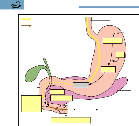
432 9 The Digestive System
|
Proteins |
|
Gullet (esophagus) |
|
|
|
|
|
Amino acids and |
|
|
|
dipeptides or tripeptides |
|
|
|
|
|
Stomach |
|
|
|
(gaster) |
|
|
|
Pepsinogen |
|
|
|
HCl |
|
|
|
Pepsins |
Duodenum |
Peptides |
|
|
|
|
||
|
|
Trypsin |
|
Aminopepti- |
Chymotrypsin |
Pancreas |
|
dases and |
|
|
|
carboxypepti- |
|
|
|
dases |
|
Portal vein |
liver |
|
|
||
|
|
Absorption in the jejunum |
|
Fig. 9.31 |
Diagrammatic representation of protein digestion |
||
taken up actively into the epithelial cells of the small intestine and from there reach the bloodstream. About 10 % of the ingested proteins arrive undigested in the large intestine, where they are broken down by bacteria.

Summary 433
Summary The Digestive System
Metabolism, Digestion, Energy Requirements
Nutrients (carbohydrates, proteins, fats) are split enzymatically in the various segments of the digestive tract into components that can be absorbed. They are then taken up by the mucous membrane of the small intestine. Through the bloodstream they reach the cells of the body, where in the presence of oxygen they undergo combustion in the mitochondria (biological oxidation). The energy liberated during this process is transmitted through a chain of reactions (mitochondrial respiratory pathway) to be stored in the form of the universal biological fuel ATP. From the mitochondria, ATP reaches the energy-consuming areas of the cell. ATP splits off phosphate molecules as required, liberating the stored energy.
Catabolism or metabolic degradation (functional and energy metabolism): degradation of substances rich in energy into those with less energy (= dissimilation), e. g., biological oxidation, but also vital processes (e. g., muscular work, impulse conduction in nerve fibers etc.) requiring ATP (= hydrolysis of ATP).
Anabolism or metabolic synthesis (constructive metabolism): synthesis of cellular substances; energy-consuming transformation of ingested foreign materials into endogenous substances (= assimilation).
Energy Equivalent (= Physiological Caloric Value) of the Most Important
Nutrients |
|
1 g carbohydrate: |
4.2 kcal (17.6 kJ) |
1 g fat: |
9.3 kcal (38.9 kJ) |
1 g protein: |
4.1 kcal (17.2 kJ) |
Energy Requirements of the Human Organism
Basal or resting metabolism: the amount of energy required by the human body for maintenance of vital functions at complete rest in the morning, fasting, and at normal body and ambient tempera-
ture. For adult men, 1 kcal/kg body weight/hour: 70 kg body weight 1700 kcal/day. For women, 10−20 % less.
Leisure metabolism: energy metabolism during average leisure activity, i.e., without much movement: about 2300 kcal/day.

434 9 The Digestive System
Work metabolism: energy requirement during physical labor; exceeds leisure metabolism to varying degrees.
The respiratory quotient (RQ = CO2 production/O2 consumption) provides information about the type of foodstuff metabolized: carbohydrate diet, RQ = 1; mixed diet, RQ = 0.85; fatty diet, RQ = 0.7.
Dietary Components
Proteins: should make up about 15 % of the daily energy requirement. They supply amino acids (for biosynthesis of endogenous proteins); of the 20 amino acids occurring in protein, 8 are essential (cannot be synthesized in the body). Other than lysine (which is not present in many vegetable foodstuffs), all the essential amino acids are present in vegetable and animal foodstuffs.
Fats: should make up about 30 % of the daily energy requirement. They supply and store energy, and supply cholesterol (cell membranes, precursors of sex hormones), essential fatty acids (in vegetable oils: linoleic acid, α-linolenic acid (components of membrane lipds). Intake usually includes more animal fats (mainly saturated fats) than vegetable fats (mainly unsaturated fats).
Carbohydrates: should make up about 55 % of the daily energy requirement. They are the most important source of energy: monosaccharides (glucose), disaccharides (cane sugar), and polysaccharides (starch). Simple sugars are “empty calories” and overload the pancreas, because the blood sugar rises rapidly to high levels. Whole-wheat products are better (starch + vitamins + fiber).
Vitamins: cannot be synthesized by the body (exception: vitamin
D3). They do not supply energy and are not structural material but rather biocatalysts. They are classified into fat-soluble (A, D, E, K)
and water-soluble (C, B1, B2, B3, B5, B6, folic acid, B12, biotin, and inositol) vitamins. Function: B vitamins and vitamin K are components of coenzymes for carbohydrate, fat, and protein metabolism (phylogenetically old, because they are indispensable for every cell), while vitamins A, D, E, and C are necessary to maintain certain organ functions (phylogenetically younger, demonstrable only in higher invertebrates; vitamin D only in vertebrates).
Minerals and trace elements: vital inorganic substances that are not metabolized. Trace elements = minerals with a daily requirement

Summary 435
of 100 mg (chromium, iron, fluorine, germanium, iodine, cobalt, copper, manganese, molybdenum, nickel, selenium, silicon, vanadium, zinc, tin; total weight in the body is 8−9 g, of which iron comprises 4−5 g). Minerals (macroelements) = minerals of which the daily requirement is 100 mg (calcium, magnesium, phosphorus, sodium, potassium, chlorine, sulfur). Calcium is present in the largest proportion in the body at 1.5 kg (99 % in the skeleton).
Antioxidants (free radical scavengers): vitamins A ( -carotene), E, and C as well as the trace elements selenium, manganese, zinc, and molybdenum inactivate free radicals (highly reactive unstable molecules with unpaired electrons) that are metabolic by-pro- ducts. Radicals are formed in the body during all reactions involving oxygen, but are reinforced by noxious substances and UV and radioactive radiation. The consequences might include malformed cells (cancer), arteriosclerosis, premature aging.
Active substances in plants: chemical compounds that protect plants and humans from damaging environmental influences. Every plant contains several thousand active substances, most of which have not yet been studied and which might be active only in combination with each other.
Fiber: indigestible vegetable carbohydrates that stimulate intestinal activity and increase the sensation of satiety.
The Digestive Organs
Divided into those of the head (oral cavity with salivary glands, oropharynx, and hypopharynx) and those of the trunk (esophagus; stomach; small bowel = duodenum + jejunum + ileum; large bowel = caecum + appendix + ascending, transverse, descending, and sigmoid colon + rectum; liver; pancreas).
The Oral Cavity
Ends posteriorly in the oropharyngeal isthmus (palatal arches with uvula). Function: to take up food and divide it into smaller pieces, and add saliva.
The tongue: muscular transporting organ; assists in chewing and sucking. The dorsum of the tongue carries sensory organs (papillae) for touch, pressure, temperature, and pain sensation, and taste papillae for sour, salty, bitter, and sweet.

436 9 The Digestive System
The teeth: second dentition of 32 permanent teeth—8 incisors (cutters), 4 canines (tear and retain), 8 premolars (chew and grind), 12 molars (chew and grind). Dental formula for each half of each jaw: I2/2 C1/1 P2/2 M3/3. Milk teeth (first dentition) has 20 teeth—8 incisors, 4 canines, 8 milk molars. Structure of tooth: crown (above the gum; coated with enamel), root (in the alveolus; coated with cementum), and neck (transition from enamel to cementum). Cementum and enamel cover the dentine. The dental cavity with the dental pulp lies inside the tooth. Vessels and nerves reach the dental cavity through the root canal. The junction of dentine and pulp is lined with odontoblasts.
Salivary glands: exocrine glands; they produce 1−1.5 liters of saliva/day. Function: increase lubrication of food after chewing, initiate splitting of carbohydrates (amylase). They are divided into bilateral small (in the mucosa of the lips, cheeks, tongue, and palate) and large salivary glands (parotid, submandibular, and sublingual glands), with ducts draining into the oral cavity.
The Pharynx
Part common to the digestive and respiratory tracts: nasopharynx (junction with the nasal cavity), oropharynx (junction with the oral cavity; crossing of the respiratory and digestive pathways), and hypopharynx (junction of the laryngeal opening and esophagus). At the junction of the nasal and oral cavities: tonsils of Waldeyer’s tonsillar ring (midline pharyngeal tonsils [adenoids], palatine tonsils, midline lingual tonsils, lymphatic tissue of the lateral pharyngeal wall). During swallowing, the larynx is closed by the epiglottis (producing a momentary respiratory pause), so that no food can reach the larynx. The initiation of the act of swallowing (contraction of the floor of the mouth) is voluntary. This triggers the swallowing reflex (involuntary, coordinated by the swallowing center of the medulla).
The Esophagus
Function: moves the food from pharynx to stomach by peristalsis; length: 25−30 cm. Cervical and thoracic parts run behind the trachea. After piercing the diaphragm (esophageal hiatus), it enters the abdomen and opens into the stomach. Large boluses can become wedged

Summary 437
in the esophageal constrictions (cricoid cartilage, aortic arch, esophageal hiatus) and cause pain.
The Stomach
Lies directly under the diaphragm; volume about 1200−1600 ml. It is divided into inlet (cardia: esophagus stomach), fundus, body, pyloric antrum, and pylorus (circular sphincter muscle: stomach duodenum). Function: chemical maceration and liquefaction of the food by gastric juice (pH 1.5; 2−3 l/day; water, mucus, pepsin, hydrochloric acid). Its secretion is triggered by sensory input (vagus nerve) and by entry of food into the stomach stimulating secretion of tissue hormones from the gastric mucosa (gastrin). Tissue hormones from the duodenal mucosa also inhibit (secretin) or stimulate (gastrin) secretion of gastric juice as needed. The gastric mucosa forms longitudinal folds. Several gastric glands drain into punctiform pits in the folds, where their cells secrete hydrochloric acid (parietal cells) and pepsinogen (chief cells). The necks of the glands contain neck cells that produce mucus (protection against autodigestion). In contrast to the rest of the gastrointestinal tract (circular and longitudinal muscle layers), the wall of the stomach is made up of three muscle layers (additional inner oblique muscle layer for kneading movements).
The Small Intestine
Total length 3−5 m; divided into duodenum (25−30 cm, begins distal to the pylorus), jejunum, and ileum. Function: degradation by pancreatic enzymes of food into compounds that can be absorbed (e. g., glucose, amino acids, fatty acids), and absorption of nutrients. Food is moved by peristalsis (alternating contraction and relaxation of longitudinal and circular muscle). To facilitate absorption, the mucosa of the small bowel (especially the jejunum) includes folds (Kerckring’s folds), villi (projections on the folds), and crypts (crypts of Lieberkühn = depressions between the villi). The villi are lined by simple columnar absorbing epithelial cells with microvilli (total absorbent surface formed by all the surface extensions: 120 m2) and mucus-forming goblet cells. Each villus contains a network of capillaries by which the nutrients reach the bloodstream, a central lymph vessel (removal of chylomicrons), and the arteriole and venule supplying the villus. The

438 9 The Digestive System
mucosa also contains collections of lymphoid follicles (especially dense in the mucosa of the appendix and the ileum = Peyer’s patches): part of specific immunity.
The Large Intestine
Total length 1.5−1.8 m; ileocecal valve at junction of ileum and large bowel. Large bowel is divided into caecum (outpouching below the ileocecal valve), vermiform appendix (specific immunity), colon (four segments: ascending, transverse, descending, and sigmoid colon), and rectum (15 cm with ampulla, anal canal, and anus = distal end of gastrointestinal tract). Function: reabsorption of salt and water (the mucosa therefore consists mainly of mucus-forming goblet cells; surface extension only by crypts) and decomposition of indigestible food remnants by bacteria. External appearance of the large bowel: longitudinal muscle (taeniae) as well as constrictions and sacculations (by contraction of the circular muscle). The intestinal content is mixed and moved toward the rectum by alternating contraction and relaxation of the longitudinal and circular muscle. Closure of the anal canal: internal smooth muscle (= involuntary) and external striated (= voluntary) sphincter and the puborectalis muscle (most important muscle for closure, part of the musculature of the pelvic floor).
The Pancreas
Consists of head, body, and tail of the pancreas. The head is surrounded by the duodenum, into which drains the pancreatic duct (usually together with the common bile duct). The pancreas lies behind the stomach and is an exocrine gland secreting ca. 2 liters of alkaline (high bicarbonate content) pancreatic juice a day. The juice contains the most important digestive enzymes: lipases (fat), proteases (proteins), amylases (carbohydrates). Secretion is regulated by sensory input (vagus nerve) and the food itself (tissue hormones secretin and pancreozymin/cholecystokinin). The endocrine activity of the islet apparatus secretes insulin and glucagon (regulation of blood sugar).
The Liver
Lies directly under the diaphragm, to which it is partially adherent. Divided into left and right lobes (division on the surface by the fal-

Summary 439
ciform ligament) and two smaller lobes, the quadrate and caudate, both on the visceral side. In the visceral area of the right hepatic lobe the vessels and nerves enter and exit (porta hepatis): portal vein, hepatic artery proper (enter); bile duct (exits); lymph vessels and nerves (enter and exit). Weight: 1500−2000 g. Function: exocrine gland (secretes bile: bile salts for the emulsification of fats); the largest metabolic and detoxifying organ (nutrients absorbed in the intestines reach the liver through the portal vein). The liver is subdivided into hexagonal liver lobules (diameter 1−2 mm) (structure: hepatic epithelial cells, central vein, hepatic sinusoids, cholangioles), between which lie the portal canals, each with a branch of the portal vein and the hepatic artery as well as a biliary ductule. Blood flows through the portal vein into the central veins of the hepatic lobules, leaves the liver through the hepatic veins, and eventually drains into the inferior vena cava. The secreted bile flows through the biliary ductules to the common hepatic duct and the cystic duct into the gallbladder.
The Gallbladder
The reservoir for the bile (30−50 ml). When needed, the concentrated gallbladder bile is transported back through the cystic duct and the common bile duct (choledochous duct) to the duodenum at the head of the pancreas (usually together with the pancreatic duct). During digestive quiescence, bile flow into the duodenum is prevented (sphincter at the distal end of the common bile duct).
Digestive Processes
Fat Digestion
Splitting of fats begins in the stomach (10−30 %) by lipase from the palatine part of the tongue, followed by emulsification (bile acids) and enzymatic splitting by pancreatic lipase and phospholipase A2 in the duodenum. Fatty acids, monoglycerides, cholesterol, and phospholipids are absorbed in the form of fat droplets (micelles) by the mucosa of the small intestine, where the individual components together with proteins are immediately transformed in part into chylomicrons (mode of transportation of alimentary fats to the cells of the body). These bypass the liver and reach the blood by lymphatic pathways. Only short-chain fatty acids reach the bloodstream directly and are carried by the portal vein to the liver and thence to the cells of the body.

440 9 The Digestive System
Carbohydrate Digestion
The splitting of carbohydrates begins in the mouth with salivary amylase (starch oligosaccharides, disaccharides). In the small intestine there is further splitting by pancreatic amylase, as well as by glycosidases and disaccharidases from the mucosa of the small intestine, into monsaccharides (e. g., glucose, fructose). Absorption is by the mucosa of the small intestine and from there into the bloodstream, through the portal vein into the liver, and eventually into the cells of the body.
Protein Digestion
Begins only in the stomach. Proteins are denatured by acid gastric juice and split by pepsin (pepsinogen from the chief cells of the gastric glands pepsin by the action of hydrochloric acid from the parietal cells) into polypeptides and peptides. In the duodenum (neutral milieu) there is further cleavage by trypsin and chymotrypsin (pancreas) into tripeptides and dipeptides, part of which are absorbed. Carboxypeptidases and aminopeptidases (pancreas, mucosa of the small intestine) continue cleavage into amino acids, which are also absorbed (active transport systems), enter the bloodstream, and reach the cells of the body through the portal vein and the liver.