

Lection 14. Neuroendocrinology circadian rhythms
Temporal endocrine structure (tes)
It can be defined as a combination of predictable hormonal changes that are time-related. The definition covers a range of rhythmic frequencies and at any level: organs and tissues, cells and subcellular structures. Regarding their frequency, endocrine rhythms may be circadian, ultradian and infradian. In this context we will pay particular attention to the endocrine circadian time structure (ECTS).
The ECTS is closely dependent on some areas of the hypothalamus. In the report “Neural Control of Pituitary Gland” in 1955, Harris stated that all the blood reaching the anterior hypophysis via the portal system had initially been in contact with the eminence of the hypothalamus by means of a primary capillary plexus. Later studies defined the morphofunctional characteristics of the monoaminergic system and reported the capacity of the hypothalamus to produce releasing hormones or factors (RH) and inhibiting hormones or factors (IH).
The basis of neuroendocrinology were established, concentrating particularly on the study of the neural control of hormonal secretion and also dealing with the interactions between the monoaminergic system and the behaviour of the subject (psychoneuroendocrinology) and, on a wider scale, the relationship between the rhythmic endocrine activity of the body and neural activity (chrono- neuroendocrinology).
The hypothalamic neuropeptides (RH, IH) known so far at the hypothalamic level are shown in Fig. 1.
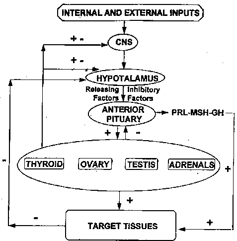
Fig. 1. - Neuroendocrine network indicating the relationship among its components: the central nervous system; hypothalamus, anterior pituitary and target glands and tissue; +: Stimulatory effect; Inhibitory effect.
Long and short loop feedback link together the various components: central nervous system, hypothalamus, anterior pituitary and target glands and tissues. “Both these feedback systems are essentially closed-loop systems, which allow self-regulation and avoid overshoot in the production of any secretory product. Such systems also respond to exteroceptive and interoceptive stimuli, though they are apparently not involved in the function of the hypothetical “clock(s)” which regulates the circadian periodicity of many pituitary and releasing hormone concentrations. Such periodicity persists in the absence of target organ gland secretions and exhibits phase characteristics similar to those seen in intact subjects, but at higher hormonal levels, which reflect the absence of target organ feedback processes”.
Thyrotropin releasing hormone (TRH). - This was the first RH to be isolated; its activity is not completely clear but it is thought to stimulate the release of prolactin (PRL) and thyrotropin (TSH). TRH, T4 and T3 in turn act directly on the anterior pituitary gland to control the secretion of TSH.
Luteinizing releasing hormone (LH-RH). - Both natural and synthetic LH-RH provoke the release of LH and FSH. Although it is defined a releasing factor, it has also been shown capable of hormonal synthesis.
Growth releasing hormone (GH-RH). - GH secretion regulation is provided not only by GH-RH but also by somatostatin, which has an inhibiting action on the release of the hormone.
Prolactin releasing hormone (PRH). - Together with the TRH, which releases PRL, there is a PRH, which seems to act independently from TRH. Prolactin secretion regulation is carried out by an inhibitory hormone, prolactin inhibiting factor (PIF).
The release of adrenocorticotropic hormone is induced by a corticotropin releasing hormone or factor (CFR).
Recently, for a further insight into the neuroendocrine regulation of anterior pituitary function, in women with functional hypothalamic amenorrhea (FHA), serum LH, FSH, cortisol, GH, PRL, TSH concentrations were measured simultaneously at frequent intervals for 24 h. The 24 h secretory pattern of each hormone except TSH was altered in the women with FHA. Compared to normal women, those with FHA had a 53% reduction in LH pulse frequency and an increase in the mean LH interpulse interval; LH pulse amplitude was similar. The 24 h integrated LH and FSH concentrations were reduced. Pituitary hormone increases the response to the simultaneous i.v. administration of GnRH, CRF, GHRH and TRH. The response was normal also in the group of women with FHA, except for a blunted PRL response to TRH.
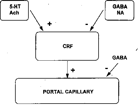
Fig. 2. - Suggested model for the control of corticotropin releasing hormone (CRH) from the hypothalamus + : Stimulatory effect; Inhibitory effect.
Neurotransmitter regulation of pituitary hormone secretion
No central nervous system areas have been reported as being able to specifically regulate the release of a pituitary hormone. Fig. 2 shows a possible model for CRF regulation by the hypotalamus. The CRF neuron ending at a portal capillary gets in contact with a receptor for a 5-HT pathway that releases CRF after stimulation of the cholinergic neuron. One may conclude that the final common pathway to CRF release is cholinergic (Ach). One of the two cholinergic pathways is placed between the 5-HT pathway and the CRF cell, suggesting that this may be used to control the circadian rhythm. The CRF neuron could be under the inhibitory control of both a noradrenergic (NA) pathway and GAB A inhibitory neuron. Fig. 2 summarizes the state of knowledge and the controversies regarding the neurotransmitter regulation of pituitary hormone secretion. The neurotransmitter may act on the RH, since receptors for neurotransmitters have been found in the pituitary gland. For PRL, an inhibitory action of the DA on the pituitary gland has been reported.
Central neural mechanisms and endocrine rhythms regulation
Three alternative models have been suggested for the mammalian circadian oscillating system as illustrated in Fig. 3. In the first and simplest model there is a driving oscillator (DO) that is an active cellular unit capable of maintaining a self sustaining oscillation with its own independent driving influence.
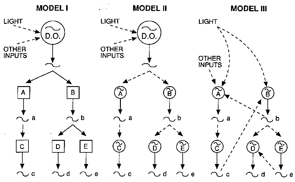
Fig. 3. - Representation of three alternative models for the mammalian circadian oscillating system. DO: driving
oscillators (see text).
By light and other inputs the oscillation is transmitted to subordinate centers A and B and from these to others, C, D and E. In this system, the destruction of the driving oscillator would bring about the loss of all the subordinate circadian rhythmic functions, while the phase-shift of the driving oscillator would lead to the same consequences, that is the dephasing of the subordinate centers. The destruction of a single connection between the DO and any one of the subordinate centers leads to the loss of that single circadian rhythmic function.
The second model consists of a hierarchically organized set of oscillators. The DO will entrain secondary (A and B) and tertiary (C, D and E) oscillators but unlike the first model, if the DO (S) is or are eliminated, the secondary oscillating mechanisms will be able to maintain their own autonomous rhythm.
The third model is a multioscillator system organized in a non-hierarchical manner. There is no DO and the different centers can interact with each other and with external inputs.