

Tumours of lungs
Distinguish between bening and malignant tumours of lungs.Bening tumours are divided into 2 groups after localization (intrabronchial and intrapulmonar) and after a histological structure (epithelium are adenomas, papiloma, unepithelium, - gammartoma, lypoma, chondroma, myoma, neuroma, angioma); they are rare and do not have a practical value.
Malignant tumours are divided into primary and secondary, or metastatic. Among malignant tumours often is a cancer.
Lungs cancer
The lungs cancer develops unnoticed, almost nothing can be find out itself, that is why 60-70% patients come to the doctor already with the started forms of disease. By the clinical symptoms of lungs cancer on the first stages of disease are rapid fatigueability, weight loss, pain in breasts, cough, sometimes at cough is a sputum patient notices the veins of blood. At the laboratory analysis of blood promoted ERS is marked, anaemia.
A cancer in lungs is almost always developed from the epithelium of bronches; large bronches often struck: main and lobar, rarer segmentar and yet rarer shallow periferial. From lobar bronches the bronches of superior lobe of right are mostly struck lungs.
From plenty of the offered classifications of this disease the most widespread is the classification of Y.M.Socolov (1956), who distinguishes such forms of lungs cancer:
Primary:
1) Central (endo-, exo-, peribronchial);
2) Peripheral (spherical and cavital);
3) Untypical forms (mediastinal, apex (Penkosta) and miliar carcinosis).
Secondary:
1) Metastases.
A central cancer of lungs develops from the epithelium of main, lobar or segmentar bronches. In one cases a tumour grows mainly in the road clearance of bronchus - endobronchial, in other – exobronchial and peribronchial round a bronchus, germinating in contiguous pulmonary tissue.
The clinical symptoms of central cancer appear in that stage of the development of tumour, when violation of the external respiratory, drainage function of bronchus, inflammatory reaction of adjoining tissues, are present.
The roentgenological pattern of central cancer consists of displays of tumour, signs of violation of bronchus communicating, complications which arise up in connection with progresive growth of tumour and its metastases. The decline of transparency of lobe is marked and all the lungs, which increase lung pattern appears on a background, narrowing of intercostal intervasal, something is promoted position of diaphragm cupula and insignificant displacement of mediastinal organs, mainly to the esophagus (at contrasting his examination) and trachea.
At endobronchial cancer tumour of obturate educations of bronchus which brings an origin over of hypoventilation or atelectasis and development of concomitant inflammation. A node of tumour on the first stages of the development can be expressly marked off from adjoining tissue. However, to the extent of progress of tumour process he loses clear scopes and begins to spread outside a bronchus in lungs tissues. Computer and ordinary tomography finds out a tumour, that obturue road clearance of bronchus.

Fig.17.1. Endobronchial cancer. Degrees of bronchus obturation and symptoms, that it is accompanied.

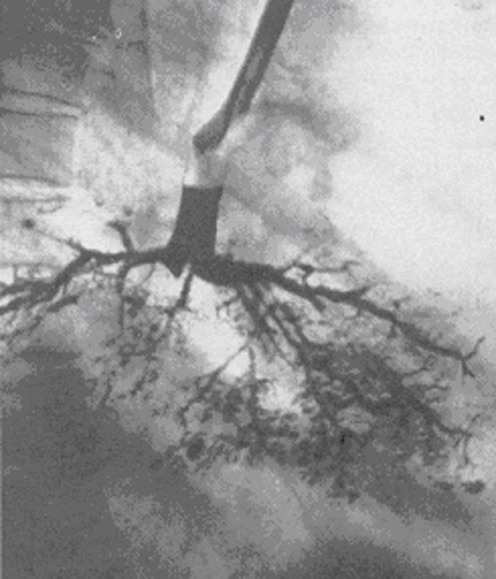
Fig.17.2 Atelectasis of the left lungs as a result of obturation of left main bronchus, bronchogramic obturation of the under lobar bronchus by a cancer tumour (symptom of bronchus amputation).
At the exobronchial type of growth a tumour appears only then, when its sizes more than diameter of large pulmonary vessels. At the beginning the development of the bronchial communicating tumour violations. Only later, during infiltration of wall of bronchus, and then increase of tumour there are too a few centimetres, there are violations of ventilation (hypoventilation, valvular emphysema, and atelectasis). Lungs roots are extended, unstructured, and hard.

Fig.17.3 A central exobronchial cancer of the left lung
At the peribronchial type of growth tumours appear thick muffs round bronches and vessels. The increased lung pattern, which in next grows into rough hard which fan-shaped walk away from a root in pulmonary tissue, is determined. Educations of bronches remain clock-houses, signs of violation of ventilation lungs do not appear. Computer and ordinary tomography finds out the bulge of bronchial walls and visualised tumour mass, bronchographa is the concentric even narrowing of road clearance of bronches on a considerable draught, related to the bulge of their wall.
A peripherial cancer arises out of epithelium of mucus shell of subsegmentar bronches and bronchioles. The clinical symptoms of this form of cancer depend on situating of tumour node in relation to a thoracic wall or large bronches. Growing in of tumour in pleura causes pain in the area of thorax. When a tumour gets to the large bronchus, a cough appears, sputum, hemoptysis.
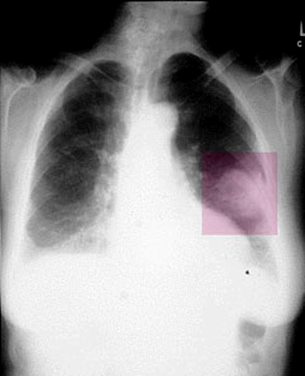
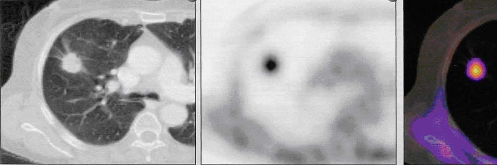
Fig.17.4 The peripheral cancer is on a sciagram, CT PAT.
Roentgenological and CТ-examination finds out a peripheral cancer as a node a diameter 2-4 sm and anymore, as a rule, in the superior lobes of lungs, more often business. The important signs of peripheral lungs cancer are: 1) wrong spherical form, visualised on CT and roentgenological multiaxial examination of patient; 2) determined on roentgenograms, ordinary and computer tomography heterogeneous structure of tumour shade which consists of separate nodes; 3) unequal, hilly, on separate areas unclear, the contours of shade are smeared; 4) pulmonary tissue is unchanged round shade of cancer node; 5) presence on occasion "paths" to the root lungs, predefined the shallow metastatic pouring out, or germination of tumour along lymphatic vessels (cancer lymphangoitis); 6) regional defects of walls of bronches which are determined on occasion at bronchography; 7) bulge of pleura, joint, accretion, or pleural exudate which are observed at the near placing of pleural tumour; 8) cavity of wrong form with unequal edges, without a horizontal level which appears at disintegration of tumour and appears on a sciagram, CT and ordinary tomogramas.
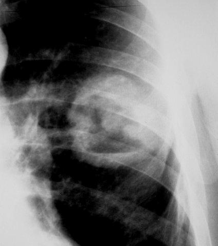
Fig.17.5 Cavitar form of cancer
The mediastinal cancer of lungs is the display of metastatic of primary cancer tumour, which is more often placed at the level of bifurcation of trachea, medial wall of main or lobar bronches. There is metastasing in tracheobronchial and paratracheal lymphatic nodes. Clinically characterized by the compression syndrome of defeat of mediastinum (syndrome of superior vena cava, compression of esophagus, turning and diaphragmal nerves), in particular by the edema of fase and neck. A roentgenological pattern characterizes the presence of large shade from one side, which closes shade of root. Hyperplasy of staggered lymphatic nodes appears on CT-skans.

Fig.17.6 mediastinal cancer
An apex cancer (cancer Penkosta) is a cancer of superior lobe lungs. His early germination in ribs and involvements the process of nervous interlacements stipulates a clinical pattern: pain in a humeral joint, thorax, and also Gorner`s syndrome (of superior eyelid, enophtalm, narrowing of pupil). However many symptoms of already started process which spread outside lungs. The roentgenological pattern of apex cancer characterizes homogeneous shade in the area of apex lungs. Characteristic is destruction of back segments of one-two ribs, and also transversal sprouts of a few vertebrae.

Fig.17.7. Cancer Penkosta
Miliar carcinomatous (carcinosis) is not indiform of tumour growth, but is only one of the forms of his metastatic - hematogenous skidding of tumour mews. A primary tumour node is small and does not appear during roentgenological and CТ-examinationes. Plural shades appear on rent-genogramas and computer tomogramas; by a size to 3 mm. Shades increase afterwards, anymore in inferior lobes. A lung pattern is not traced.
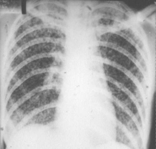
Fig.17.8 Miliar carcinosis