

Атлас по рентгенологии травмированных собак и кошек / an-atlas-of-radiology-of-the-traumatized-dog-and-cat
.pdf312 Radiology of Musculoskeletal Trauma and Emergency Cases
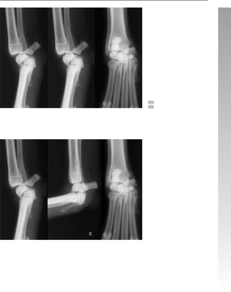
Case 4.27
4

Forefoot 313
Signalment/History: “George” was a 6-year-old, female Irish Setter with a history of unknown injury to the right foot.
Physical examination: Palpation of the digits showed firm soft tissue masses around the 2nd and 5th digits on the right foot.
Radiographic procedure: Radiographs were made of both feet.
Radiographic diagnosis: On the right foot, chronic intraarticular fractures affected the distal interphalangeal joint of the 2nd digit and the same joint on the 5th digit. The fragments (white arrows) had a smooth border without any signs of active repair processes. The fracture lines were indistinct, an indication of chronic trauma. Periosteal new bone was especially prominent on the 2nd digit. Soft tissue swelling was minimal.
A similar pattern of chronic change was noted on the left foot, though with much less severe change.
Differential diagnosis: Exclusion of infectious and neoplastic lesions was the main differential problem. In this dog, the features were rather specific for chronic trauma with the fracture lines and fracture fragments being identified. The periosteal response was adjacent to the injured joint and did not suggest either an inflammatory or neoplastic lesion.
Treatment/Management: Having found an injury due to old trauma, the choice of treatment was limited. Amputation could be considered if one of the lesions was causing a particular clinical problem for the dog.
Comments: Injury of this type is more likely in the 2nd and |
4 |
5th digits. |

314 Radiology of Musculoskeletal Trauma and Emergency Cases
Case 4.28
4
Day 1
Signalment/History: “Beaver”, a 5-year-old, female Labrador Retriever, had fallen 4 meters from a roof onto the ground. Injury to both forefeet was evident. In addition, a pneumothorax required immediate treatment.
Physical examination: Palpation of the feet produced marked instability suggesting fracture/luxation. Dyspnea was pronounced.
Radiographic procedure: Radiographs of the thorax and the cranial portion of the thoracic spine were done at admission. The former were made because of the dyspnea and the latter because of a suspected segmental instability noted on the thoracic studies. After nine days in the clinic, the dog stabilized and both feet were radiographed including stress radiographs.
Radiographic diagnosis (day 1, cranial thoracic spine):
A collapse of the T4–5 disc space with minimal malalignment of the segments was note on both projections.
Radiographic diagnosis (day 9, feet): Fracture/luxation of the carpometacarpal joints on the left caused extensive instability as evidenced by a palmar displacement of the head of the 2nd metacarpal bone. Also note the lateral displacement of the metacarpal bones indicating the severity of the injury.
Fracture/luxation of the intercarpal joints on the right resulted in a palmar displacement of the distal row of carpal bones and hyperextension. The fractures appeared to be limited to small chip and avulsion fragments.
Treatment/Management: Pancarpal arthrodesis and partial carpal arthrodesis were attempted.
Despite the collapse of the T4–5 disc space (arrow), the neurologic examination was thought to be normal and consequently, the spinal subluxation was not treated. The finding of the spinal injury did indicate the requirement for cage rest for a period of time after the trauma.
Outcome: Radiographs were made eight months following the corrective surgeries, at which time both surgeries were clinically and radiographically considered healed with the anticipated arthrodeses.
Comments: The minimal periosteal new bone seen on the distal aspect of the accessory carpal bones is compatible with time being nine days post-trauma.
Forefoot 315
4
Day 9

316 Radiology of Musculoskeletal Trauma and Emergency Cases
Case 4.29
4
Signalment/History: “O.J.” was a 7-week-old, female Labrador Retriever puppy noticed by the owner to be lame on the right forelimb.
Physical examination: Pain was not evident on examination; however, she was an excited, hyperactive puppy. She was definitely lame when jumping around the examination room.
Radiographic procedure: A study was done of the right and left forefoot
Radiographic diagnosis: Complete fractures of the proximal portions of the 2nd and 3rd metacarpal bones were noted (arrows).
Treatment/Management: The fractures were treated by splinting.
Outcome: The metacarpal fractures healed within two weeks, which is within the expected time considering the apparent low energy of the trauma and the young age of the patient.

Forefoot 317
Day 1 |
4 |
|
|
|
|
Case 4.30 |
|
|
|
Signalment/History: “Muffie” was a 7-year-old, female Miniature Poodle who had been struck by a car and injured her left forelimb.
Physical examination: Severe soft tissue injury characterized the open, comminuted fractures in the left foot.
Radiographic procedure: Two views were made of the distal left forelimb.
Radiographic diagnosis (day 1): The original radiographs were made with the foot in a thin bandage and were diagnostic of a severe “degloving” type of injury with the abrasion removing a portion of the distal radius and ulna, part of the carpal bones, and the proximal part of the metacarpal bones. The injury is on the dorsal surface and all of the soft tissues on the extensor surface are missing.
Differential diagnosis: Detection of bone infection cannot be made at the time of an injury, but such an open lesion must always be considered as being infected.
Treatment/Management: A second radiographic study was made five weeks later following only treatment of the soft tissue injury.
Week 5
Radiographic diagnosis (week 5): At this time, the distal ulna had become atrophic as characterized by “penciling” (arrow). The remaining bones had less density, although the study was made with the splint in position somewhat compromising the determination of bone density. It remained difficult to identify any changes typical of bone infection, but it had to be assumed that it was present.
Treatment/Management: Carpal arthrodesis was performed with good results fusing the radius, carpal bones, and metacarpal bones.
Outcome: Unfortunately, a marked dorsal angulation plus varus deformity resulted leaving the dog with a leg that could be used for little more than a support.
Comments: Interestingly, infection was successfully controlled by antibiotic therapy and did not interfere with the arthrodesis.

318 Radiology of Musculoskeletal Trauma and Emergency Cases
Case 4.31
4

|
Pelvis 319 |
|
|
Signalment/History: “Kalu”, a 1-year-old, male Labrador |
4.2.1.6 Pelvic limb injury |
|
|
Retriever had suffered an acute lameness in the left forelimb |
Pelvis |
|
|
six weeks previously. The limb had been splinted for ten days. |
The os coxae, or the hipbone, is composed of the ilium, ischi- |
|
|
Physical examination: Point tenderness over the proximal |
um, pubis, and the acetabular bone. Fusion of these bones re- |
|
|
sults in the creation of the os coxae including the acetabulum |
|
||
sesamoid bones on the palmar surface of the 2nd digit on the |
that ultimately receives the femoral head. The two hipbones |
|
|
left. Similar tenderness was noted in the same area of the 2nd |
join at the pelvic symphysis to form the pelvis. The pelvic |
|
|
and 3rd digits on the right |
symphysis consists of the pubic symphysis and the ischial sym- |
|
|
|
physis. The ischial symphysis contains a separate small triangu- |
|
|
Radiographic procedure: Multiple views were made of |
lar ossification center caudally. With age, the ischial symphysis |
|
|
both forefeet. |
ossifies though in smaller dogs, the pubic symphysis often re- |
|
|
|
mains cartilaginous. The pelvis attaches to the sacrum at the |
|
|
Radiographic diagnosis: Multiple non-union fractures of |
sacroiliac joints. They are a combined synovial and cartilagi- |
|
|
the proximal sesamoid bones on the plantar surface of the 2nd |
nous joint; the cranial portion of which is radiolucent because |
|
|
and 3rd digits on the right and the 2nd and 5th digits on the left |
of the presence of a fibrocartilage plate, whereas the caudal |
|
|
were to be seen (arrows). |
portion often undergoes bony fusion with age. The pattern of |
4 |
|
|
degeneration is breed/size dependent. Visualization of the |
||
Differential diagnosis: The differential diagnosis for lesions |
sacroiliac joint on the ventrodorsal radiographs is dependent |
|
|
of this type included three specific entities: (1) congenital bi- |
on the conformation of the iliac wings and often is not sym- |
|
|
partite sesamoid bones, (2) fractured sesamoid bones, and (3) |
metrical as seen on radiographs made of a malpositioned pa- |
|
|
degenerative changes resulting in fragmentation and soft tissue |
tient. |
|
|
mineralization. |
Injuries of the pelvis are unique because of its anatomic struc- |
|
|
|
|
||
Treatment/Management: Because the clinical signs were |
ture (Table 4.5). The resulting pattern of injury to the bony |
|
|
on the left, that foot was operated and the fragmented |
pelvis can be thought of as that expected with a disruption of |
|
|
sesamoid bones were removed from the 2nd and 5th digits. |
a “box” or “ring” in which one side has been fractured. To |
|
|
|
permit the displacement of one fragment, additional fractures |
|
|
Comments: The use of a plastic paddle to assist in position- |
must be present and three separate fractures are often identi- |
|
|
ing the foot resulted in a shadow of reduced tissue density |
fied as having occurred together. A common pattern of injury |
|
|
across the feet. |
involves ipsilateral fractures of the body of the ilium, the body |
|
|
|
of the pubis, and the ischiatic table. This effectively frees a seg- |
|
|
|
ment of the pelvis containing a hip joint. Another common |
|
|
|
pattern involves fractures of the body of the ilium on one side, |
|
|
the body of the opposite pubis, and the opposite ischiatic table. This also frees a segment of the pelvis including a hip joint. Often fractures affect the pelvic symphysis and these cannot be identified either on lateral radiographs due to a lack of fragment displacement or on the VD radiograph because of superimposed coccygeal segments and a rectum filled with fecal material. These are referred to as fractures in the “floor of the pelvis”.
Injury to the sacroiliac joints is somewhat dependent on age, since the caudal portion of the joints ossifies with age and the joints become stronger. In the younger patient, the joints can luxate rather easily often resulting in luxation of one sacroiliac joint plus ipsilateral fractures in the pubis and ischium. A common injury in the cat is the luxation of both sacroiliac joints as the only injury freeing the bony pelvis to shift cranially, the result of pulling by the rectus abdominis muscles. Any injury to the sacroiliac joints should prompt a careful search for a fracture line that enters the sacrum; this is more commonly found in the older patient because of the bony fusion of the joints.
Careful inspection of the acetabulum and femoral head is important, since fractures that enter the hip joint affecting the articular surface have great clinical importance because if the

320 Radiology of Musculoskeletal Trauma and Emergency Cases
fracture is not anatomically reduced, a post-traumatic arthrosis will develop. The fracture may only affect the margin of the acetabulum or may pass through the hipbone, or the fracture may involve the opposite articular surface with fragmentation of the femoral head.
Avulsion fractures in the pelvis can occur in the immature patient resulting in a separation of the centers of ossification in the ilial crest and in the ischiatic tuberosity. Because these fractures do not affect weightbearing bones and are not articular, they are not usually treated, though they are a source of pain.
Table 4.5: Radiographic signs of pelvic trauma
1.pattern of fractures or luxations because of a “box” or “ring” configuration (Cases 4.34, 4.35, 4.36, 4.37 & 4.41)
a.ilium, pubis, and ischium on the same side
b.ilium on the one side, and pubis and ischium on the opposite side
c.pelvic symphysis plus other fractures (Case 4.27)
d.both sacroiliac joints with cranial displacement of the bony pelvis (Case 4.45)
e.sacroiliac joint, pubis and ischium on the same side (Cases 4.32, 4.63 & 4.128)
f.sacroiliac separation (Cases 4.195 & 4.107)
g.sacroiliac joint on the one side, and pubis and ischium on the opposite side (Case 4.41)
Injuries to the tail can be assessed on the radiographs of the pelvis, though they are best seen on the lateral view, since the rectal contents often prevent the detection of fracture/luxations near the sacrococcygeal junction. The nature of the frac-
4 ture uncommonly tells of the severity of the injury to the cauda equina contained within the segments.
Injury to the lumbosacral junction has a particular importance and is discussed with lesions of the lumbar spine (Chap. 4.2.2.3).
2.avulsion of the ilial crest or ischial tuberosity (Cases 4.42, 4.106 & 4.132)
3.sacrococcygeal fracture/luxation (Case 4.38)
4.coccygeal fracture/luxation (Case 4.56)
5.unique patterns
a.fracture pattern leading to a narrowing of the pelvic canal (Cases 4.33, 4.56, 4.99, 4.103, 4.102 & 4.112)
b.pelvic injury including an acetabular fracture (Cases 4.34, 4.36, 4.37, 4.40 & 4.107)
c.pelvic injury including a sacral fracture (Cases 4.39, 4.49 & 4.93)
6.patterns of soft tissue injury
a.intrapelvic hemorrhage
b.rupture of the urethra or bladder neck (Case 4.33)
c.change in position of the fecesor air-filled rectum (Cases 4.108 & 4.112)
d.failure to identify the prostate gland because of hemorrhage or urine (Case 4.104)
e.failure to identify the urinary bladder because of rupture
f.subcutaneous emphysema (Case 4.62)
g.peritoneal fluid
h.displaced urinary bladder (Case 4.108)
i.rectal diverticulum (Case 4.108)

Pelvis 321
Case 4.32
Signalment/History: “Dog” was a 2-year-old, female mixed breed that had jumped from the back of a moving truck and was unable to walk normally after the accident.
Physical examination: She would not bear weight on the left pelvic limb in the examination room. Palpation of the pelvic region produced pain especially when moving the left pelvic limb. Crepitus was not detected.
Differential diagnosis: A pelvic fracture was suspected.
Radiographic procedure: Both VD and lateral views were
made.
4
Radiographic diagnosis: Fractures of the left hemipelvis involved the pubis (black arrow) and ischium, and were located just caudal and medial to the acetabulum. The comminuted fracture entered the caudal aspect of the acetabular roof as viewed on the lateral projection (white arrow) with a single bony fragment being identified. Displacement of the fracture fragments caused only minimal narrowing of the pelvic canal.
Treatment/Management: Because of the slight displacement of fragments and involvement of the caudal, non- weight-bearing portion of the acetabular roof, “Dog” was successfully treated with cage rest.
Outcome: Fracture healing in a young dog occurs quickly and he was exercising normally within 3 weeks.
Comments: It appeared as though the rule of “three pelvic fractures” was broken in this case. The third site of trauma apparently was the undetected injury to the left sacroiliac joint that provided the movement necessary to free the hemipelvis. Often sacroiliac injury is extensive; however, in this patient, the injury was minimal and without displacement.
The flexed view for the pelvis was used in this case because it was much less painful to position the pelvic limbs in flexion than attempting to extend what may have been a limb with a fractured femur or move bony fragments associated with an injured hip joint.
Note how the left-sided fractures resulted in a medial displacement of the left hemipelvis and a collapse resulting in the obturator foramen appearing smaller on the radiograph.