

5. The Major Histocompatibility Molecules Bind Antigenic Peptides
The major histocompatibility complex (MHC) is a large genetic complex with multiple loci. The MHC loci encode two major classes of membrane-bound glycoproteins: class I and class II MHC molecules. As noted above, TH cells generally recognize antigen combined with class II molecules, whereas TC cells generally recognize antigen combined with class I molecules.
MHC molecules function as antigen-recognition molecules, but they do not possess the fine specificity for antigen characteristic of antibodies and T-cell receptors. Rather, each MHC molecule can bind to a spectrum of antigenic peptides derived from the intracellular degradation of antigen molecules. In both class I and class II MHC molecules the distal regions (farthest from the membrane) of different alleles display wide variation in their amino acid sequences. These variable regions form a cleft within which the antigenic peptide sits and is presented to T lymphocytes (see Figure 5).
Different allelic forms of the genes encoding class I and class II molecules confer different structures on the antigen-binding cleft with different specificity. Thus the ability to present an antigen to T lymphocytes is influenced by the particular set of alleles that an individual inherits.
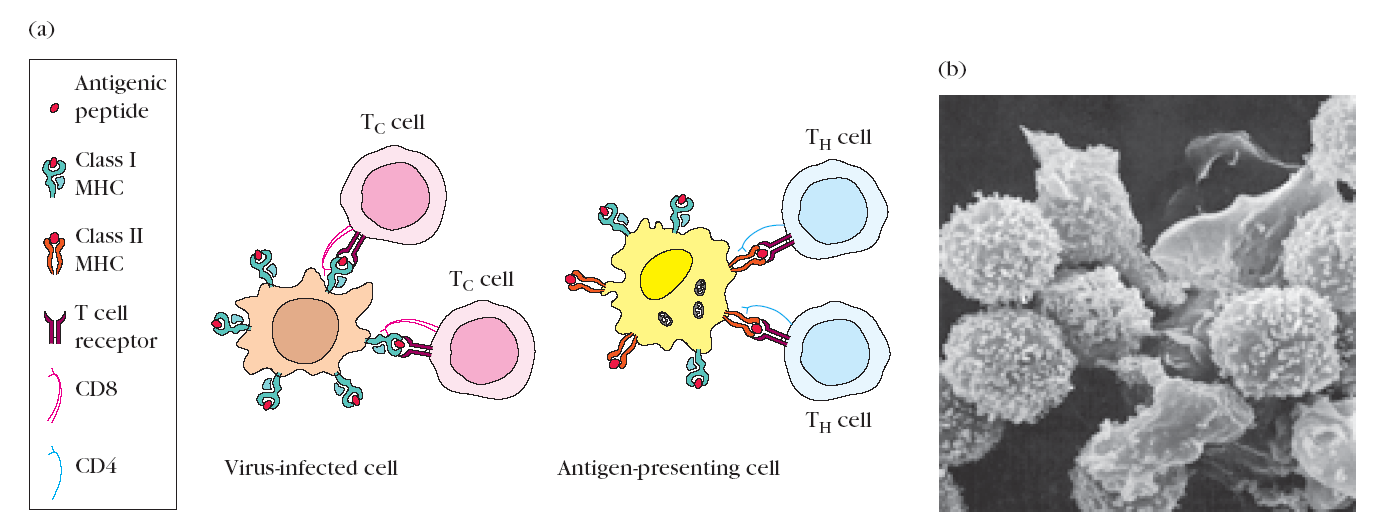
Figure 5. The role of MHC molecules in antigen recognition by T cells. (a) Class I MHC molecules are expressed on nearly all nucleated cells. Class II MHC molecules are expressed only on antigenpresenting cells. T cells that recognize only antigenic peptides displayed with a class II MHC molecule generally function as T helper (TH) cells. T cells that recognize only antigenic peptides displayed with a class I MHC molecule generally function as T cytotoxic (TC) cells. (b) This scanning electron micrograph reveals numerous T lymphocytes interacting with a single macrophage. The macrophage presents processed antigen combined with class II MHC molecules to the T cells. [Photograph from W. E. Paul (ed.), 1991, Immunology: Recognition and Response, W. H. Freeman and Company, New York; micrograph courtesy of M. H. Nielsen and O. Werdelin.]
6. Complex Antigens Are Degraded (Processed) and Displayed (Presented) with mhc Molecules on the Cell Surface
In order for a foreign protein antigen to be recognized by a T cell, it must be degraded into small antigenic peptides that form complexes with class I or class II MHC molecules. This conversion of proteins into MHC-associated peptide fragments is called antigen processing and presentation. Whether a particular antigen will be processed and presented together with class I MHC or class II MHC molecules appears to be determined by the route that the antigen takes to enter a cell (Figure 6).
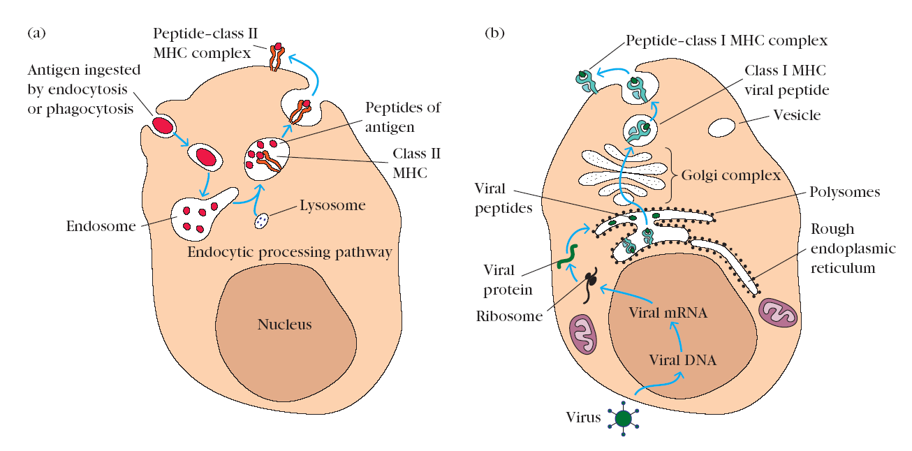
Figure 6. Processing and presentation of exogenous and endogenous antigens. (a) Exogenous antigen is ingested by endocytosis or phagocytosis and then enters the endocytic processing pathway. Here, within an acidic environment, the antigen is degraded into small peptides, which then are presented with class II MHC molecules on the membrane of the antigen-presenting cell. (b) Endogenous antigen, which is produced within the cell itself (e.g., in a virusinfected cell), is degraded within the cytoplasm into peptides, which move into the endoplasmic reticulum, where they bind to class I MHC molecules. The peptide–class I MHC complexes then move through the Golgi complex to the cell surface.
Exogenous antigen is produced outside of the host cell and enters the cell by endocytosis or phagocytosis. Antigenpresenting cells (macrophages, dendritic cells, and B cells) degrade ingested exogenous antigen into peptide fragments within the endocytic processing pathway. Experiments suggest that class II MHC molecules are expressed within the endocytic processing pathway and that peptides produced by degradation of antigen in this pathway bind to the cleft within the class II MHC molecules. The MHC molecules bearing the peptide are then exported to the cell surface. Since expression of class II MHC molecules is limited to antigen-presenting cells, presentation of exogenous peptide–class II MHC complexes is limited to these cells. T cells displaying CD4 recognize antigen combined with class II MHC molecules and thus are said to be class II MHC restricted. These cells generally function as T helper cells.
Endogenous antigen is produced within the host cell itself. Two common examples are viral proteins synthesized within virus-infected host cells and unique proteins synthesized by cancerous cells. Endogenous antigens are degraded into peptide fragments that bind to class I MHC molecules within the endoplasmic reticulum. The peptide–class I MHC complex is then transported to the cell membrane. Since all nucleated cells express class I MHC molecules, all cells producing endogenous antigen use this route to process the antigen. T cells displaying CD8 recognize antigen associated with class I MHC molecules and thus are said to be class I MHC restricted. These cytotoxic T cells attack and kill cells displaying the antigen–MHC class I complexes for which their receptors are specific.