

Учебники / Pocket Guide To The Ear
.pdfOtitis Externa |
39 |
|
|
In the full-blown florid stage, the patient presents with a swollen, draining, tender canal. Touching the tragus or pulling the auricle backward elicits severe pain. Swelling narrows the lumen of the canal, sometimes to a pinpoint. In addition, the infection may spread through the fissures in the anterior cartilage to the parotid gland and adjacent skin, causing parotid cellulitis and localized adenopathy.
Treatment of this condition has traditionally centered on topical therapy with eardrops, namely, combinations of neomycin, polymyxin, and hydrocortisone (Cortisporin, or its generic substitute). In recent years, additional tools have become available. Often, the canal is so swollen shut that drops will not penetrate. The new Pope ear wicks are easily inserted without too much trouble and are then soaked with the topical preparation. They soften and expand when moistened and stay in place so that the medication can work “around the clock.” The drops are applied several times a day, and the wick may be removed a few days later.
Quinolone eardrop preparations containing Floxin or Cipro have now become available, although the traditional Cortisporin is still effective. In addition, the Pseudomonas-killing quinolones may be administered orally in severe cases if the patient is old enough. Analgesics should not be forgotten—this infection, when severe, ranks with kidney stones and acute gout for pain intensity. The patient should keep the head elevated at home and expect two or three more days of hard times, even with good treatment.
Anecdotally, I have seen a number of patients in the past with this disease who had been treated for days with only oral amoxicillin or cephalosporins. It should be emphasized that Pseudomonas is the vastly predominating pathogen and that it will not respond to these antibiotics.
Acute Localized Otitis Externa
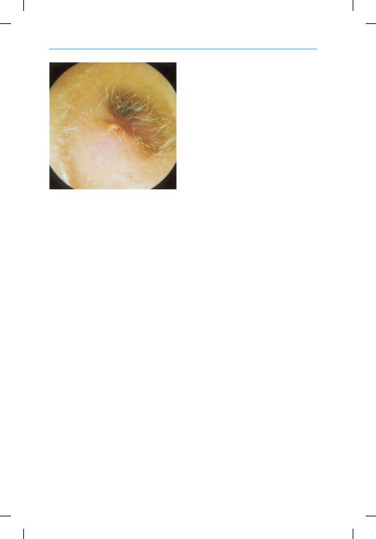
This disorder, a different disease from the diffuse type, also presents with a very painful ear. It is otherwise known as a furuncle of the canal. The infection is localized in an obstructed sebaceous gland or hair follicle out near the meatus. A tender red, raised pustule is readily seen occluding the meatus (Fig. 4.6). Staphylococcus aureus is the usual offender here, and appropriate oral antibiotics such as cephalosporins or amoxicillin/ clavulanate, as well as topical neomycin preparations, are indicated. When bulging and soft, incision and drainage at the most fluctuant point with a #11 blade will benefit.
Menner, A Pocket Guide to the Ear © 2003 Thieme
All rights reserved. Usage subject to terms and conditions of license.
40 4 Disorders of the External Ear
Fig. 4.6 Furuncle of the external meatus.
(Source: Sanna M, Russo A, DeDonato G. Color Atlas of Otoscopy. Stuttgart: Thieme; 1999)
Malignant Otitis Externa
Malignant otitis externa is also known as necrotizing otitis externa or skull base osteomyelitis in its full-blown form. Obviously, the second name is the gentlest one, but the other two imply its ominous characteristics. This type of infection is typically seen in elderly diabetics or immunocompromised patients. It can spread from the external canal to cause osteomyelitis in the temporal bone with potentially fatal complications. Characterisically, the patient presents with an external earache similar to other forms of external otitis. However, examination of the ear shows something different. The canal may be swollen and tender, but a small area of red granulation tissue is seen posteroinferiorly in the canal at the junction of cartilage with bone, one-third inward. This finding, plus the type of patient described, points to the diagnosis.
When suspected, ENT consultation for aggressive treatment should ensue. The organism is almost always Pseudomonas, but a culture should be done. Biopsy of the granulation can rule out neoplasm. Treatments include appropriate topical and systemic antibiotics and aggressive debridement. Medical management of the diabetes and/or immunodeficiency can also improve the prognosis. Conventional CT scans and bone scans can image the areas of involvement. Even with appropriate aggressive treatment, devastating bone infection and death can occur.
Menner, A Pocket Guide to the Ear © 2003 Thieme
All rights reserved. Usage subject to terms and conditions of license.

Otitis Externa |
41 |
|
|
 Summary of Acute External Infections
Acute diffuse otitis externa (“swimmer’s ear”) is much more common than the localized furuncle, and should be recognized by its diffuse swelling and tenderness. Some clinicians err in prescribing oral antibiotics that will not work on Pseudomonas, the vastly predominating organism. Topical antibiotic drops, with wick insertion if the canal is swollen shut, are the mainstay of treatment. Oral quinolones, if the patient is old enough, may be used in severe cases. A culture should be done if there is any doubt about what you are dealing with, and pain medication should not be forgotten. An obvious localized furuncle has staph as its cause. Treatment with topical drops and antistaphylococcal oral antibiotics is indicated.
The primary practitioner can diagnose and treat these infections, even  with the insertion of a wick if the canal is swollen. However, granulation
 tissue in the canal of an elderly or diabetic patient portends malignant
 otitis externa and warrants early ENT consultation.
Mycotic Otitis Externa
This disorder is also known as fungal otitis externa or chronic diffuse otitis externa. It differs from the previously mentioned infections in that it is not quite so painful, but more indolent, yet persistent. The usual complaints are of blockage, thick drainage, dull pain, and itching. These infections occur more often than most clinicians expect, and they are often treated inappropriately with antibiotic drops. The most notable finding on ear examination is the presence of “matter”—thick moist debris—in the canal (Fig. 4.7).
Fig. 4.7 Fungal otitis externa. (Source: Hughes GB, Pensak ML. Clinical Otology. New York: Thieme; 1977)
Menner, A Pocket Guide to the Ear © 2003 Thieme
All rights reserved. Usage subject to terms and conditions of license.

42 4 Disorders of the External Ear
Aspergillus niger is the most common infection, and its exudate appears as part black, part light-gray “wet blotter paper” in the deeper portion of the canal, even against the TM. Other species of Aspergillus appear tan or yellow-orange and also locate themselves deeply. Aspergillus is more apt to cause pain than itching. When the canal is cleared of the exudate, the underlying skin is red and raw. This organism is a mold that may be picked up from the environment, wherever molds may grow.
Candida albicans and other Candida species are also frequent pathogens. Their exudates tend to be flocculent and white or creamy in appearance, and itching is a notable complaint, in addition to blockage. It is often seen in antibiotic-treated or immunocompromised (including diabetic) individuals. Other fungi, such as actimomyces and phycomycetes, may also rarely occur.
One diagnostic point—the fungi mentioned have a mild musty odor, or none at all, whereas Pseudomonas has a characteristic sweet, musty smell, and Staph. aureus and Proteus are downright putrid. In any external infection, a culture for both bacteria and fungi should be done if there is doubt about the organism. Sometimes bacteria and fungi coexist, especially Pseudomonas with Aspergillus.
Treatment of these fungal infections keys on complete removal, so that no spores are present for regrowth. If there is no perforation of the TM, gentle irrigation and suction may be the best way to clear the canal. (Caution: irrigating an infected ear is not usually recommended, but with fungal infections it has not caused problems in my experience, as long as the canal is suctioned dry.) The ear can then be insufflated with a large amount of nystatin (Mycostatin) powder, which comes in containers that facilitate this. Clotrimazole (Lotrimin) drops are another recommended therapy. Antibiotics or steroids do not help and may even promote growth, especially with Candida. In fact, fungal otitis may be a complication of overtreatment with Cortisporin-type preparations. Oral antifungals, such as fluconazole (Diflucan) can be considered in refractory cases.
Menner, A Pocket Guide to the Ear © 2003 Thieme
All rights reserved. Usage subject to terms and conditions of license.

Other Chronic External Ear Disorders |
43 |
|
|
 Summary
Mycotic otitis externa, synonymous with chronic diffuse otitis externa, is characterized by milder pain than the acute infections. Patients complain of chronic moisture, blockage, thick drainage, itching, and mild discomfort. A characteristic finding is the presence of thick matter in the canal, without severe swelling or tenderness. Aspergillus and Candida are the usual culprits, but a culture for all organisms should be done if there is any doubt. Successful treatment hinges on complete removal of the debris, followed by the topicals mentioned in the text.
The primary practitioner can make the diagnosis, but usually referral to ENT is needed for thorough cleansing. Even then, there is a tendency for recurrence and persistence, and repeat cleanings and topical applications may be necessary. Oral systemic antifungals, such as Diflucan, might also be considered in refractory cases.
Other Chronic External Ear Disorders
Some patients suffer from another disorder, chronic stenosing otitis externa. These individuals have repeated infections; sometimes cultures are positive for bacteria or fungi, and sometimes there is no identifiable pathogen. The dermatoses may be involved. The external canal itches, drains repeatedly, and becomes chronically swollen, with progressively severe narrowing of the lumen. This problem responds temporarily to office cleansing and wick/eardrop insertions, but it is often relentless. Severe cases may eventually need surgery to widen the canal. Canalplasty with skin grafting, or even limited mastoidectomy, can be performed to open the canal and regain the hearing.
Other individuals have problems with ongoing or recurrent acute otitis externa without the complication of stenosis. These patients often create their own problems and should be cautioned regarding the cause and prevention of external otitis. Many individuals feel the need to dowse their ears daily with water in the shower, and then vigorously clean with applicators. The old adage about “nothing smaller in the ear than your elbow” is not bad advice. Gentle removal of cosmetically visible cerumen in the meatus is all that should be done. In the population at large, the vast majority of ears are self-cleaning—only 5% or so have problematic cerumen buildups.
Prophylaxis for external otitis in swimmers, however, is a valid consideration. Insertion of alcohol/acetic acid drops before or after swimming may prevent infection. In fact, an effective, nontoxic, “all-purpose” remedy for all forms of external otitis is the propylene glycol/acetic acid eardrop
(e.g., Vosol). The desired effect of drying and lowering pH can also be com-
Menner, A Pocket Guide to the Ear © 2003 Thieme
All rights reserved. Usage subject to terms and conditions of license.

44 4 Disorders of the External Ear
bined with steroids, if indicated, in certain preparations (e.g., Vosol HC). The steroids are effective for itching, but may aggravate fungal infections.
Cerumen Accumulations and Keratoses
Cerumen exists for several reasons. It protects the skin of the EAC from water penetration; its low pH discourages microbial growth; and it traps foreign material, carrying it outward by migration. It is produced in the outer third of the canal, where the gland/hair units are located.
In normal individuals, the skin of the entire canal migrates very slowly and steadily from the inside outward. Studies with ink dots have shown that epithelial migration actually starts near the center of the TM and proceeds all the way out, at a rate of about 2 mm a month! In the outer third, cerumen migrates together with the epithelium and eventually sloughs, carrying foreign material with it.
In individuals with repeated cerumen impactions (about 5% of the population), analysis of the cerumen shows high amounts of keratin. Apparently, migration of the skin and its sloughing pattern are abnormal—the epithelium and cerumen tend to roll up in a ball. This is readily appreciated when cleaning ears in the office—many times one will see desquamating segments of epithelium that still cling to the midcanal after most of the cerumen has been removed.
Irrigation, as described in Chapter 2, is the simplest way for most clinicians to clean problem ears. Wax softeners may help the process. Sometimes, suction or curetting is needed to remove troublesome buildups and/ or epithelial debris, and this is where specialized equipment and skills come into play.
Patients with recurrent accumulations often ask what they can do to clean their own ears and avoid periodic office visits (which may be needed as much as two or three times a year in some individuals). First, these patients should be advised not to use cotton swabs in their ears, as they will often aggravate a buildup by blindly packing it in. Wax softeners like Debrox or Ceruminex are often helpful for the impaction-prone individual, to be used in each ear once or twice a week. Self-irrigating kits are available at pharmacies as well. These aids may help some patients, but failure of removal or infection may be complications.
A rarer buildup in the ear is keratosis obturans. This is a mass of squamous epithelium accumulating in large whorls that are difficult to remove. It can erode through the skin of the bony canal and then erode bone itself, causing pain and draining infections. It is tenacious, and removal often requires the “headlight and two-hands” approach, using hooks, curettes, and alligator forceps. Thus, an ENT referral might be necessary. Individuals
Menner, A Pocket Guide to the Ear © 2003 Thieme
All rights reserved. Usage subject to terms and conditions of license.

Trauma and Foreign Bodies |
45 |
|
|
with this problem should be seen at frequent intervals, perhaps every six months, for cleaning. Chronic, untreated cases of this disorder may show up with huge excavations into the bone of the canal wall, usually inferiorly or posteriorly.
A related entity is cholesteatoma of the external canal. This entity differs in that the epithelial accumulation tends to be deeper, near the TM. It is seen more often in older individuals and is usually unilateral.
Discussing cerumen problems raises an important point. Occasionally a clinician will see a patient (usually a child) with a fever and an earache and will not be able to see the drum due to cerumen or other debris. If this cannot be removed at the time, it is reasonable to go ahead and treat empirically for an ear infection. However, if the problem recurs, and the blockage cannot be removed, ENT referral is indicated.
Trauma and Foreign Bodies
Abrasions and Lacerations of the External Canal
This type of trauma to the external canal is most often self-inflicted by an individual with a cotton applicator or a hairpin while attempting to remove wax. The patient may feel pain or notice blood welling up, and thus present to the office. The examiner sees an ear full of blood and the question arises, “Is there a perforation of the drum?” Suctioning out the blood is the only way to get a good look, but often it is too copious and tenaciously clotted, even for a specialist with good equipment. In these cases, it is best to leave things alone and treat the patient for a week with antibiotic drops to prevent infection and loosen the blood. Avoidance of water in the ear should be advised.
A follow-up visit will then give more information. Most of the time, only an abrasion or laceration of the canal is present—these bleed very easily, but usually heal with no complications. It is important to see the patient in follow-up until things are completely healed, however. The cotton-tip swab may have abraded a small carcinoma of the canal to cause the bleeding! If a perforation has occured, continued water avoidance and initial observation are indicated. Perforations are discussed more fully in Chapter 5.
Incidentally, this raises the question of whether neomycin-polymyxin eardrops should be used if a tympanic perforation is present. Much has been said in recent years about their potential ototoxicity. Be advised that most otolaryngologists have been using these drops routinely for years, even in middle ear surgery. Less than a handful of cases of ototoxicity (assuming entry of the drops into the inner ear via a hole in the oval or
Menner, A Pocket Guide to the Ear © 2003 Thieme
All rights reserved. Usage subject to terms and conditions of license.

46 4 Disorders of the External Ear
round window) have been reported in literature. However, now that other nontoxic antipseudomonal drops such as Cipro and Floxin are available, these can be used to play it safe, keeping in mind the much greater cost.
A bloody traumatized canal can be initially evaluated and treated with antibiotic drops by the primary physician. ENT follow-up within a few days would be appropriate.
Foreign Bodies
Objects lodged in the canal occur most often in young children, mentally handicapped adults, or temporarily mentally handicapped adults. If the foreign bodies are soft, such as the end of a cotton swab or furniture stuffing, removal with alligator forceps is usually not difficult. More often, however, they are solid round or oval objects, such as beads. These are more difficult. Do not attempt instrumental removal unless you feel you can “get around behind” the object to pull it out (Figs. 4.8a, b). Many times, a well-meaning attempt with a suction tip or forceps will lodge the object deeper in, beyond the bony hump of the anterior canal wall. A general anesthetic may then be necessary for removal, especially with an uncooperative young child. With cooperative patients, the best instrument for removal of solid objects, is the Day hook, a straight, thin, metal probe with a right-angle turn of about 2 mm on the end. The end is inserted just past the object and turned 908 to retrieve it. Also, irrigation, as with cerumen impactions, may be successful in some cases.
Special mention must be made of insects. Live ones in the ear can be terrifying to the patient because the fluttering and crawling is relatively loud, and skin of the bony canal is very sensitive to light touch. Mineral oil, wax softener, or dish detergent, all being mild and viscous, can be in-
Figs. 4.8a, b Removal of rigid foreign body from the external canal. An attempt to remove a foreign body with simple forceps (a) usually displaces the object deeper and may cause middle ear damage. Using a hook (b) is the effective way.
(Source: Becker W, Naumann HH, Pfaltz CR. Ear, Nose, and Throat Diseases. Stuttgart: Thieme; 1994)
Menner, A Pocket Guide to the Ear © 2003 Thieme
All rights reserved. Usage subject to terms and conditions of license.

Trauma and Foreign Bodies |
47 |
|
|
stilled in the canal to kill the insect quickly. Then, removal can be done with alligator forceps or possibly irrigation. A few days of antibiotic drops are then a good idea.
Primary and emergency physicians should exercise great discretion when removing foreign bodies. If there is any doubt about one’s ability to extract a foreign body, an ENT specialist should be consulted. The inert ones may wait a day or two. Live insects should be killed immediately, as described above.
Auricular Trauma
Sharp and blunt trauma to the external ear might result in contusions, hematomas, lacerations, or even disruption of the cartilaginous framework. Appropriate surgical intervention is certainly indicated, and antibiotic coverage for Pseudomonas and staph is recommended to prevent perichondritis. Hematoma and perichondritis have been discussed earlier in this chapter.
Temporal Bone Fracture
Also termed basal skull fracture, this injury occurs with severe head trauma and characteristically presents with blood oozing from the external meatus. Hearing loss, vertigo, and facial paralysis may be present. Patients with this type of trauma are usually severely injured in other ways as well, and are hospitalized by the neurosurgeon after presenting to the emergency room. Occasionally however, a stoic individual will not seek acute care. Sometimes, a cerebrospinal fluid (CSF) leak is present, with steady dripping of clear fluid from the ear.
Initial treatment is bed rest, with the head elevated, and observation. Prophylactic antibiotics should be given, even if there is no apparent CSF leak. CT imaging will identify the fracture. Two types—longitudinal and transverse fractures—may occur, each with a different pattern of damage. (A more detailed discussion can be found elsewhere.) Permanent hearing and vestibular damage can result. Facial paralysis, if immediate and not delayed, warrants surgical exploration with decompression and/or repair.
Frostbite of the Auricle
This type of damage from prolonged exposure to cold is likely to affect the ear first. The auricle is quite vulnerable due to its exposed location, superficial blood supply near the skin’s surface, and lack of sensitivity. At first, the involved skin is pale and numb. As warming occurs, the affected areas become hyperemic and painful and may even blister.
Menner, A Pocket Guide to the Ear © 2003 Thieme
All rights reserved. Usage subject to terms and conditions of license.

48 4 Disorders of the External Ear
Treatment should be with gradual warming in a cool room. Direct heat, massage, or snow application is not recommended, as it will simply aggravate tissue trauma. “Hands-off” observation is the rule, although prophylactic antipseudomonal antibiotics are indicated for severe injuries. Eventual necrosis and loss of tissue may occur, but even then, delineation by sloughing gives a better result than premature surgery, unless gross infection is present.
Regarding the last three categories of trauma, the physician involved depends on the severity of the injury. Obviously, temporal bone fractures and severe frostbite require ENT consultation in a timely fashion, whereas minor auricular trauma can be repaired by the capable family or ER physician, taking care to prescribe antibiotics to prevent perichondritis.
Tumors of the External Ear
Bony Tumors
The most frequently seen tumors of the EAC, at least in northern climates, are the bony ones—exostoses and osteomas. Exostoses are sessile rounded bony projections in the inner two-thirds, often seen on the floor of the canal anteriorly and posteriorly, although they may be based superiorly as well. The inferior ones tend to be more external, flatter, and broader, whereas the superior ones are often deeper, smaller, and more rounded (Fig. 4.9). Their epithelial covering is normally smooth and unremarkable in appearance. The cause is usually cold-water swimming over several years.
Fig. 4.9 Multiple exostoses of the external canal.
(Source: Sanna M, Russo A, DeDonato
G. Color Atlas of Otoscopy. Stuttgart: Thieme; 1999)
Menner, A Pocket Guide to the Ear © 2003 Thieme
All rights reserved. Usage subject to terms and conditions of license.