

Учебники / Pocket Guide To The Ear
.pdfNeuralgias Involving the Ear |
29 |
|
|
 Summary
The key point to make about TMJ syndrome is that it is the most common cause of multiple ear symptoms in patients with normal ears, and that it is often misdiagnosed as a eustachian tube or middle ear disorder. The patient usually presents with unilateral or bilateral fullness in or near the ear, and ache may be present. Thorough evaluation of the TM, its mobility, and hearing can rule out ear disease. A positive history of bruxism, coupled with findings of joint tenderness or crepitance, supports the diagnosis. Patient counseling and anti-inflammatory medicines, along with possible dental consultation and nighttime appliance fitting, are the mainstays of treatment. The primary practitioner can diagnose and initiate treatment for TMJ syndrome, with elective referral to an ENT or dental specialist for confirmation of the diagnosis and further options.
Neuralgias Involving the Ear
Webster defines “neuralgia” as a severe pain along the course of a nerve or in its distribution. Obviously, this is a concise and accurate description. In diagnosing neuralgia, there are few physical findings to support one’s impression. Clinical suspicion is aroused mostly by the patient’s history—the description of the type of pain and its location. Typically, neuralgia pains are severe and lancinating, lasting only a few seconds to a half-minute, and are variable in frequency. Often, there is tenderness to light touch. The duration of the disorder is also variable. Some neuralgias are short-lived, accompanying a viral illness, and others are chronic and disabling, lasting months to years. Notably, almost all are unilateral.
The cause of most neuralgias is uncertain, although viral or postviral neuropathy may play a role. It has recently come to light that sometimes direct nerve compression is involved. Trigeminal neuralgia, for example, has been cured by surgically alleviating a vessel’s compression of the nerve root near the brainstem. In addition, ablation procedures, by nerve section or toxic injection, have been done successfully for years. These surgical successes certainly support an anatomic basis for the disorder.
As discussed in Chapter 3, innervation of the external and middle ear arises from multiple roots, namely C2 and C3, and cranial nerves V, VII, IX, and X. Thus, several neuralgia syndromes include ear pain. Diagnosis of the following neuralgias is often by exclusion—one must rule out other reasons for the pain. However, the severity of the pain, sensitivity in the area of nerve distribution, and unilaterality are all contributing considerations.
Menner, A Pocket Guide to the Ear © 2003 Thieme
All rights reserved. Usage subject to terms and conditions of license.

30 4 Disorders of the External Ear
Trigeminal neuralgia, also known as tic douloureux, is the most wellknown. It involves one or more of the three divisions of cranial nerve V, usually the lower two. The lancinating pains are typically triggered by light touch, or even the wind! The pain distribution may include the ear, presumably via the auriculotemporal branch. Occipital neuralgia occurs in the distribution of this branch of C3 in the posterior scalp and mastoid regions. Often the patient even complains of “hair” tenderness in these areas.
Extremely rarely, glossopharyngeal neuralgia, arising from cranial nerve IX, is experienced as pain in the posterior oropharynx, tonsil, or base of tongue, with radiation to the ear (via Jacobson’s nerve). Also rare, sphenopalatine neuralgia arises from the ganglion of the same name in the fossa behind the maxillary sinus, with nerve relays from V and VII. It manifests itself as a unilateral pain in the maxillary, orbital, and temporal regions, with ear pain as well.
Treatment for all these disorders depends on the chronicity of the symptoms and the certainty of the diagnosis. Acutely, one should consider the usual pain medications, if all other causes are ruled out. More chronically, carbamazepine (Tegretol) is effective, although the side effects of drowsiness and rare severe reactions (bone marrow depression) warrant great caution. Monitoring the complete blood count, before and during use, is indicated. Oxcarbamazepine (Trileptal) is a newer compound without the above side effects, although neuralgia is not yet listed in its indications.
 Summary
Most importantly, neuralgias are severe pains, often lancinating and accompanied by tenderness to light touch. They may be acute or chronic,  and an exact cause is seldom known. To make the diagnosis, all other
 causes of the pain should be ruled out. We must emphasize here again
 that when unexplained ear pain is a symptom, malignant tumors elsewhere in the respiratory tract, especially in the pharynx and larynx, may
  cause referred ear pain. These should be ruled out by a full ENT exam-
 ination, if at all suspected. Traditionally, Tegretol is a popular treatment, to be prescribed with care. Trileptal is proving to be a safe and effective
  replacement. The primary practitioner can diagnose and treat these
 disorders, with referral to an ENT specialist or neurologist if there is
any doubt.
Menner, A Pocket Guide to the Ear © 2003 Thieme
All rights reserved. Usage subject to terms and conditions of license.

Hematoma of the Auricle |
31 |
|
|
Hematoma of the Auricle
This problem occurs most often in young athletes, particularly wrestlers or football players who practice without their headgear. However, any severe blunt trauma to the auricle can cause a hematoma at any age. The vascular anastamoses of the auricle make subperichondrial accumulation of blood, with recurrences and lack of reabsorption, a likelihood. Usually, the hematoma occurs on the superolateral surface, centered over the scapha and upper concha (Fig. 4.1). If left untreated, fibrosis and even calcification can develop in time, causing the classic “cauliflower ear” deformity.
Incision and drainage should be done aseptically to avoid the dreaded complication of perichondritis, which will be discussed next. Antipseudomonal antibiotics should be prescribed. Evacuation of the hematoma may be carried out by making an incision, or two parallel incisions, and inserting a rubber drain. Pressure dressings are applied and the drain removed several days later. A follow-up visit to rule out recurrence should also be made.
Another method of treatment is aseptic needle aspiration, using an 18gauge needle after numbing the skin with a tiny-needle lidocaine injection. After aspiration, a cotton wad soaked with collodion is form-fitted over the area and held in place until it dries. Over it, an additional small
Fig. 4.1 Hematoma of the auricle. (Source: Becker W, Naumann HH, Pfaltz CR. Ear, Nose, and Throat Diseases. Stuttgart: Thieme; 1994)
Menner, A Pocket Guide to the Ear © 2003 Thieme
All rights reserved. Usage subject to terms and conditions of license.

32 4 Disorders of the External Ear
pressure dressing is taped into place on the auricle. The patient is instructed to hold it there firmly for 20 minutes or so and to keep the dressing on for a few days. With either technique, there may be recurrences that need repeat drainage procedures, but success usually eventuates with careful treatment, observation, and avoidance of the activity that brought on the hematoma.
The primary practitioner or emergency physician, if comfortable, may perform the second of these two procedures on an initial encounter, but ENT follow-up is recommended as recurrences are likely.
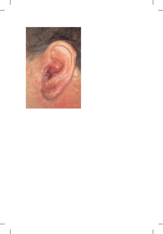
Perichondritis of the Auricle
This devastating infection occurs most often as a result of trauma, with penetration of the skin and a contaminated wound. Another possible cause is iatrogenic injury, i.e., ear surgery. The auricle becomes hot, red, swollen, and tender after the contaminating injury (Fig. 4.2).
When perichondritis is suspected, aggressive treatment is necessary. The organism is usually Pseudomonas aeruginosa, although Staphylococcus aureus may be involved. If there is evidence of fluctuance from pus, drainage should be carried out, of course with a culture. Appropriate antibiotics (antipseudomonal, if not cultured otherwise) should be administered,
Fig. 4.2 Acute perichondritis of the auricle.
(Source: Becker W, Naumann HH, Pfaltz CR. Ear, Nose, and Throat Diseases. Stuttgart: Thieme; 1994)
Menner, A Pocket Guide to the Ear © 2003 Thieme
All rights reserved. Usage subject to terms and conditions of license.
Congenital Disorders of the External Ear |
33 |
|
|
Fig. 4.3 Poor end result after treatment of perichondritis of the auricle.
perhaps even intravenously in the hospital, with close observation and warm moist dressings. The quinolones, as well as the aminoglycosides, such as tobramycin, are effective against Pseudomonas and staph. A severe infection, which begins and stays localized under the perichondrium, often results in necrosis of the cartilage and eventual fibrosis with a permanent severe auricular deformity (Fig. 4.3).
A condition simulating perichondritis can be encountered—the allergic insect bite reaction. Typically, the patient is seen during the summer months with rapid onset of a swollen, warm, itching, pink auricle. The culprits are often gnats, and an obvious insect bite is not necessarily seen. The absence of gross skin injury and predominance of itching, rather than pain, favors this diagnosis rather than perichondritis. In these cases, antihistamines and topical steroids are the indicated treatments. If there is any doubt about the diagnosis, precautionary antipseudomonal antibiotics should be used.
If the primary physician suspects full-blown perichondritis with its characterisitc red, swollen, tender auricle, hospitalization for IV antibiotics and early ENT consultation are indicated.
Congenital Disorders of the External Ear
Microtia
The scope of this text will not include embryology nor a detailed discussion of all of the types of external ear deformities that can occur. The most severe deformity is microtia, which is immediately noticeable at birth.
Menner, A Pocket Guide to the Ear © 2003 Thieme
All rights reserved. Usage subject to terms and conditions of license.

34 4 Disorders of the External Ear
Here, there is nothing more than a few deformed nubbins of tissue where the auricle should be. Less severe deformities of the auricle may also be encountered. These external defects are often accompanied by stenosis or atresia of the external canal, as well as by middle ear anomalies.Occasionally, they accompany syndromes with other craniofacial defects. With complete occlusion of the canal, the conductive hearing loss is very large, on the order of 50–60 dB HL.
Sometimes the other ear is normal and there is no urgency for treatment of the deformed ear, although a hearing aid should be placed at a young age for the sake of good bilateral hearing. A child with deformities of both ears should have a hearing aid placed as soon after birth as possible to gain speech input. The specialist should get involved early. CT scans will show the extent of the defect, and surgery can be done on the more correctable ear before the school years. The procedure is extremely specialized, with risk for complications and failure. Regarding the cosmetic deformity of the auricle, multiple procedures, or simply a prosthesis, may be indicated. Obviously, referral to a “super-specialist,” who deals with such cases often, is recommended.
Preauricular Cyst
A less severe congenital condition, the preauricular cyst and/or sinus tract, may occur just anterior to a normally formed external ear (Fig. 4.4). This
Fig. 4.4 Infected preauricular cyst. (Source: Hughes GB, Pensak ML. Clinical Otology. New York: Thieme; 1997)
Menner, A Pocket Guide to the Ear © 2003 Thieme
All rights reserved. Usage subject to terms and conditions of license.

Noncongenital Cysts and Keloids of the Auricle |
35 |
|
|
usually presents as a small fistula in the skin anterior to the helix at the upper tragus. A number of people have only a punctum here as an embryonic remnant with no clinical problems. However, the associated sinus tract can develop a dilated cyst with repeated infection and abscess formation. An acute abscess should be treated by drainage through an incision as close to the punctum as possible. In problem cases, surgical excision, with complete removal of the tract, is the answer. Care must be taken to avoid the upper branches of the facial nerve.
First Branchial Cleft Cyst
These cysts occur just beneath the lobule of the auricle and may be mistaken for parotid gland tumors. They often have a sinus tract and tiny fistula that empties into the floor of the EAC. Thus, when infected, they may present with swelling below the ear and drainage into the canal. Even though these are “benign cysts,” a skilled specialist should do the surgery. The tract is closely associated with the facial nerve, which may be injured as a surgical complication.
Noncongenital Cysts and Keloids of the Auricle
Epidermal Cysts
Two cystic conditions may be encountered externally, each in a different location. Epidermal inclusion cysts, traditionally known as sebaceous cysts, are usually located low in the postauricular crease. They represent backed-up oil glands and occur in individuals with oily skin and acne. These patients tend to have them behind both auricles and in other facial areas as well. Their usual content is cheesy sebum, but at times they may swell up and abscess, often infected with staph. If they are infected, the treatment is an antistaphylococcal antibiotic. Often the infection will resolve, but incision and drainage may be necessary. Troublesome recurrences can be surgically excised, when not infected, taking care to remove the entire cyst lining. Even then, they may reappear.
Epidermal cysts of the lobule can occur within the epithelialized tract of an ear-piercing site. They present with swelling, weeping, and repeated infection. If conservative treatment with antibiotics and cleansing fails, surgical excision, with removal of the entire epithelialized lining, may be necessary. The defects in the lateral and medial skin of the lobe are sutured and the patient is doomed to use conventional earrings here and find another site for body piercing.
Menner, A Pocket Guide to the Ear © 2003 Thieme
All rights reserved. Usage subject to terms and conditions of license.

36 4 Disorders of the External Ear
Keloids
At the same piercing site on the lobe, keloids (nodular hypertrophic scars) may develop in keloid-prone individuals, who are usually dark-skinned. These can be an extremely difficult problem, with growth to incredible size. They can recur even when excised completely. Triple therapy—exci- sion, postoperative steroid injections, and irradiation—may be necessary.
Skin Disorders of the External Meatus
The three major “dermatoses” of the external ear are seborrheic dermatitis, eczema, and psoriasis. They have some overlapping characteristics and often affect the same areas, namely, the external canal, its meatus, and the concha. Sometimes adjacent regions, such as the lobule and postauricular areas, are affected. They seldom extend deeper than the outer onethird of the canal. Dermatologists refer to all three as the papulosquamous disorders. Patients afflicted with these disorders complain of itching and weeping of the external canal. Occasionally, there is pain if inflammation or superinfection is present.
Seborrheic Dermatitis
This is the most prevalent of the three dermatoses affecting the external ear. It presents as a diffuse scaliness, with a pink or orange discoloration of the skin, in and around the external meatus. Often the involved skin is greasy, but other times it is simply dry and flaky. The lesion may be seen behind the auricle as well, along with other locations on the face, especially on the forehead between the eyes and lateral to the nose. It occurs more often in the older adult population. Dandruff (seborrheic dermatitis of the scalp) often accompanies it. Treatment centers around mild topical steroids, as well as selenium sulfide shampoo. When the latter is applied to the scalp for the dandruff, it may be applied to the ears as well. Sometimes yeast accompanies it, which responds to topical ketoconazole cream or shampoo.
Eczematoid Dermatitis
Eczema of the meatus and surrounding structures may affect any age group. It may be “familial atopic dermatitis,” “acquired-irritant,” or “allergic” eczema. The lesions usually start as small blisters, which itch intensely and are scratched away, leaving skin that becomes “lichenified” with exaggerated striations and scales. Weeping of sticky clear fluid is often present. Allergy to topical irritants, such as fabrics, soap, hair coloring, or hair spray, as well as other environmental allergens, may be causative. Neomycin allergy from eardrops can cause an acute eczema (Fig. 4.5), aggravating
Menner, A Pocket Guide to the Ear © 2003 Thieme
All rights reserved. Usage subject to terms and conditions of license.
Noncongenital Cysts and Keloids of the Auricle |
37 |
|
|
Fig. 4.5 Eczematoid neomycin reaction.
(Source: Hughes GB, Pensak ML. Clinical Otology. New York: Thieme; 1997)
a condition you may be already treating! In addition, food allergy has been implicated—eggs, milk, cheese, chocolate, and nuts head the list.
Unfortunately, many times there is no identifiable allergen. Bacterial superinfection, especially with staph, may complicate the picture. A culture will help if infection is suspected. Treatment also hinges on topical steroids, oral or topical antihistamines for the itching, antibiotics if indicated by culture, and of course, avoidance of the allergen, if known.
Psoriasis
This affliction of the external ear has some similarities to seborrheic dermatitis but tends to be more localized and patchy. It is also thicker, with a superficial white “micaceous” scale. Underneath, the skin is often deep red and tends to bleed if the scales are peeled off. Patients with psoriasis of the ears usually have the lesions elsewhere and have probably already been diagnosed. The other favorite body sites for the lesions are the extensor surfaces of the extremities. Warm water soaks, which soften the scales, are an easy remedy for accessible lesions, as well as the topical steroids or vitamin D ointment. Dermatologists are more qualified to treat this and should be involved.
Menner, A Pocket Guide to the Ear © 2003 Thieme
All rights reserved. Usage subject to terms and conditions of license.

38 4 Disorders of the External Ear
 Summary
The dermatoses of the external ear usually present with itching and weeping. Topical steroids are a mainstay of treatment in these scaly skin disorders. Dermatologists often advise the use of weak steroid preparations to avoid thinning or ulceration of the skin. A culture for both bacteria and fungus might grow out a pathogen, which may be treated. The primary practitioner can recognize and treat these dis-
 orders. ENT or dermatology referral can be made for persistent cases,
 especially if psoriasis is suspected.
Otitis Externa
We will now discuss several forms of external otitis. At this point, it is important to introduce two key clinical concepts. The first is that with an acute earache the presence of tenderness helps to distinguish between otitis externa and otitis media. If you are called on the phone at an inconvenient time by a mother whose child is screaming with an earache, ask her to pull backward on the auricle or press on the tragus. This will hurt if the problem is external, but will not if only the middle ear is infected. At least you can get a feel for the cause, and perhaps the treatment.
This leads to the second point. The bacterial organisms causing Otitis externa are usually Pseudomonas, Staphylococcus, Proteus, Enterobacter, or other Gram-negatives. On the other hand, the bacterial offenders for acute otitis media are usually Pneumococcus, Haemophilus influenza, and Moraxella catarrhalis, the ones that are often seen in acute sinusitis or other bacterial respiratory infections. This generalization is quite reliable and implies a different treatment for each entity.
Acute Diffuse Otitis Externa
This condition, a well-known painful infection of the canal, is otherwise known as swimmer’s ear. Water immersion is not always the cause, but the disease occurs most often in warm, humid conditions. Moisture in the ear, even from perspiration, plays a causative role. Local trauma to the canal is also a precipitating factor. Abrading a wet, macerated canal with a cotton swab to clean it or scratch an itch is often the initiating insult, implanting bacteria under the epithelium. The darkness of the canal, its warmth, high pH, and moisture all promote microbial growth. Pseudomonas causes this acute infection almost exclusively, although staph and others may rarely be involved. The bacteria go on to infiltrate, growing beneath the epithelium; then more itching ensues, progressing to soreness.
Menner, A Pocket Guide to the Ear © 2003 Thieme
All rights reserved. Usage subject to terms and conditions of license.