

Учебники / Pocket Guide To The Ear
.pdfBasic Equipment |
9 |
|
|
the terms “positive” and “negative” is often recommended, but these can be ambiguous. Documenting “air conduction is greater than bone” (or vice-versa) leaves the least room for doubt.
Tuning forks of 256, 1024, and 2048 Hz are also available and can be useful in many ways. For example, most conductive losses are severest in the lower frequencies. The 256-Hz fork may pick up abnormalities that the 512-Hz one would miss; thus it is the most sensitive one for conductive losses, especially when using the Rinne test. All the frequencies can be used to compare a patient’s right ear to his/her left ear, or to the examiner’s ear, for rough assessment. Individuals with advanced high-frequency loss from either noise damage or aging may show impairment at 1024 or 2048 Hz.
Other simple assessments can be made. The light rubbing of dry fingers is in the 3000 Hz range, as is the ticking of a watch. The sound intensity, when near the ear, is probably 25 dB HL or less. These are rudimentary but valuable screenings for high-frequency loss.
Cerumen Removal Devices
Irrigation of the external canal for cerumen removal can be done with any of several devices that can introduce a stream of water under moderate pressure. I mention the WaterPik as a good option, although it is not medically intended for this purpose. It is commercially available at a reasonable price, but it can be cumbersome and messy. In addition, it can generate huge pressure; be careful to use the lowest settings. Ear syringes are also available.
Here is some general advice for irrigation. Use body-temperature water to avoid caloric-induced vertigo. Clean tap water is fine; adding peroxide may help the loosening process but is probably not necessary. Have the patient hold a receptacle beneath the ear to catch the water. Pull the auricle posteriorly with the free hand to straighten the canal. To avoid trauma, aim the stream posterosuperiorly at the canal wall and not directly at the TM; this tends to force the water around and out, without applying severe pressure on the drum. Even with 20 years of experience, I perforated a patient’s thin, scarred drum several years ago using proper technique. A good rule would be to never irrigate an ear when the patient has a history of perforation or scarring. Also, one should stop irrigating if the patient complains of pain.
A frequently encountered problem is firm or gummy cerumen that will not budge. In this case, postpone further clearing efforts and use a wax softener, such as Debrox or Cerumenex. Some physicians use a stool softener, docusate sodium, and claim it works effectively. Softening could be done during the office visit by having the patient lie down for 15 minutes
Menner, A Pocket Guide to the Ear © 2003 Thieme
All rights reserved. Usage subject to terms and conditions of license.

10 2 Equipment and Ear Examination Methods
with an earful of softener, after which the ear would be re-irrigated. The patient could also be sent home with instructions to use the drops tid for a few days before the next visit. Usually, the ear can then be cleared. If not, a referral would be appropriate.
Advanced Equipment
The purchase of two more pieces of equipment may be considered to further expand a clinician’s office diagnostic abilities:
1.An Audioscope
2.A tympanometer
The Audioscope is a hand-held device used to screen hearing. It resembles an otoscope in size and shape. The one I have, patented by Welch Allyn, can test hearing at 500 Hz and 1, 2, and 4 KHz, at levels of 20, 25, or 40 dB HL. It requires a charger and periodic returns to the manufacturer for calibration, but it is a most valuable screening device. One can look through its otoscope speculum to examine the ear as well, though the orifice is quite small, and its main use is to make sure you are aiming the device at the TM rather than the canal wall. One could consider having a more elaborate and expensive audiometer in the office, but if an audiologist is available, a referral is probably more appropriate.
Fig. 2.1 Equipment for ear evaluation. Clockwise, from upper left: WaterPik, Audioscope, tuning forks, otoscope with pneumatic bulb, specula, wax softener.
Menner, A Pocket Guide to the Ear © 2003 Thieme
All rights reserved. Usage subject to terms and conditions of license.

Advanced Equipment |
11 |
|
|
A tympanometer is a device that assess compliance of the TM and middle ear pressure. It plots a curve that indicates middle ear fluid, pressure abnormalities denoting eustachian tube dysfunction, or abnormal mobility of the drum due to scarring or ossicular chain problems. It is an especially useful item for screening the pediatric population, the little ones who are difficult to examine or test audiometrically. A pressure probe is placed just like a speculum in the outer canal, and compliance readings are taken in a short period of time. These assess TM mobility in a much more sensitive way than the pneumatic otoscope. As a specialist, I enjoy matching my impressions from the office examination with the “tympanogram.” Drawbacks of the device include air-seal difficulties, errors in probe placement, and false readings due to cerumen impactions. Tympanometry curves will be discussed in Chapter 5.
Finally, ENT specialists have equipment for direct ear examination that expands their capablities beyond most nonspecialists. By using a light source on the head, they are able to use both hands to examine an ear—or nose and throat, for that matter. The illumination can be from an electrical headlight or a traditional head mirror with an indirect light source on a stand. With the use of bright, direct light from the vantage point of the eyeball, one is able to look “down the pipe” into narrow, deep spaces. Head-mounted lighting enables the specialist to use a magnifier with an ear speculum in the nondominant hand, and to work with instruments in the dominant hand. Half-circle “operating” lenses allow one to use various curettes, loops, or hooks to remove cerumen or foreign bodies under magnification. “Alligator” forceps and suctions may also be used to remove debris or secretions. Irrigation with pinpoint spray devices, or insufflation of medications, can also be done. These require more equipment, both small and large. The electrical pumps and their associated apparatus are usually contained in a console, which takes up space and is expensive. Hardware of this type is available through various ENT equipment companies.
Menner, A Pocket Guide to the Ear © 2003 Thieme
All rights reserved. Usage subject to terms and conditions of license.

12 2 Equipment and Ear Examination Methods
 Summary
Emphasis must be placed on the pneumatic otoscope for diagnosis. The
 mobility of the drum is a key factor in diagnosing TM retraction, middle
ear fluid, and TM perforation. Tuning forks, using the Weber and Rinne
 tests, can provide a good idea about conductive (outer or middle ear)
 hearing loss versus sensorineural (inner ear) loss. Tuning forks of varied
 frequencies (along with a ticking watch or rubbing of fingers) can provide additional information about hearing, especially when used to
  compare a patient’s hearing to the examiner’s. Cerumen removal is of-
 ten necessary and can be facilitated by commercial wax softeners or stool softeners. Debris of any type in the canal can impair diagnosis, thus necessitating more sophisticated equipment, or perhaps even ENT referral. The Audioscope and the tympanometer are two other
  more elaborate pieces of equipment that one might have in the office
 to screen hearing and middle ear pressure.
Menner, A Pocket Guide to the Ear © 2003 Thieme
All rights reserved. Usage subject to terms and conditions of license.

13
3 Useful Anatomy and Function
This chapter describes the structure of the ear and discusses its pertinent physiology and innervation. I have limited the coverage to what is most important to the practitioner, while making points of clinical interest along the way. The anatomy and physiology of the ear are quite complex for such a small structure of the body, but reading through these pages will provide a good background for the clinical chapters. In this and the following chapters on diagnosis and treatment, we progress anatomically from the outside inward.
The External Ear
The Auricle
The external ear consists of the outer portion, the auricle (or pinna), and the external auditory canal, leading to, but not including, the tympanic membrane (TM). Figure 3.1 shows an auricle with its landmarks labeled.
The auricle is composed of skin overlying an irregular framework of fibroelastic cartilage; it “gathers” sound and directs it into the canal. With the external canal, it actually acts as a resonator to slightly amplify lower frequency sounds. It also helps to localize sounds, especially in conjunction with the other ear. Innervation of the auricle occurs through the greater and lesser auricular nerves (from cervical roots C3 and C2) pos-
Fig. 3.1 The auricle and its landmarks:
1. Helix; 2. Scapha; 3. Antihelix; 4. Concha;
5. Crus of helix; 6. External auditory meatus;
7. Tragus; 8. Antitragus; 9. Lobule.
(Source: Becker W, Naumann HH, Pfaltz CR. Ear, Nose, and Throat Diseases. Stuttgart: Thieme; 1994)
Menner, A Pocket Guide to the Ear © 2003 Thieme
All rights reserved. Usage subject to terms and conditions of license.

14 3 Useful Anatomy and Function
teriorly, and the auriculotemporal branch of cranial nerve V anteriorly. The skin and vascular channels on the outer surface are somewhat adherent and “brittle,” making the auricle susceptible to traumatic hematoma formation that might lead to “cauliflower ear” if not treated. The innervation is not particularly sensitive, especially in the lobule. To prove this, pinch yourself anywhere on the auricle; then compare the pain with the same insult to other body sites. This insensitivity facilitates ear piercing, but unfortunately masks frostbite pain.
The External Auditory Canal
The external auditory canal (EAC) consists of a cartilaginous outer onethird and a bony inner two-thirds. Figure 3.2 shows its relationship with deeper structures. The entire length of the canal is a little over 3 cm from the external meatus to the TM. From the outside in, the outer third is directed slightly posteriorly, whereas the inner two-thirds is directed anteroinferiorly and has an anterior bony hump that occludes a view of the very front portion of the TM. Because of this curvature, the canal can best be straightened, as for eardrop insertion, by pulling the auricle backward. The skin of the external canal changes greatly from the outside in. In the outer cartilaginous segment, it is thick and contains hairs and numer-
Fig. 3.2 The external auditory canal and its relationships:
l. Cartilaginous part; 2. Parotid gland; 3. Bony part; 4. Lateral wall of epitympanum (attic); 5. Mastoid antrum; 6. Attic (epitympanum); 7. Temporomandibular joint fossa; 8. Facial, vestibular, and auditory nerves (VII and VIII); 9. Eustachian tube; 10. Bone of tympanic ring; 11. Fissure of Santorini in cartilage of external canal.
(Source: Becker W, Naumann HH, Pfaltz CR. Ear, Nose, and Throat Diseases. Stuttgart: Thieme; 1994)
Menner, A Pocket Guide to the Ear © 2003 Thieme
All rights reserved. Usage subject to terms and conditions of license.

The Middle Ear |
15 |
|
|
ous glands, both sebaceous and cerumen-producing. These three elements exist together as “apopilosebaceous” units. The skin lining the inner bony two-thirds is very thin, with little hair and no glands, but it is well-inner- vated and very sensitive to touch, in contrast to the skin of the auricle. The bony portion of the external canal is also known as the tympanic ring.
The “fissures of Santorini” (discontinuities in the cartilage of the outer canal) enable infection or neoplasm to easily spread down and forward into the adjacent parotid gland. Hence, a patient with a severe external otitis can develop cellulitis and parotitis adjacent to the ear. The outer third of the canal can be very narrow in width in some patients, limiting good access to the inner two-thirds and the TM. However, these individuals usually have normal-caliber bony canals further in. In general, the external canal is about 9 mm in height and 7 mm in width (due to the anterior bony hump).
The EAC is innervated by branches from four nerves: cranial nerves V (anteriorly), VII, IX, and X. The last three, innervating most of the canal, send their branches into the brainstem via the nervus intermedius and Arnold’s nerve. The relays of Arnold’s nerve near the nucleus ambiguus explain why stimulation of the canal during cleaning will often produce a cough reflex.
It is significant that other areas innervated by these four cranial nerves can transmit referred pain to the ear. Examples include post-tonsillectomy otalgia (ear pain) via cranial nerve IX or, more ominously, otalgia from malignancies in the tonsil, hypopharynx, or supraglottic larynx via cranial nerves IX and X. Several years ago I was referred a patient whose only complaint was a left earache, not even during swallowing. On laryngoscopy, he had a massive squamous cell carcinoma of the left hypopharynx! Finally, be advised that otalgia may even be referred from organs in the chest— disease of the heart, lungs, great vessels, and esophagus may all cause ear pain via the vagus nerve (X).
The Middle Ear
The Tympanic Membrane and its Landmarks
The middle ear begins with the TM. This is the most important structure for the clinician diagnostically, because its appearance, and what can be seen through it, are by and large the only anatomic clues to what goes on inside (Figs. 3.3 and 3.4). The most visible landmark beneath the TM is the manubrium, or handle, of the malleus, the lower portion of this first ossicle. The upper portion of the manubrium has a visible prominence, seen near the top of the TM, called the short process. The bottom end of the handle, at the
Menner, A Pocket Guide to the Ear © 2003 Thieme
All rights reserved. Usage subject to terms and conditions of license.

16 3 Useful Anatomy and Function
Fig. 3.3 Normal tympanic membrane of right ear:
1. Pars flaccida; 2. Short process of the malleus; 3. Handle (manubrium) of the malleus; 4. Umbo; 5. Supratubal recess; 6. Eustachian tube orifice (just to the right of the light reflex); 7. Hypotympanic air cells; 8. Stapedius tendon; c. Chorda tympani; I. Incus; P. Promontory; o. Oval window; R. Round window; T. Tensor tympani; A. Annulus. (Source: Sanna M, Russo A, DeDonato G. Color Atlas of Otoscopy. Stuttgart: Thieme; 1999)
center of the TM, ends in the umbo, the “apex” of the drum (the TM is actually somewhat conical in shape, from the periphery toward the center). Thus, the umbo, the deepest central depression, has a circular appearance.
The examining light usually reflects back most brightly from the TM in a triangular shape, located anteroinferiorly to the umbo, the light reflex. When a TM is dulled by acute or chronic infection, or thickening for other reasons, the light reflex may be decreased or absent, but this is not an infallible diagnostic criterion. A light reflex can still be seen in many abnormal middle ears, especially those with clear fluid effusions.
Most of the TM (the pars tensa) has three layers: outer squamous epithelium, middle fibrous tissue, and inner cuboidal epithelium. Above the short process of the malleus is a variable and irregular small portion of the drum called the pars flaccida, which contains no fibrous middle layer, only an outer squamous and inner mucosal layer. The pars flaccida is clinically significant as a site for possible cholesteatoma formation. The same applies for the marginal posterosuperior portion of the pars tensa, which may also be
Menner, A Pocket Guide to the Ear © 2003 Thieme
All rights reserved. Usage subject to terms and conditions of license.

The Middle Ear |
17 |
|
|
Fig. 3.4 Structures of the right ear after removal of the tympanic membrane: f. Facial nerve bulge; j. incudostapedial joint. See also key to Fig. 3.3.
(Source: Sanna M, Russo A, DeDonato G. Color Atlas of Otoscopy. Stuttgart: Thieme; 1999)
deficient in the middle fibrous layer in some disease states. Cholesteatoma, a squamous ingrowth, is discussed in more detail later.
Posterosuperiorly, the long process of the incus is frequently visible, deep to the drum, as it inserts into the stapes head. Going backward, the stapedius tendon may be seen as well. The vertical long process of the incus and the horizontal stapedius tendon can often be seen as a faint white backward-tilted “V.” Overlying and above them, just adjacent to the margin of the drum, the white band of the chorda tympani nerve may also be seen through the drum. This arises from the facial nerve (cranial nerve VII) and courses forward between the malleus and incus to eventually innervate the anterior tongue, receiving taste sensation. Not well seen through the TM, the stapes, the smallest bone in the body, transmits sound from the incus into the oval window via its footplate. It is shaped like a stirrup and is directed medially toward the inner ear. The round window niche and the bony bulge of the promontory are structures of the medial wall of the middle ear. The visibility of all these landmarks varies with individual anatomy and scarring of the drum.
The periphery of the pars tensa, bordering the innermost external canal, contains a dense white fibrous ring called the annulus. Variably present is
Menner, A Pocket Guide to the Ear © 2003 Thieme
All rights reserved. Usage subject to terms and conditions of license.
18 3 Useful Anatomy and Function
the vascular strip of vessels, not visible in Figure 3.3, but very prominent in Figure 5.5. This is seen on the upper drum just posterior to the manubrium, giving off vessels that course down the TM just behind the manubrium, as well as posteriorly, running along the peripheral annulus. These vessels may become bright red and inflamed in the early stages of acute otitis media. However, they may also be somewhat prominent in the normal individual.
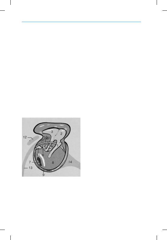
The Ossicles and their Function
Here the ossicles and contents of the middle ear will be discussed in more detail. Figure 3.5 depicts the position of middle ear and related structures. The TM, the bone of the tympanic ring, and outer (cortical) mastoid bone have been removed in this depiction.
The ossicles—the malleus, incus, and stapes—transmit sound vibrations from the large tympanic membrane into the small oval window, at a huge mechanical advantage. This system is really an “impedance-matching” one that enables sound waves in the air, at relatively low energy, to transfer sound waves into the fluid-filled medium of the inner ear, which offers higher resistance to sound flow. A fish, for example, has no need for a mid-
Fig. 3.5 Middle ear and related structures:
1. Manubrium of malleus, near the short process above; 2. Head of malleus; 3. Body of incus; 4. Long process of incus; 5. Stapes footplate; 6. Stapedius tendon; 7. Round window; 8. Promontory; 9. Fibrous annulus of the TM; 10. Chorda tympani nerve; 11. Pyramidal eminence leading to stapedius muscle and tendon; 12. Location of lateral semicircular canal bulging within the mastoid antrum (this cavity is covered over here); 13. Vertical portion of facial nerve; 14. Eustachian tube.
(Source: Becker W, Naumann HH, Pfaltz CR. Ear, Nose, and Throat Diseases. Stuttgart: Thieme; 1994)
Menner, A Pocket Guide to the Ear © 2003 Thieme
All rights reserved. Usage subject to terms and conditions of license.