Учебники / LECTURE NOTES ON Diseases of the Ear, Nose and Throat Bull 2002
.pdf
82 |
Chapter 21: The Nasal Septum |
|
|
DEVIATION OF NASAL SEPTUM
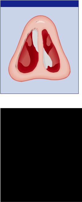
Fig. 21.1 ‘S’-shaped deviation of the nasal septum with hypertrophy of the right middle turbinate.
Fig. 21.2 The dorsal line of the nasal septum has been marked and is displaced to the left, causing external nasal deformity in addition to nasal obstruction.

The Nasal Septum 83
SUBMUCOUS RESECTION OF THE SEPTUM
(a) |
(b) |
|
(c) |
|
|
|
Fig. 21.3 Submucous resection of the septum. (a) Incision through the mucoperichondrium. (b) Elevation of muco-perichondrial flaps on either side of the septal skeleton. (c)The displaced cartilage and bone has been resected, allowing the septum to resume a midline position.
Where more severe symptoms are present, correction of the septal deformity is justified (though never essential).
Submucous resection (SMR)
SMR (Fig. 21.3) is the operation of choice for mid-septal deformity when the caudal septum is in a normal position. It is to be avoided in children, because interference with nasal growth will occur, leading, in turn, to collapse of the nasal dorsum.
Under local or general anaesthetic, an incision is made 1 cm back from the front edge of the cartilage through the muco-perichondrium, which is elevated from the cartilage. The incision is then deepened through the cartilage and the muco-perichondrium on the other side is elevated.
Deflected cartilage and bone are removed with punch forceps and the two mucosal flaps are allowed to fall back into the midline.
The nose is packed gently for 24 h to maintain apposition of the flaps and the patient may go home after 2 days.
Septoplasty
Septoplasty is the operation of choice (i) in children, (ii) when combined with rhinoplasty, and (iii) when there is dislocation of the caudal end of the

84Chapter 21: The Nasal Septum
septal cartilage. The essential features of septoplasty are a minimum of cartilage removal and careful repositioning of the septal skeleton in the midline after straightening or removing spurs and convexities.
It may be performed in conjunction with midor posterior-septal resection. It avoids the drooping tip and supra-tip depression seen sometimes after SMR and causes less interference with facial growth in children.
Complications of septal surgery
1Post-operative haemorrhage, which may be severe.
2Septal haematoma, which may require drainage.
3Septal perforation — see below.
4External deformity — owing to excessive removal of septal cartilage, allowing the nasal dorsum to collapse from lack of support. It can be very difficult to correct.
5Anosmia — fortunately rare, but untreatable when it occurs.
SEPTAL PERFORATION
AETIOLOGY
Perforation of the nasal septum is most common in its anterior cartilaginous part and may result from the following conditions:
1postoperative (particularly SMR);
2nose-picking (ulceration occurs first, perforation later);
3trauma;
4Wegener’s granuloma;
5inhalation of fumes of chrome salts;
6cocaine addiction;
7rodent ulcer (basal cell carcinoma);
8lupus;
9syphilis (the gumma affects the entire septum and nasal bones, with resulting deformity).
SYMPTOMS
Symptoms consist of epistaxis and crusting, which may cause considerable obstruction. Occasionally, whistling on inspiration or expiration is present. Frequently, the subject is symptom-free.
SIGNS
A perforation is readily seen and often has unhealthy edges covered with large crusts.

The Nasal Septum 85
INVESTIGATION
In any case where the cause is not clear, the following should be carried out:
1full blood count and ESR to excludeWegener’s granuloma;
2urinalysis, especially for haematuria;
3chest X-ray;
4serology for syphilis;
5if doubt remains, a biopsy from the edge of the perforation is taken.
TREATMENT
Septal perforations are almost impossible to repair. If whistling is a problem, enlargement of the perforation relieves the patient’s embarrassment.
Nasal douching with saline or bicarbonate solution reduces crusting around the edge of the defect, and antiseptic cream will control infection.
If crusting and bleeding remain a problem, the perforation can be closed using a silastic double-flanged button.

CHAPTER 22
Miscellaneous
Nasal Infections
Acute coryza
The common cold is the result of viral infection but secondary bacterial infection may supervene. Its course is self-limiting and no treatment is required other than an antipyretic, such as aspirin. The prolonged use of vasoconstrictor nose drops should be discouraged, owing to their harmful effect on nasal mucosa (rhinitis medicamentosa).
Nasal vestibulitis
Both children and adults may be carriers of pyogenic staphylococci, which can produce infection of the skin of the nasal vestibule. The site becomes sore and fissured and crusting will occur. Treatment, which needs to be prolonged, consists of topical antibiotic/antiseptic ointment and systemic flucloxacillin. Always take a swab for culture and sensitivity.
Furunculosis
Abscess in a hair follicle is rare but must be treated seriously as it can lead to cavernous sinus thrombosis. The tip of the nose becomes red, tense and painful. Systemic antibiotics should be given without delay, preferably by injection. Drainage may be necessary but should be deferred until the patient has had adequate antibiotic treatment for 24 h. In recurrent cases, diabetes must be excluded.
Chronic purulent rhinitis
Chronic purulent nasal discharge may occur, especially in children.The discharge is thick, mucoid and incessant and often resistant to treatment. In such cases, a nasal swab may show the presence of Haemophilus influenzae, which should be treated with a prolonged course of antibiotics (amoxycillin, cotrimoxazole).
It is necessary to exclude immunological deficiency, cystic fibrosis and
86

Miscellaneous Nasal Infections 87
ciliary abnormality in such cases of chronic rhinitis, as well as more obvious causes, such as enlarged adenoids, foreign body or allergic rhinitis.
Atrophic rhinitis (ozaena)
Fortunately now uncommon in Western society, this disease is still seen occasionally. The nasal mucosa undergoes squamous metaplasia followed by atrophy, and the nose becomes filled with evil-smelling crusts, the stench of which is detectable even at a considerable distance. Such a patient will be ostracized and children will be abused by their peers.
The aetiology of atrophic rhinitis is unknown. Various forms of treatment have been tried. In the early stages, meticulous attention to sinusitis and nasal hygiene may be helpful. In the more established case, the use of 50% glucose in glycerine as nasal drops seems to reduce the smell and crusting.
Various surgical measures have been devised, the most reliable of which is closure of the nostrils, using a circumferential flap of vestibular skin. After a prolonged period of closure, recovery of the nasal mucosa may occur and the nose can be reopened (Young’s operation).

CHAPTER 23
Acute and
Chronic Sinusitis
MAXILLARY SINUSITIS
Anatomy and physiology
The maxillary antrum is pyramidal and has a capacity in the adult of approximately 15 mL. Above it lies the orbit. Behind it is the pterygo-palatine fossa containing the maxillary artery. Inferiorly hard palate forms the floor and lies close to the roots of the second premolar and the first two molar teeth. Medially the antrum is separated from the nose by the lateral nasal wall made up of the middle and inferior turbinate bones, each with a corresponding recess or meatus below it (Fig. 23.1).
The ethmoidal sinuses form a honey-comb of air cells between the lamina papyracea of the orbit and the upper part of the nose. An upward extension forms the fronto-nasal duct draining the frontal sinus.
The openings of the sinuses under the middle turbinate form the ostiomeatal complex and it is now recognized that abnormality of this area leads to failure of sinus drainage and thence to sinusitis.
Abnormalities may be structural, as with a large aerated cell blocking the ostial openings, or may be functional such as oedema, allergy or polyp formation.The key to treatment of sinusitis lies in recognition of the abnormality and its correction by surgery or medication.
ACUTE INFECTION
AETIOLOGY
Most cases of acute sinusitis are secondary to:
1common cold;
2influenza;
3measles, whooping cough, etc.
In about 10% of cases the infection is dental in origin, as in:
1apical abscess;
2dental extraction.
88

Sinusitis 89
RELATIONSHIPS OF MAXILLARY ANTRUM
Infra-orbital nerve
Superior dental nerve
Cribriform plate
Ethmoid sinus
Nasal septum
Middle turbinate
Inferior turbinate
Maxillary antrum
Fig. 23.1 The anatomical relationships of the maxillary antrum.
Occasionally, infection follows the entry of infected material, as in:
1diving — water is forced through the ostium, into the sinus;
2fractures;
3gunshot wounds.
SYMPTOMS
1The patient usually has an upper respiratory tract infection, or gives a history of dental infection or recent extraction.
2Pain over the maxillary antrum, often referred to the supra-orbital

90 |
Chapter 23: Acute and Chronic Sinusitis |
|
|
Fig. 23.2 Coronal CT scan showing left-sided ethmoidal and
maxillary sinusitis.
region.The pain is usually throbbing and is aggravated by bending, coughing or walking.
3 Nasal obstruction — may be unilateral if unilateral sinusitis is present.
PATHOLOGY
The causative organisms are usually streptococcus pneumoniae, Haemophilus influenzae or Staphylococcus pyogenes. In dental infections, anaerobes may be present.
The mucous membrane of the sinuses becomes inflamed and oedematous and pus forms. If the ostia are obstructed by oedema, the antrum becomes filled with pus under pressure — empyema of the antrum.
SIGNS
1Pyrexia is usually present.
2Tenderness over the antrum and on percussion of the upper teeth.
3Mucopus in the nose or in the nasopharynx.
4There may be dental caries or an oro-antral fistula.
5X-ray shows opacity or a fluid level in the antrum (Fig. 23.2).
Three important rules
1Swelling of the cheek is very rare in maxillary sinusitis.
2Swelling of the cheek is most commonly of dental origin.
3Swelling of the cheek as a result of antral disease usually indicates carcinoma of the maxillary antrum.

Sinusitis 91
TREATMENT
1The patient should be off work and should rest.
2An appropriate antibiotic should be started after taking a nasal swab. Amoxycillin (to take account of Haemophilus) is a good first-time treatment.
3Vasoconstrictor nose drops, such as 1% ephedrine or 0.05% oxymetazoline, will aid drainage of the sinus.
4Analgesics.
In most cases, resolution of acute maxillary sinusitis will occur, but on occasion antral wash-out will be necessary to drain pus.
Chronic sinusitis
Most cases of acute sinusitis resolve but some progress to chronicity. This is particularly likely to happen if there is an abnormality of the anatomy, allergy, polyps or immune deficit.
SYMPTOMS
1Patients with chronic maxillary sinusitis usually have very few symptoms.
2There is usually nasal obstruction and anosmia.
3There is usually nasal or postnasal discharge of mucopus.
4Cacosmia may occur in infections of dental origin.
SIGNS
1Mucopus in the middle meatus under the middle turbinate.
2Nasal mucosa congested.
3Imaging shows fluid level or opacity, or mucosal thickening within the sinus.
TREATMENT
Medical
A further course of treatment with antibiotics, vasoconstrictor nose drops and steam inhalations is worthwhile, as it may produce resolution.
Functional endoscopic surgery
Developments in endoscopic instruments allow inspection of the sinus ostia and interior of the antrum. Ostial enlargement and removal of polyps and cysts can be performed. The ostio-meatal complex under the middle turbinate is opened up and allows more physiological drainage of the antrum than inferior meatal antrostomy.
