

Учебники / Middle Ear Surgery
.pdf56 12 Materials for Ossicular Chain Reconstruction
late the frequency average and postoperative air-bone gap. Some authors have only reported extrusion but not exposure of the prosthesis. Furthermore, the case mix amongst these reports has been different. It is therefore difficult to draw any conclusions from the literature as to which ossiculoplasty material gives the best results.
The present chapter is a literature review of the various ossiculoplasty materials. In order to make the comparison between different reports meaningful, the author considered both extrusion and exposure of the prosthesis as “prosthesis related complications”. A postoperative air-bone gap of 20 dB or less was used as the criterion for “success following ossiculoplasty” since this has been described by the majority of authors. Particular attention was given to the size of the population, the length of the follow-up period and the average frequency for reporting of hearing results.
Of the many ossiculoplasty materials that have been described, not many have stood the test of time. In Fig. 12.1, a time line with the specific year each material was first reported in clinical use is shown [3 – 17]. Some materials were more popular than others, at least for a period. Some are still being used. The specific materials highlighted on the time line are given a more detailed review.
In 1957, Hall and Rytzner first described the technique of repositioning or autotransplanting the incus remnant in ossicular reconstruction [4]. The patient’s incus is not always available for repositioning, and Farrior first advocated the use of cortical bone as an ossicular substitute [6]. In 1966, House and associates suggested the use of homograft incus in ossiculoplasty [8]. Since all these bone grafts are basically “dead” bone, their long-term fate has been the subject of many investigations. These investigations were based on the histopathology of a small number of implants retrieved at revision surgery, therefore biasing the histological findings [18 – 22]. On the whole, these reports indicated that these “dead” bone grafts underwent new bone formation and remodelling by a slow process of “creeping substitution”. They maintained their morphologic structure even when the bone was “dead”. However, resorption by rarefying osteitis occurred in an environment of chronic suppurative otitis media, similar to their living counterpart.
It is generally agreed that ossicular bone grafts are less likely to be extruded from the ear than alloplastic materials; however, ankylosis can occur between the bone graft and the adjacent bony structures, such as the facial canal. The functional results reported by several authors are listed in Table 12.1. These are mainly reports from the 1970s and 1980s, when the three-frequency average of 0.5, 1 and 2 kHz was commonly used in reporting results. It is therefore difficult to compare these results with more recent reports concerning alloplastic materials (Tables 12.2 – 12.6). The “success” rate of bone PORP (partial ossicular reconstruction prosthesis) was between 50 % and 90 % and that of bone TORP (total ossicular reconstruction prosthesis) was between 35 % and 81 % (defined by a postoperative air-bone gap of 20 dB or better).

12 Materials for Ossicular Chain Reconstruction |
57 |
|
|
Table 12.1. Comparison of the functional results of ossicular bone grafts between different clinical reports
ABG air-bone gap,
4 freq. 4-frequency average of 0.5, 1, 2 and 3 kHz,
3 freq. 3-frequency average of 0.5, 1 and 2 kHz
Author (year) |
No. |
Follow-up |
Materials |
Postop. ABG |
Postop. ABG |
|
cases |
period |
|
20 dB |
> 20 dB |
|
|
|
|
PORP |
TORP |
|
|
|
|
|
|
Wehr (1978) |
262 |
> 1 year |
Homologous |
90 % (freq. |
76 % (freq. |
[23] |
|
|
ossicle |
not specified) |
not specified) |
Austin (1972) |
207 |
> 1 year |
Autologous and |
83 % (freq. |
71 % (freq. |
[24] |
|
|
homologous |
not specified) |
not specified) |
|
|
|
ossicle |
|
|
Vartiainen |
246 |
2 years |
Autologous |
50 % (3 freq.) |
35 % (3 freq.) |
(1985) [25] |
|
|
ossicle and |
|
|
|
|
|
cortical bone |
|
|
Glasscock |
69 |
> 1 year |
Autologous and |
65 % (3 freq.) |
|
(1975) [26] |
|
|
homologous |
|
|
|
|
|
ossicle |
|
|
Penington |
216 |
> 1 year |
Autologous and |
Combined |
|
(1978) [27] |
|
|
homologous |
PORP and |
|
|
|
|
ossicle and |
TORP = 85 % |
|
|
|
|
cortical bone |
(3 freq.) |
|
Gersdorff |
103 |
> 1 year |
Homologous |
78 % (freq. |
56 % (freq. |
(1989) [28] |
|
|
cortical bone |
not specified) |
not specified) |
Vercruyysse |
60 |
1 year |
Homologous |
|
81 % (3 freq.) |
(2002) [29] |
|
|
ossicle |
|
|
Siddiq (2004) |
24 |
> 4 years |
Autologous |
71 % (4 freq.) |
|
[30] |
|
|
ossicle |
|
|
|
|
|
|
|
|
The introduction of alloplastic materials in ossiculoplasty took place in the 1950s. Early implant materials include vinyl acrylic [3], polyethylene [5], PTFE (polytetrafluoroethylene) [7] and metal [9]. The early results of these materials were so disappointing that by the time of the Fourth Shambaugh-Shea Workshop on Middle-Ear Surgery in 1971, there was general agreement that these solid plastic and metallic implants had no place in the surgical treatment of chronic otitis media [31].
Plastipore was the first alloplastic material that was commercialized and used worldwide [11]. It is a semi-soft white sponge of high-density polyethylene. Although the short-term results were encouraging, there was an unacceptable long-term extrusion rate. Smyth reported a 12 % extrusion rate at 5 years and Portmann reported a 30 % extrusion rate at 2 years when the Plastipore prosthesis was placed in direct contact with the tympanic membrane [32, 33]. Kerr examined 52 Plastipore prostheses removed at revision surgery. He noticed that multinucleated foreign body giant cells were present in large numbers in both types together with histological evidence of breakdown of the prostheses [34].
Many authors tried to reduce the extrusion of Plastipore by placing a cartilage disk over the prosthesis. This modification of technique has reduced the

58 12 Materials for Ossicular Chain Reconstruction
extrusion rate to less than 10 %, at least in many medium-term studies [35 – 39]. In spite of many other alloplastic materials being introduced since then, Plastipore is currently still being favoured by some otologists [38, 39]. Table 12.2 lists the prosthesis extrusion/exposure rates and the functional results reported by different authors. From two separate reports that have provided long-term follow-up data (3 and 4 years) on Plastipore, the success rate of Plastipore PORP (partial ossicular reconstruction prosthesis) was 59 % and 80 % and that of Plastipore TORP (total ossicular reconstruction prosthesis) was 27 % and 44 %. Both studies involved interposition of cartilage between the tympanic membrane and the head plate of the prosthesis.
Aluminium oxide ceramic (Al2O3) ossicular prosthesis was introduced into clinical practice by Jahnke and Plester in 1979 [12], and at least from the relatively few clinical reports in the literature the results have been comparable to those of Plastipore (Table 12.3) [40, 41]. Like Plastipore, cartilage interposition is recommended between the prosthesis and the tympanic membrane. Furthermore, a piece of perichondrium needs to be placed between the footplate and the Al2O3 TORP to reduce the risk of perforation of the footplate. It is not entirely clear why this material was never accepted widely amongst otologists, when there was no major concern over its biocompatibility.
Ceravital was another alloplastic material that became very popular in the 1980s before it was eventually withdrawn from the market. It is a glass ceramic material composed of the oxides of silicon, calcium, phosphorus, sodium,
Author (year) |
No. |
Follow-up |
Extrusion/ |
Postop. ABG |
Postop. ABG |
|
|
|
cases |
period |
exposure |
20 dB |
> 20 dB |
|
|
|
|
|
PORP |
TORP |
|
|
|
|
|
|
|
Smyth (1983) |
116 |
5 years |
12 % |
|
|
|
[32] |
|
|
|
|
|
|
Portmann et al. |
146 |
2 years |
30 % |
|
|
|
(1984) |
[33] |
|
|
|
|
|
House et al. |
1040a |
2 months |
3.7 % |
69 % (4 freq.) |
61 % (4 freq.) |
|
(2001) |
[39] |
|
|
|
|
|
Bayazit et al. |
156a |
< 1 year |
4.2 % |
63 % (3 freq.) |
43 % (3 freq.) |
|
(1999) |
[38] |
|
|
|
|
|
Brackman et al. |
1042a |
> 6 months |
7 % |
53 % (freq. not |
33 % (freq. not |
|
(1984) |
[37] |
|
|
|
specified) |
specified) |
Jackson et al. |
141a |
> 1 year |
10 % |
43 % (freq. not |
49 % (freq. not |
|
(1983) |
[36] |
|
|
|
specified) |
specified) |
Mangham et al. |
53a |
3 years |
|
59 % (3 freq.) |
27 % (3 freq.) |
|
(1990) |
[2] |
|
|
|
|
|
Slater et al. |
37a |
4 years |
1.3 % at |
80 % (3 freq.) |
44 % (3 freq.) |
|
(1997) |
[35] |
|
|
6 months |
|
|
|
|
|
|
|
|
|
Table 12.2. Comparison of the functional results and extrusion rates of Plastipore ossicular prostheses between different clinical reports
a indicates that cartilage disks were used to interpose between the prosthesis and tympanic membrane
ABG air-bone gap, 4 freq. 4-frequency average of 0.5, 1, 2 and 3 kHz, 3 freq 3-fre- quency average of 0.5,
1 and 2 kHz

12 Materials for Ossicular Chain Reconstruction |
59 |
|
|
Table 12.3. Comparison of the functional results and extrusion rates of aluminium oxide ossicular prostheses between different clinical reports
Author (year) |
No. |
Follow-up |
Extrusion/ |
Postop. ABG |
Postop. ABG |
|
cases |
period |
exposure |
20 dB |
> 20 dB |
|
|
|
|
PORP |
TORP |
|
|
|
|
|
|
Yamamoto et al. |
173 |
> 1 year |
7 % |
66 % |
53 % |
(1988) [41] |
|
|
|
|
|
Plester et al. |
112 |
2 years |
|
Combined PORP and TORP = |
|
(1981) [40] |
|
|
|
64 % (freq. not specified) |
|
|
|
|
|
|
|
Table 12.4. Comparison of the absorption and extrusion rates of Ceravital ossicular prostheses between different clinical reports
a Bone paste was not used between the prosthesis and tympanic membrane
Author (year) |
No. |
Follow-up period |
Absorption |
Extrusion/ |
|
cases |
|
rate |
exposure rate |
|
|
|
|
|
Portmann et al. (1984) [33] |
50 |
1 year (mean) |
0 |
8 % |
Babighan (1985) [44] |
70 |
> 1 year |
0 |
8 % |
Blayney et al. (1986) [43] |
128 |
2 years (mean) |
1.6 % |
3 % |
Gersdorff et al. (1986) [45] |
53 |
2 years (mean) |
0 |
4 % |
Niparko et al. (1988) [46] |
37 |
3 years (mean) |
0 |
3 % |
Reck (1988) [13] |
1056 |
7 years (maximum) |
0.7 % |
|
Mangham et al. (1990) [2] |
39 |
3 years (mean) |
5 % |
|
Austin (1985)a [47] |
|
1 year |
|
29 % |
Brewis (2003) [48] |
25 |
6.5 years (mean) |
36 % |
12 % |
|
|
|
|
|
potassium and magnesium [42]. It is bioactive and when implanted bonds to adjacent bone [43]. The first clinical report on the use of Ceravital ossicular prosthesis was by Beck in 1983 [13].
Several authors have reported good shortand medium-term results with Ceravital ossicular prostheses with reference to hearing gain and extrusion rate [2, 13, 43 – 47]. In clinical trials with longer follow-up, it became apparent that some prostheses eventually became absorbed. The absorption rate was quoted as being 0.7 %, 1.6 % and 5.1 % in three different reports (Table 12.4) [2, 13, 43], but a high proportion of patients in these trials were lost to the long-term fol- low-up. There was enough concern for the manufacturer to stop the production of Ceravital prostheses. In a more systematic long-term follow-up study of up to 14 years, the incidence of absorption of Ceravital was found to be 36 %, and only 16 % of the implanted ears maintained a good audiological outcome [48].
The most popular alloplastic material to date is hydroxyapatite (HA) or calcium triphosphate. It is a bioactive ceramic that resembles the mineral matrix of bone. The clinical results of HA in middle ear reconstruction were first reported by Grote in 1981 [14]. It is still being used worldwide and has so far stood the test of time. The prosthesis extrusion/exposure rate is generally observed to be less than 10 % (Table 12.5) [39, 49 – 52]. However, it is surprising that there are only a few long-term follow-up studies in the literature. Grote observed 4 % extrusion in a long-term study using HA. Using a postoperative air-bone gap of 20 dB or less as “success”, he reported 84 % success for PORP and 64 % success for TORP [52]. The only exception to the relatively “favour-

60 12 Materials for Ossicular Chain Reconstruction
Author (year) |
No. |
Follow-up |
Extrusion/ |
Postop. ABG |
Postop. ABG |
|
cases |
period |
exposure |
20 dB |
> 20 dB |
|
|
|
|
PORP |
TORP |
|
|
|
|
|
|
House (2001) [39] |
127 |
3 months |
7.9 % |
57 % (4 freq.) |
39 % (4 freq.) |
Wehr (1994) [49] |
42 |
1 years |
30 % |
89 % (3 freq.) |
78 % (3 freq.) |
Murakami (1995) |
106 |
2 years |
7.5 % |
62 % (freq. not |
56 % (freq. not |
[50] |
|
|
|
specified) |
specified) |
Shinohara (2000) |
106 |
> 5 years |
16 % |
|
|
[51] |
|
|
|
|
|
Grote (1996) [52] |
170 |
> 3 years |
4 % |
84 % (freq. not |
64 % (freq. not |
|
|
|
|
specified) |
specified) |
|
|
|
|
|
|
Table 12.5. Comparison of the functional results and extrusion rates of hydroxyapatite ossicular prostheses between different clinical reports
ABG air-bone gap, 4 freq.
4-frequency average of 0.5, 1, 2 and 3 kHz, 3 freq. 3-frequency average of 0.5, 1 and 2 kHz
able” results of HA was a long-term study by Shinohara, who reported a prosthesis extrusion/exposure rate of 16 % [51]. One of the difficulties in comparing the results of various clinical reports is that the surgical techniques differ amongst surgeons, with some surgeons using cartilage sheets over the prosthesis to prevent extrusion [53].
The biocompatible nature of HA allows the head plate of the ossicular prosthesis to be placed directly in contact with the tympanic membrane. In order to make the HA prosthesis more user-friendly, various composite HA prostheses have been subsequently developed. These prostheses consist of an HA head plate and a malleable shaft made from other materials, so that they can be trimmed easily with a scalpel. These materials include Plastipore [54], Polycel (thermal-fused Plastipore), PTFE, Flex-HA (a mixture of HA and silastic) and HAPEX (HA reinforced polyethylene composites). Some prostheses even incorporate a stainless steel core in the shaft to aid sound conduction [55]. The various combinations resulted in an explosion of the number of designs of composite HA prostheses, making direct comparison of results between different surgeons even more difficult. On the whole, these composite HA prostheses also had a mediumand long-term extrusion rate of less than 5 %, but the success rate varies greatly between reports.
Ossicular prostheses made from Bioglass were first introduced by Merwin in 1986 [15]. Although commercially available, they have not gained worldwide popularity and hence the number of clinical reports is relatively small. Ossicular prostheses made from carbon were also tested in a clinical trial by Podoshin in 1988. In spite of their biocompatibility, carbon ossicular prostheses are not available commercially because the material is brittle and difficult to handle [16].
Furthermore, in a small clinical study using carbon fibre reinforced carbon as the ossicular prosthesis, Blayney et al. reported a 40 % extrusion rate at 9 months and a further 8 % with inflammatory responses around the implant [56].

12 Materials for Ossicular Chain Reconstruction |
61 |
|
|
Titanium (Ti) was established as an excellent biocompatible material by Branemark in the 1970s [57]. It was first introduced as an alloplastic material for ossiculoplasty in 1993 [17]. The material is light and strong, allowing many possibilities in the prosthetic design. The shaft of the prosthesis can be thin and yet rigid. All the current designs have an open head plate rather than a solid plate. This allows surgeons to observe the position of the bottom end of the prosthesis during the placement of the prosthesis. The bottom end of the Ti PORP has a better fit over the stapes head due to its “claw-like” design instead of a “cup-like” design. A piece of cartilage needs to be interposed between the head plate and the tympanic membrane to prevent extrusion. Like hydroxyapatite, Ti ossicular prostheses are being marketed worldwide. As new designs of Ti prosthesis are developing all the time, it makes the direct comparison of clinical results in the literature more difficult; in particular some manufacturers are producing composite prostheses using Ti for the head plate and hydroxyapatite for the shaft. On the whole, medium-term follow-up reports on Ti ossicular prostheses indicate that the extrusion rate is less than 5 % and the “success” rate is comparable to that of the hydroxyapatite prosthesis (Table 12.6) [17, 58 – 60].
In summary, the most popular materials used in ossiculoplasty at the present time are bone, Plastipore, hydroxyapatite and titanium using a large number of different designs of prosthesis. Many are composite prostheses combining two or three different alloplastic materials. Prostheses with a Plastipore or titanium head plate require the interposition of a cartilage disk between the prosthesis and the tympanic membrane, whereas bony or hydyroxyapatite prostheses can be placed directly in contact with the tympanic membrane. It is impossible to determine which is the best prosthesis, as there is a paucity of prospective randomized trials and systematic long-term follow-up studies. Long-term surgical failure is often due to the pathology in the middle ear cleft rather than being prosthesis related.
Table 12.6. Comparison of the functional results and extrusion rates of titanium ossicular prosthesis between different clinical reports
ABG air-bone gap, 4 freq. 4-frequency average of 0.5, 1, 2 and 3 kHz
Author (year) |
No. cases |
Follow-up |
Extrusion/ |
Postop. ABG |
Postop. ABG |
|
|
|
|
period |
exposure |
20 dB |
> 20 dB |
|
|
|
|
|
PORP |
TORP |
|
|
|
|
|
|
|
Begall et al. |
528 |
(14 |
6 months |
4 % |
|
|
(2000) [58] |
hospitals) |
|
|
|
|
|
Wang et al. |
124 |
|
> 1 year |
0 % |
Combined PORP and TORP = |
|
(1999) [59] |
|
|
|
|
59 % (3 freq.) |
|
Zenner et al. |
114 |
|
> 1 year |
|
70 % (at 2 kHz) |
60 % (at 2 kHz) |
(2001) [60] |
|
|
|
|
|
|
Dalchow et al. |
1304 |
|
> 6 months |
1 % |
Combined PORP and TORP = |
|
(2001) [17] |
|
|
|
|
76 % (4 freq.) |
|
|
|
|
|
|
|
|

62 Chapter 13
13 Antrotomy and Mastoidectomy
Holger Sudhoff, Henning Hildmann
Definition
The procedure of antrotomy involves the exposure of the antrum, exhibiting the posterior part of the body of the incus.
The procedure of mastoidectomy includes the removal of the mastoid cells, exposing the sinus dural angle, the bony shell of the sigmoid sinus, the middle fossa and the posterior fossa, the skeletonization of the labyrinth and the opening of the antrum to the epitympanum. This extreme exposure as described is seldom necessary in clinical practice and is modified according to the pathology.
Indications
Indications are acute mastoiditis and its complications, occult mastoiditis in children, removal of long-term middle ear effusion and of pathologic tissue in middle ear surgery, chronic otitis media with persistent discharge, neoplasms of the mastoid, part of the approach to the labyrinth, and inner auditory canal, and for cochlear implantation.
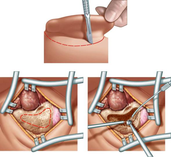
The skin incision is done approximately 1 cm postauricularly behind the posterior auricular sulcus by pulling the auricle anteriorly (see Fig. 13.1). In very young children the inferior portion of the incision is placed more laterally so as not to injure the facial nerve. The development of the mastoid tip is still incomplete. The surgeon pulls the auricle anteriorly, incising the skin and subcutis from the superior to the inferior portion. Afterwards the scalpel has to be used under permanent contact to the bone. The middle finger of the hand not holding the scalpel is placed in the ear canal. This helps to feel the position of the incision in relation to the ear canal.
The soft tissue is completely removed by a raspatory. Henle’s spine and the temporal line are exposed as landmarks. The mastoid tip should only be exposed if this area has to be opened. In this case the tendinous insertions of the sternocleidoid muscle should be sectioned.
Two self-retaining retractors with sharp edges are inserted to expose the mastoid cortex (Fig. 13.2). The approach is marked by the red dashed line.
The opening should be wide to allow a good overview over the surgical field also if a complete antrotomy is not intended (Fig. 13.3). After opening the cortical bone the first landmarks and structures at risk are the sigmoid sinus and the dura of the medial fossa. Due to the late development of the mastoid tip
13 Antrotomy and Mastoidectomy |
63 |
|
|
Fig. 13.1
Fig. 13.2 |
Fig. 13.3 |
and the growth of the tympanic bone, the lateral position of the facial nerve must be respected especially in younger patients.
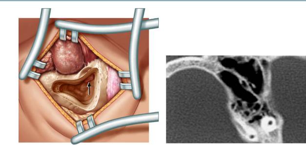
After the delineation of the antrum the aditus ad antrum is enlarged with a curette from its medial to the lateral aspect. Attention is paid not to dislodge the incus (Fig. 13.4). This procedure reduces the probability of a recurrent mastoiditis. Landmarks such as the lateral semicircular canal and the short process of the incus have to be identified. Children aged 3 – 4 years may already have a well pneumatized mastoid.
64 13 Antrotomy and Mastoidectomy
Fig. 13.4 |
Fig. 13.5 |
In older children and adults large burrs reduce the risk of inadvertent damage to vital structures. It is pivotal not to drill holes but to have a large area approach to the mastoid. The linea temporalis and Henle’s spine serve as landmarks. The thin bone of the tegmen tympani is skeletonized, leaving the dura covered with bone. Körner’s septum may exist deep in the mastoid within the air cells of the pneumatized mastoid (see computed tomogram in Fig. 13.5). If present, it is a solid wall separating the superficial mastoid bone from the deeper cells.
The sigmoid sinus and the lateral semicircular canal are identified. The lateral semicircular canal and the medial wall are the landmarks with which to identify the facial nerve and the attic. In badly pneumatized mastoids the dura over the tegmen tympani can be used for orientation. In the antrum it is preferable to use a diamond burr. Strict attention must be paid to the incus to avoid its dislodgment or acoustic trauma to the inner ear. The antrum is blocked straight after its exposure with Curaspon pieces to prevent the intrusion of bone dust into the epitympanic and middle ear spaces. Bone dust may lead to a bony closure of the eustachian tube or the windows or to a fixation of the ossicular chain. The fossa incudis is identified by removing bone in the zygomatic arch overlying the antrum.
Mastoidectomy comprises modelling of the sinus dura angle, the bony covering of the sigmoid sinus and the posterior fossa. The posterior meatal wall is thinned. The attic is opened to visualize the head of the malleus and the body of the incus. This is impossible in a sclerosed mastoid and a low middle fossa.
13 Antrotomy and Mastoidectomy |
65 |
|
|
Fig. 13.6
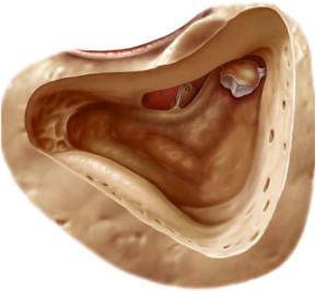
A wide exposure facilitates the orientation. A cutting burr is used at the mastoid tip until the digastric ridge is visualized (Fig. 13.6).
Identification of the lateral semicircular canal and the mastoid portion of the facial nerve: For the less experienced it is helpful to identify the facial nerve in its mastoidal course by drilling the bone over the nerve until it is seen along with the remaining thin layer of bone. A diamond burr larger than the diameter of the nerve should be used to reduce the danger of injury. The drilling must be performed parallel to the course of the nerve.
In treatment of mastoid disease causing intracranial complications, each mastoid cell and piece of diseased tissue should be removed. The dura of the middle and posterior cranial fossa are partly uncovered. In case of granulations, bone should be removed further until whitish healthy mucosa appears. With suspected sinus thrombophlebitis or thrombosis, the sigmoid sinus should be exposed and checked for patency by puncture. Thrombophlebitis normally requires no additional surgical treatment other than complete mastoidectomy. Only in cases of sepsis, extension of the thrombus or pulmonary complications ligation of the jugular vein should be performed in addition to anticoagulation and antibiotic therapy.
The surgical treatment of any brain abscess should be done by or in cooperation with the neurosurgeon. Abscesses adjacent to the temporal bone are more easily reached from the site of origin of the disease and punctured from the mastoid (see computed tomogram and magnetic resonance images in Figs. 13.7, 13.8a, b by courtesy of PD Dr. Randolf Klingbiel, Department of Neuroradiology, Charit´e, Berlin).