

Учебники / Middle Ear Surgery
.pdf46 10 Cartilage Palisade Tympanoplasty
no evidence of cartilage resorption, and there was no extrusion of the prosthesis.
All procedures revealed a statistically significant difference between the preoperative ABG and the postoperative ABG (p < 0.001, t-test for paired samples) at all frequencies. The overall preoperative four-frequency pure-tone average (PTA) ABG was 34.4 dB; the postoperative PTA-ABG was 18.1 dB. In the PORP group, the preoperative PTA-ABG was 28.3 dB hearing level (HL), and at 54 months it was 16.8 dB HL. In the TORP group, the average PTA-ABG was 40.5 dB HL, and at 54 months it was 19.4 dB HL. The differences between PORP and TORP were not statistically different. At 54 months, the postoperative results for TORP with CWU and TORP with CWD were 18.1 dB and 20.7 dB, respectively, and for PORP with CWU and PORP with CWD they were 15.4 dB and 18.8 dB, respectively.
According to the two-tailed Student’s t-test for independent samples to compare functional results in the groups, the average ABG improved at all frequencies, showing a significant statistical difference in favour of CWU procedures (p < 0.05) for 0.5 kHz and 1 kHz. Speech reception scores showed no significant changes in the threshold for 224 of 362 procedures. Speech discrimination scores, determined in 151 of 362 patients at 40 dB above the speech reception threshold, showed no significant decrease (i.e. 10 %) in any of the patients. We observed no significant differences between the functional and anatomic results according to sex or ear sides.
Postoperative complications included two cases of complete hearing loss, one of them associated with the fracture of the stapes footplate in an ear with tympanosclerosis, and five cases of sensorineural hearing loss. These seven cases were considered in the audiologic calculations.
Conclusions
The cartilage palisade procedure (partial or total) was used in 362 cases. The basic differences between our modification and the original procedure described by Hermann [1, 2] consist of using two to four palisades and taking care that one has a broad contact with the ossicular reconstruction [3]. Handling and lacing the grafts needs some training. In our experience, obtaining, measuring, and positioning of the cartilage strips lengthens a routine surgical procedure by approximately 15 – 20 min.
Tympanic closure was achieved in 98.3 % of our cases, the reperforation rate being 1.7 %. Cartilage seems to be an appropriate graft for the reconstruction of subtotal or total perforations, providing rigidity and stability. The support given by both the intratympanic cartilage and the prosthesis prevented retraction of the membrane in the majority of our patients’ ears. Retraction pockets (2.5 %) or recurrent cholesteatoma (2.2 %) may have been due to improper use of cartilage; for example, the number or size of cartilage fragments may have been insufficient to obliterate the epitympanum, the attic or the antrum, or the
10 Cartilage Palisade Tympanoplasty |
47 |
|
|
reconstruction with cartilage may have developed only partial retractions in unsupported areas.
Wiegand [5] reported excellent anatomic results, with a perforation rate similar to ours in more than 600 cases. Milewski [6] obtained similar results after 5 years of follow-up with the use of one piece (“plate”) of cartilage for tympanic closure, and Schulte et al. [7] obtained good anatomic and functional results using irradiated rib cartilage graft. Amedee et al. [8] used cartilage composite grafts, also with satisfactory results. These reports are consistent with our results. Furthermore, our study demonstrates persistent results after a long-term follow-up.
From a functional point of view, in our series, PORP and CWU proved to be predictors of better sound transmission, although the results were not significantly different from those of TORP or CWD.
Preliminary results with the same surgical technique, but using a titanium prosthesis instead (PORP and TORP), showed much better results in the hearing outcome [9]. Thus, 43 % of the patients were in the hearing group 0 – 10 dB, and more than 80 % had a gap of 0 – 20 dB after more than 1 year of follow-up [9]. The better outcome may be attributed to the low weight of the titanium prosthesis of about 4 mg, which would offer a lower resistance in sound transmission compared to hydroxyapatite, Ceravital or bioceramics, which have an overall weight of about 60 mg.
Following our results, sound transmission seems to be improved when a piece of cartilage is in broad contact with the prosthesis. This did not seem to be an obstacle to sound wave transmission at the frequencies measured. We therefore emphasize that the prosthesis or the autologous ossicle used for reconstruction should be situated columella-like in an upright position, in contact with the strip of cartilage. In vitro studies on the mechanical properties of tympanic membrane transplants demonstrate that the acoustic behaviour of cartilage slices smaller than 500 μm (0.5 mm) is almost the same as that of the normal tympanic membrane [10]. Therefore, the assumption that the tympanic membrane must be thin and mobile to allow good sound transmission should be reconsidered.
Our mean follow-up of 54 months (range 36 – 107 months) allows us to claim that the results are stable [11]. The palisade technique allowed functional recovery in CWD procedures as well, although there was a statistically significant difference between the functional results of the groups undergoing CWD and CWU at the two frequencies mentioned.
In totally reconstructed tympanic membranes, it is not possible on postoperative checking of the middle ear cleft to assess whether there is either a collection of mucus or a persistence of cholesteatoma. In the latter case, the tympanic membrane tends to lateralize and prior good sound transmission may worsen. In unclear cases a CAT scan in “w” mode may help distinguish between scar and cholesteatoma.

48 10 Cartilage Palisade Tympanoplasty
Complications such as sensorineural hearing loss are not believed to have resulted from the use of cartilage grafts or the ossicular replacement prosthesis.
Our 15-year experience with the modified cartilage palisade technique is summarized in the indications shown in Table 10.1. The greatest benefit has been achieved in total and subtotal perforations. Our results with a long-term follow-up corroborate our preliminary results [4].
The study shows the reader long-term results of the cartilage palisade technique for tympanoplasties, revealing anatomic and functional benefits that remain stable after long-term follow-up. Used properly, cartilage is able to prevent new retraction pockets and recurrent cholesteatomas. Palisades were used instead of cartilage plates because the palisades adapt more easily to the varying anatomy of the middle ear. Placed close to each other they are as resistant as plates. Gaps between the cartilage stripes should be avoided since squamous epithelium may enter and develop a cholesteatoma. A thorough obliteration of the anterior epitympanum, the supratubaric fossa, and the attic is strongly recommended to avoid recurrent cholesteatoma. Supporting cartilage fragments are important when the anterior aspect of the tympanic membrane needs to be reconstructed in order to avoid medialization leading to reperforation. In badly aerated middle ear clefts the stripes may suffer a partial retraction. Prosthesis can protrude through the graft. The use of cartilage on an alloplastic graft reduces considerably the tendency of protrusion but does not always prevent it. Revision surgery after palisade reconstruction is more difficult because the cartilage changes its consistency, becomes brittle and frequently shows fibrous adhesions to promontory. To avoid “fracturing” of the plates a broad tympanomeatal flap is recommended. Sometimes a reuse of the original reconstruction is impossible, and the old graft has to be discarded and replaced completely. For prosthesis replacement or repositioning, fibrous adhesions between the cartilage and the “old” prosthesis need to be sectioned.
Table 10.1. Indications for cartilage tympanoplasty
Total and subtotal perforations Perforations with tympanosclerotic plaques Perforations within atrophic membranes
Revision surgery for failed myringoplasty or tympanoplasty type I Anterior and inferior perforation with tubal discharge
Retraction pockets
Partially or completely atelectatic tympanic membranes Tympanic adherences
Revision surgery for failed tympanoplasties and tympanomastoidectomies
Chapter 11 |
49 |
|
|
11 Ossicular Chain Reconstruction
Holger Sudhoff, Henning Hildmann
The pathology and degree of ossicular chain destruction in chronic otitis media and cholesteatoma are very variable. Therefore there is no single strategy applicable to all different conditions. Wullstein in 1968 accomplished the classification of tympanoplasty into five types based on the necessary technique for reconstruction. In 20 % – 25 % of cases of chronic suppurative otitis media, the ossicular chain is involved, and in cholesteatoma cases 80 % need chain reconstruction, either because the pathology has destroyed the ossicular chain or parts of it, or the chain has to be separated by the surgeon. Also in malformation surgery the chain reconstruction surgery follows the same principles.
The extent of the destruction differs. Overall smaller defects have better prognosis. The ossicles can be reused in about 50 % of cholesteatomas and in 80 % of cases with chronic otitis media.
For reconstruction, organic and alloplastic material can be used as described in a later chapter.
Of the organic materials available, the patient’s own ossicles, parts of the incus or the head of the malleus are the most frequently used reconstruction materials. However, they must be free of cholesteatoma, which means without viable keratinocytes. The long-term stability of the use of these materials has been proven. They are especially useful when the stapes has remained intact. Contact with the surrounding bone may lead to osseous or fibrous ancylosis and may result in a recurrent conductive hearing loss. In revisions it can be difficult to separate the transposed ossicles from the head of the stapes.
Cartilage can be used to bridge short distances, for instance for stapes elevation. Long cartilage reconstructions between the stapes footplate and the tympanic membrane tend to necrotize because the cartilage does not contain vessels and depends on nutrition by diffusion. Therefore it tends to become unstable over time.
Bone is easily available from the mastoid plane. Animal experiments have shown that the cortical bone tends to remodel and change its preformed shape. Compared to the ossicles it is a material of second choice.
Alloplastic material is used as plastics, ceramics metals and compound grafts. It is extensively discussed in the next chapter by Yung.
All alloplastic materials are generally costly. Most synthetic materials are easy to remove; however, titanium implants may develop intensive interaction

50 11 Ossicular Chain Reconstruction
with bone corresponding to an ancylosis. This has to be remembered, especially in revisions. Nevertheless it is our favourite alloplastic material for prostheses.
Most operations can be completed in one stage, removing the cholesteatoma and then reconstructing the ossicular chain at the same time. A twostage procedure is indicated only in special situations. When the surgeon has doubts over whether he or she has removed a cholesteatoma completely or, in rare cases, it is not possible to remove all the squamous epithelium from the oval window or between the crura of the stapes, a second-look operation is recommended 1 (in children) or 2 years later. By that time the residual epithelium will have formed pearly masses that are much easier to locate and remove. Occasionally the epithelium will disappear in the interval, probably due to atrophic deficit. In non-compliant patients who have good postoperative hearing and most likely will not present for follow-up, it may be wise to postpone hearing improvement until a second stage.
Second look operations are not routinely done except for the situations mentioned above.
The following situations with ossicular chain defects can be distinguished:
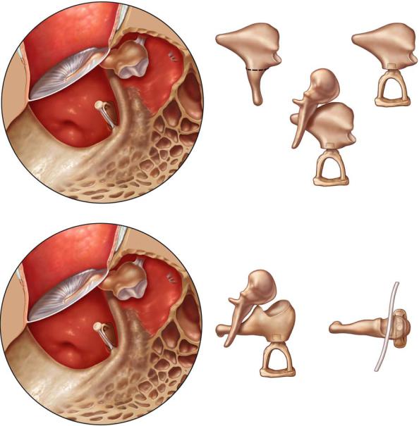
1.The incus or part of the incus is missing (Fig. 11.1).
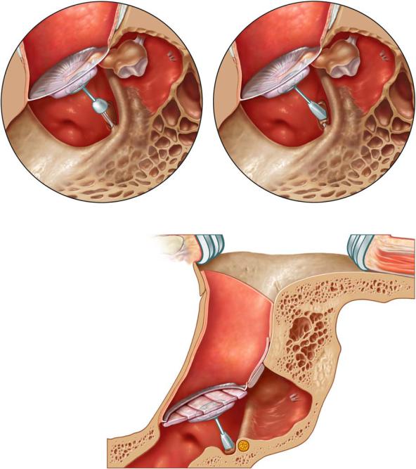
This is the most common situation requiring reconstruction. Most frequently the long process of the incus is destroyed. This allows the reuse of the incus, which can be shaped by drilling and can be placed between the stapes and malleus or the stapes and drum graft. The incus autograft is fashioned to bridge the gap between the head of the stapes and the handle of the malleus according to the anatomical situation. In the drawings the base of the long process of the incus is placed on the head of the stapes. A hole is drilled into the base of the long process and a notch into the body to receive the handle of the malleus (Fig. 11.1). Controlled force is applied to lift the handle of the malleus until the interposed incus is in a correct position. It is not always possible to place the incus between the malleus and stapes; the body may also be placed parallel to the handle of the malleus. Placing the underlay fascia graft in this situation is difficult. Depending on the anatomical situation in the middle ear, the body of the incus autograft can also be placed parallel to the handle of the malleus. The incus is leaned against the handle after placing the underlay graft. If the tip of the malleus handle is close to the promontory, distance can be gained by cutting or stretching the tensor tympani tendon. We prefer this to a partial resection of the distal part of the handle because we think that the sound transmission properties are not as good with a short handle. The incus can be stabilized by the chorda tympani (Fig. 11.2a). Ossicular bone grafts should never be in contact with the bony walls of the middle ear because ancylosis may develop. Alternatively the head of the malleus
11 Ossicular Chain Reconstruction |
51 |
|
|
Fig. 11.1
Fig. 11.2a
is utilized. If the head of the stapes has enough height, a piece of cartilage can be used for transmission. We should keep in mind, however, that cartilage depends on nutrition by diffusion and tends to necrose if used to bridge large distances.

52 11 Ossicular Chain Reconstruction
For alloplastic material, partial prostheses to be placed on the head of the stapes (partial ossicular replacement prosthesis, PORP) (Fig. 11.2b) and total prostheses to be placed on the footplate (total ossicular replacement prosthesis, TORP) are available (Fig. 11.2c). There is a large variety of designs (see Chap. 12). The surgeon, especially the beginner, should not use too many variations. He or she needs experience and dexterity with a few designs. As pointed out by Yung, several materials are in use by experienced surgeons. The drawings show titanium prostheses, because of the frequent use of this material in Europe. Other materials can be inserted similarly.
A suction tip is used to pick up the prosthesis from its packaging and place it into the middle ear. The PORP prosthesis is centred on the stapes head with its cuplike opening using the suction tip and a sickle knife.
If a TORP prosthesis is used, it can be positioned on the footplate, supported by the stapes suprastructure.
Both types of prosthesis should be covered by cartilage to prevent extrusion.
2.Only the stapes is preserved:
Generally a stapes elevation technique is used. We advise the reuse of the ossicles if possible, and stapes elevations by cartilage or titanium allografts are further alternatives. Sometimes a TORP placed between the crura of the intact stapes gives the implant a very stable position. Allografts must be covered by cartilage to prevent extrusion.
3.The malleus or handle of malleus is preserved, and the incus and stapes suprastructure are missing.
This is a rare situation. We use a titanium allograft (TORP) with a cartilage covering to prevent extrusion. If the implant can be placed under the handle of the malleus, the situation is more stable (Fig. 11.2c).
A suction tip is used to pick up the prosthesis from its packaging and place it into the middle ear. The PORP is positioned on the food plate using the suction tip and a sickle knife. To stabilize the position of the prosthesis, small pieces of Gelfoam can be used. If cholesteatoma or tympanosclerosis has covered the footplate, the bone of the footplate may be very thin. The prosthesis should be placed and later covered with cartilage on the tympanic membrane side without applying pressure to avoid perforation. A small thin piece of cartilage can be used for protection.
4.All ossicles except the stapes footplate are destroyed.
In these cases a total or subtotal perforation is generally found. Reconstruction in one stage is rather unstable but should be tried. Using cartilage palisade tympanoplasty, we obtain a good stability for the tympanic membrane and provide reinforcement for the titanium allograft. The
11 Ossicular Chain Reconstruction |
53 |
|
|
Fig. 11.2b |
Fig. 11.2c |
Fig. 11.3

54 11 Ossicular Chain Reconstruction
TORP position is stabilized by surrounding the foot with Gelfoam (Fig. 11.3).
All reconstructions have to allow enough space between the promontory and reconstructed membrane to create an aerated middle ear, if the posterior wall is preserved. An airway to the mastoid is necessary to prevent retraction and new cholesteatoma formation.
Chapter 12 |
55 |
|
|
12 Materials for Ossicular Chain Reconstruction
Matthew Yung
There are many different materials currently being used for ossiculoplasty. They can be categorized into autografts, homografts and allografts. In the 1999 Smith & Nephew Otology Catalogue alone, 54 different designs of alloplastic ossicular prosthesis were being marketed [1]!
In general, there has been a paucity of long-term comparative studies on different ossiculoplasty materials. There has been only one randomized study in the literature comparing one particular material with another (Plastipore vs Ceravital) [2]. Virtually all other reports have been either personal series of one particular type of prosthesis or comparisons of results of different prostheses based on historical data. The criteria used by different authors to describe success have varied, e.g. different methods have been used to calcu-
Vinyl-acryl |
Cortical |
PTFE |
Stainless |
Plastipore |
Ceravital |
Bioglass |
Titanium |
|
|
bone |
|
Steel |
|
|
|
(Dalchow)17 |
|
(Wullstein)3 |
(Farrior)6 |
(Austin7) |
(Palva)9 |
(Shea)11 |
(Reck)13 |
(Merwin)15 |
||
|
||||||||
(1952) |
(1960) |
(1962) |
(1969) |
(1976) |
(1983) |
(1986) |
(1993) |
1950 |
1960 |
1970 |
1980 |
1990 |
2000 |
|
|
|
|
|
|
|
|
|
|
|
|
(1957) |
(1958) |
(1966) |
(1974) |
(1979) |
(1981) |
(1988) |
Autologous |
Polyethylene |
Homologous |
Proplast |
AL2 O3 |
Hydroxyapatite |
Carbon |
incus |
|
incus |
|
Ceramic |
|
|
(Hall)4 |
(Shea)5 |
(House)8 |
(Shea)10 |
(Jahnke, |
(Grote)14 |
(Podoshin)16 |
|
|
|
|
Plester)12 |
|
|
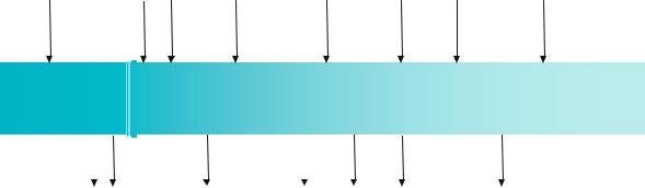
Fig. 12.1. Time line showing the specific year each alloplastic material was introduced for clinical use (the alloplastic materials in boldface are discussed at greater length in the text)