

Учебники / Middle Ear Surgery
.pdf26 7 Harvesting and Processing of Soft Tissues
Fig. 7.4
Different ways of storing the fascia until reimplantation have been tried. We prefer not to dry the fascia because we believe that vital cells improve the integration of the graft, prevent resorption of intercellular collagen fibres and later reduce the frequency of atrophic scars.
Harvesting of the temporalis fascia can be achieved by superior extension of the retroauricular approach incision. The exposure is slightly better. Larger pieces can be removed. Even vascularized flaps for reducing large cavities can be harvested when the excision is extended cranially.
Harvesting Cartilage
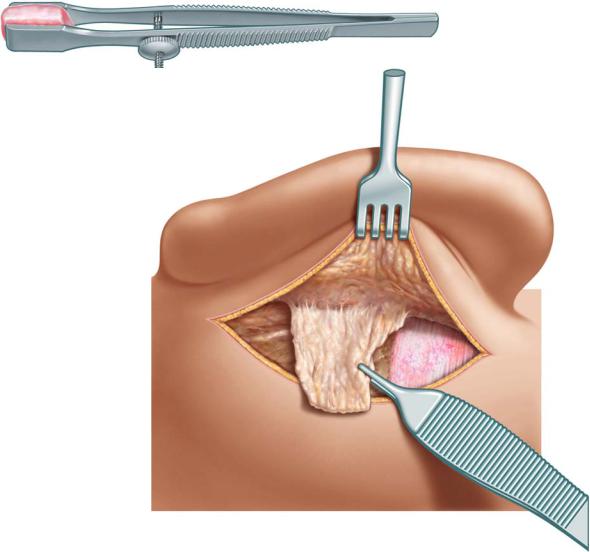
Cartilage can be harvested from: (1) the tragus, (2) the anterior crus of the helix, (3) the cavum, (4) the cymba and (5) the triangular fossa (Fig. 7.5). Mainly tragal, conchal and cymba cartilaginous grafts are used as autologous transplants in middle ear surgery. Cartilage, even in larger amounts, is more easily collected by a postauricular approach. Thick grafts may be split.
1.Tragal cartilage is mainly used when an endaural approach has been chosen. It can be exposed from the endaural incision between the tragus and the anterior crus of the helix or by a separate incision (Fig. 7.6). Generally it appears to be almost plane and even in thickness and is removed using curved scissors (Fig. 7.7). The covering perichondrium can be left attached for an island graft, used separately or exclusively.

7 Harvesting and Processing of Soft Tissues |
27 |
|
|
Fig. 7.5
Fig. 7.6
Fig. 7.7

287 Harvesting and Processing of Soft Tissues
2.Grafts from the anterior crus of the helix provide small and thin cartilage grafts with perichondrium which is useful for small posterior attic defects, when an endaural incision has been chosen.
3.Large quantities of cartilage can be gathered from the concha. The conchal cartilage varies in thickness. Therefore splitting is often necessary to avoid a bulky graft. The perichondrium is harder to remove (Fig. 7.8).
4.The cymba supplies medium sized slightly rounded grafts. The shape is favourable for the closure of larger defects in the posterior meatal wall. The covering perichondrium is easy to remove and can also be used separately as a soft tissue graft.
5.Triangular fossa: This site is seldom used. The anterior and posterior crus of the anthelix must be respected.
Preparation of Cartilage
The cartilage grafts in our opinion are easier to place as a cover of the middle ear, when applied as strips (palisades) if total or subtotal reconstruction of the tympanic membrane is intended. They adapt better to the variable anatomy of the middle ear. The grafts must be thin enough to allow air flow in the middle ear cavity and leave a sufficient distance from the promontory. To facilitate car-
Fig. 7.8
7 Harvesting and Processing of Soft Tissues |
29 |
|
|
tilage slicing, a special clamp can be used to fix a No. 15 scalpel for this procedure as illustrated (Fig. 7.9). If the distance is short, the perichondrium should be removed on the middle ear side to reduce the possibility of adhesions.
For reconstruction of the posterior wall total pieces are generally used. Sometimes, however, strips are easier to position.
Harvesting Perichondrium
Perichondrium can be taken from both sides of the cartilage for a larger graft. The cartilage must not be removed from its original site to collect perichondrium. Essentially the same procedures apply to gathering perichondrium as for cartilage (Fig. 7.10).
Fig. 7.9
Fig. 7.10

30 Chapter 8
8 External Ear Canal Surgery
Henning Hildmann, Holger Sudhoff
Surgery in the external auditory canal without surgery in the middle ear may be necessary:
1.After surgery
2.After trauma
3.Postinflammatory
4.Due to idiopathic changes
All procedures postoperatively need a wide and well aerated outer ear canal. An endaural incision is generally used. The remaining meatal skin may be removed and reimplanted as a free graft if necessary. However, the skin of the anterior tympanomeatal angle should be left untouched whenever possible to prevent blunting after surgery. If skin is missing, this area should be carefully reconstructed.
If widening of the ear canal anteriorly is necessary, the anterior bony meatal wall should be left intact. Small defects generally do not cause any problems, but larger defects cause hernias of the perimandibular tissue into the ear canal that are hard to treat.
Postoperative Changes
Postoperative Stenosis
Entrance of the Ear Canal
The entrance often becomes stenotic due to scar contraction after endaural, but especially after retroauricular, incision. The remaining ear canal is sufficiently wide.
The skin in the cavum is incised as indicated, forming a superiorly based flap that later covers the gap remaining superiorly after pulling the meatal skin outward (Fig. 8.1a, b). Suturing of this flap in the ear canal is difficult. Therefore we only use careful packing at the end of the surgery. The underlying cartilage and scar tissue must be resected until the bone is exposed. Removing bone from the posterior circumference of the ear canal gives additional space. Henle’s spine and the outer part of the tympanosquamous fissure may be prominent and should be removed.
If this does not appear to be sufficient, a pretragal pedicled flap is inserted into the endaural incision area (Fig. 8.2a, b).

8 External Ear Canal Surgery |
31 |
|
|
Fig. 8.1a
b
Fig. 8.2a
b
Postoperative Stenosis of the Canal
Weblike stenosis may be observed and may be widened with a wick. Some stenoses become so thin that they may be resected with a sickle knife after some months.
More substantial stenosis and synechia should be reoperated on. The complete scar is resected and replaced by split-thickness skin grafts. The quantity needed may be harvested from the posterior side of the auricle with a No. 10 blade.
If the anterior tympanomeatal angle is affected, it should be carefully reconstructed. A good result is dependent, as in surgery for major malformations, on the precision of the graft placement. Overlapping and folding must be strictly avoided.

32 8 External Ear Canal Surgery
Other Postoperative Changes
Annulus Cholesteatoma
This usually develops after tympanoplasty using an onlay technique for tympanic membrane closure (Fig. 8.3). The outer part of the cholesteatoma should be removed while the epithelium covering the tympanic membrane and the anterior tympanomeatal angle often forms a perfect lining and can be preserved.
Cholesteatoma of the Ear Canal
This usually develops postoperatively. Small cysts can be removed with incisions as an office procedure. Larger cysts should be removed by an endaural or postauricular approach. The underlying bone should be flattened with a diamond burr.
Lateralization
The graft may lateralize after tympanoplasty due to epithelial migration. It is more frequent after onlay techniques, but seldomly may develop spontaneously. It will result in a conductive hearing loss. An underlay tympanoplasty placing the graft under the handle of the malleus prevents recurrences. The missing epithelium, especially the anterior tympanomeatal angle, must be carefully reconstructed.
De-epithelialized Bone
After drilling without sufficient irrigation the bone may suffer heat damage. These areas often remain bare of epithelium and they may be followed up in the office. If the defect does not heal, the ear must be reoperated on, and the superficial layer of the bare bone is drilled off with sufficient irrigation and covered with fascia and a split-thickness skin graft.
Blunting
The anterior meatal wall and the tympanic membrane are positioned at an angle of about 70° to each other. This is the tympanomeatal angle. It is a “sacred” area and should not be touched if it can be avoided. Any surgery in this area carries the risk of blunting, which is scar formation. The tissue fills the tympanomeatal angle, and the angle becomes blunt. It is obvious that the sound conduction abilities are reduced. If the epithelium has to be removed or the anterior angle has to be reconstructed, for instance in malformation surgery, it must be reconstructed very carefully. Small split-thickness skin grafts from the posterior side of the auricle are used. They are sheathed with silicone.

8 External Ear Canal Surgery |
33 |
|
|
Fig. 8.3
Fig. 8.4
Trauma
Surgery should be customized depending on the nature of the injury. Fractures of the posterior wall are often associated with middle ear, inner ear or facial nerve damage. The surgery for these is described in later chapters. In any case of reconstruction the ear canal must be wide enough and well aerated. A cavity

34 8 External Ear Canal Surgery
with a wide meatoplasty is an alternative. If the ear is completely deaf, the ear canal may be obliterated if the meatal skin can be completely removed; if not, cholesteatoma formation may result.
Fractures Into the Medial Cranial Fossa
Fractures into the medial cranial fossa may need a transtemporal approach and a team approach with the neurosurgeon. Fractures of the anterior wall are often limited to the canal. In cases where they are accompanied by herniation of the perimandibular tissue, repositioning is possible but generally not successful because of the very high pressure during chewing. For smaller defects a septal or auricular cartilage placed on the mandibular side of the fractured tympanic bone can be a solution. More extended fractures (Fig. 8.4) cannot be stabilized by temporary immobilization of the jaw by the maxillofacial surgeon. It is easier to remove the posterior meatal wall in these cases and to create a cavity allowing sound and air to reach the tympanic membrane.
Post-traumatic Atresia
The prognosis for post-traumatic atresia depends on the extent of the damage. If the middle ear is shown to be aerated in the CT scan, the Eustachian tube is most likely to be affected and hence hearing improvement is not probable. However, surgery might be necessary due to cholesteatoma behind the atresia.
The ears are opened with an endaural incision. The scar forming the atresia is removed as well as bony fragments obliterating the ear canal. Defects of the posterior wall can be reconstructed with cartilage. Epithelial defects are replaced by split-thickness skin grafts.
Inflammation and Sequelae
Postinflammatory Stenosis
This condition may develop after long-standing external otitis. The subepithelial tissue is thickened and cannot be reduced by local treatment. The tympanic membrane is generally not or little affected. The epithelium is altered by infection and therapy.
Routinely we use an endaural incision. The posterior meatal skin including the subepithelial tissue is removed and replaced by spit-thickness skin grafts from the backside of the auricle after widening the ear canal by drilling in all possible directions. If the meatal skin can be reused after resecting the subepithelial tissue and the aspect of the epithelium appears fairly normal, meatal skin should be preferred. Generally a partial replacement by skin grafts is necessary. The canal must be widely exposed to provide a good aeration as prerequisite for healing. The epithelium of the anterior tympanomeatal angle should be spared to prevent anterior blunting.

8 External Ear Canal Surgery |
35 |
|
|
Fig. 8.5
Necrosis of the Floor or the External Auditory Canal
This condition appears to be a cholesteatoma of the external auditory canal generally localized in the medial part of the floor. However, it behaves differently. It seems that the epithelium has invaded the defect secondarily to bone necrosis of the floor due to an insufficient vascular supply. Using an endaural approach the irregularities need to be smoothened by a diamond burr and covered by a split-thickness skin graft.
Postinflammatory Meatal Fibrosis
This condition is supposed to develop from chronic myringitis with medial external otitis. Fibrotic scar tissue up to 1 cm thick is found under noninflamed epithelium in the medial part of the ear canal (Fig. 8.5). The fibrous layer of the tympanic membrane is generally intact or has a small central perforation. The middle ear and the ossicular chain are not affected. After circumcising the meatal skin above the blind sac, the fibrous mass is worked off the underlying bone and can generally be removed from the fibrous tympanic membrane without effort and damage. The epithelium should be carefully replaced by split-thickness skin grafts. The disease has a tendency to recur. The surgeon should take care to reconstruct the anterior tympanomeatal angle to avoid blunting.
Malignant External Otitis
This life-threatening condition may lead to extensive destruction of the ear canal and the temporal bone. It is mostly seen in elderly male diabetics. As a Pseudomonas infection it is generally treated by antibiotics. Long-term antibiotic treatment is recommended. Necrotic bone and tissue can be removed as a