

Учебники / Middle Ear Surgery
.pdf136 25 Middle Ear Trauma
affected. Persistent vertigo over 6 months might require a labyrinthectomy or vestibular nerve section. If bilateral severe sensorineural hearing loss occurs, cochlear implantation is often indicated and surgery is done when the patient becomes stable.
Leak of CSF indicates dural injury. Generally the tegmen or the mastoidal region is involved. Leaks from the internal auditory canal through the labyrinth are possible. If the tympanic membrane is ruptured, liquorrhoea through the external canal occurs. If the membrane is intact, the fluid drains through the eustachian tube and should be differentiated from a defect in the frontal skull base. In most cases the tears of the dura of the lateral skull base heal spontaneously. The ear is covered with a sterile dressing and the patient is observed. If there is concomitant infectious ear disease, especially cholesteatoma, the ear must be operated on to prevent an ascending infection. Ascending air can cause pneumatocoeles and may change the surgical concept. If the liquorrhoea persists the leak must be closed. CT mostly reveals the site of injury. Many defects can be closed via a mastoidectomy approach. The dura and the defect are exposed, bony splinters removed and the defect closed with large pieces of temporalis fascia placed on the intracranial side of the temporal bone and fixed with fibrin glue. Larger defects are stabilized with cartilage.
If necessary, especially for repair of the medial region of the upper plane of the petrous bone, a transtemporal approach can be used.
If the hearing is not preserved, the middle ear spaces can be obliterated with abdominal fat.
Haemorrhages may be life threatening and call for immediate revision. They may be due to injury of the sigmoid or the superior petrosal sinus. Sinus bleeds are low pressure bleeds and can be dealt with by drilling away surrounding bone to expose the sinus and subsequent packing. Bleeding from the carotid artery or the medial meningeal artery usually cannot be controlled by an otological approach and requires the help of an interventional radiologist.
Chapter 26 |
137 |
|
|
26 Endolymphatic Sac Surgery
Holger Sudhoff, Henning Hildmann
The aim of endolymphatic sac surgery is to expose and drain the endolymphatic sac using a transmastoidal approach as a treatment for Meniere’s disease. This disease is characterized by episodic vertigo attacks with nausea or vomiting, sudden or fluctuating sensorineural hearing loss of different degrees and tinnitus of varying intensity. A frequent symptom is the sensation of pressure or fullness in the affected ear. The pathophysiology is commonly explained by a distension of the membranous labyrinth by the endolymph, also called endolymphatic hydrops. Bilateral Meniere’s disease is rare, often occurring within a period of 5 – 30 years in the primarily unaffected ear. Preoperative evaluation should include electronystagmography, or balance test (ENG), electrocochleography (ECOG), brainstem evoked response audiometry (BERA) and magnetic resonance imaging (MRI) with gadolinium to exclude an acoustic neuroma or other brain tumours as a possible source of symptoms. In spite of today’s technical knowledge, the diagnosis of Meniere’s disease still relies mainly on the clinical symptoms: fluctuating hearing loss, tinnitus, attacks of vertigo and sense of fullness in the ear. In the beginning it is often monosymptomatic, with fullness or tinnitus in the affected ear. It may also present as a sudden hearing loss. Vertigo can present very dramatically and distressingly with insatiable emesis and may be misdiagnosed as cardiac infarction, apoplexia, intoxication or severe gastrointestinal infection. The lack of aetiological knowledge about Meniere’s disease and its difficult differential diagnosis explains the whole variety of medical and surgical treatment options. Its unresponsiveness to medical treatment is the indication for surgery. In cases with useful hearing, saccotomy is preferred before neurectomy is considered. We perform the exposure and drainage of the endolymphatic sac using a sac sialastic stent. In theory, the endolymphatic sac operation should decompress the excessive fluid within the inner ear and improve resorption of endolymph. Vertigo subsides or disappears after surgery in about 66 %, tinnitus in 50 %, improvement of hearing in 28 % and fullness in 80 % of Meniere’s cases within 3 years of surgery. Vertigo symptoms may reappear in some individuals.

138 26 Endolymphatic Sac Surgery
Surgical Procedure
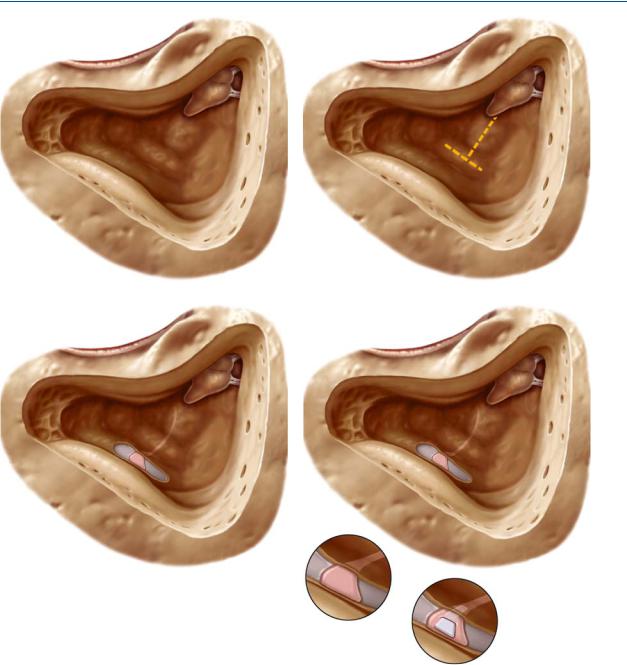
Using a retroauricular approach the mastoid is opened and the following landmarks are identified: the bony shell of the sigmoid sinus and the middle and posterior cranial fossa, the lateral and the posterior semicircular canals and the short process of the incus (Fig. 26.1). More experienced surgeons may use a less extended exposure. In any case, the sinus, the posterior fossa below the sinus and the lateral and posterior semicircular canals need to be clearly seen. The antrum is blocked after its exposure with Curaspon pieces to prevent bone dust entering the epitympanic and middle ear spaces. Bone dust may lead to a bony closure of the eustachian tube or the windows or to a fixation of the ossicular chain.
Anatomical Position of the Endolymphatic Sac
The dashed lines illustrate the direction of the lateral and the posterior semicircular canals. The arch of the lateral canal is about 3 mm more lateral than that of the posterior canal. The blue line of the posterior canal should be seen. The bone posterior to the posterior semicircular canal overlying the posterior fossa dura is removed. The distance between the posterior semicircular canal and the posterior fossa is very variable. In a well-pneumatized mastoid the endolymphatic duct leading from the sac to the vestibule can be identified. The endolymphatic sac is found below the line drawn from the lateral semicircular canal crossing the posterior canal. Avoid damage to the dura. The endolymphatic sac is situated in a shallow groove of the posterior aspect of the temporal bone as marked by the yellow lines (Fig. 26.2). It can often be identified by its change of structure of the dura of the posterior fossa. The endolymphatic duct, which is occasionally visible, has to remain intact (Fig. 26.3). The egg- shell-thin bone over the posterior fossa dura is removed with a curette. It is important to note that its superior portion of the posterior semicircular canal is closer to the posterior fossa dura than its inferior portion. The endolymphatic sac is incised with a sickle knife and is identified after the detection of a lumen. Sometimes even experienced surgeons incise the dura accidentally, mistaking it for the clearance of the endolymphatic sac (Fig. 26.4). The position of the endolymphatic sac is often assumed as being too far superiorly. In cases of Meniere’s disease it is frequently small due to fibrosis. After identifying and opening the sac, possibly also identifying the entrance to the duct, a small triangular silicone sheath is inserted into the incision. The exposed area can be covered with soft tissue. Poor pneumatization and a prominent sigmoid sinus may complicate the procedure. If the sinus is very prominent, the covering bone may be thinned and fractured.
The posterior semicircular canal may be hard to find in a sclerotic mastoid. The lateral canal can always be identified. It can be helpful to identify the blue line of the lateral canal before identifying the blue line of the posterior. The sinus can be protruding. Space can be gained by thinning the bone over the sinus and fracturing (eggshell) the bone and pushing it towards the vessel.
26 Endolymphatic Sac Surgery 139
Fig. 26.1 |
Fig. 26.2 |
Fig. 26.3
Fig. 26.4

140 Chapter 27
27 Malformation Surgery
Henning Hildmann
Surgery of Middle Ear Malformations
For surgical purposes malformations can be subdivided into minor and major malformations. This classification seems practical because minor malformations with a normal or nearly normal tympanic membrane can generally be operated on with the techniques of tympanoplasty and stapes surgery. Major malformations are associated with atresia or stenosis of the outer ear canal and need a more extensive diagnosis. The indications for surgery must be very precise and consideration should be given to fitting a temporary hearing aid depending on the individual’s age. Only cases with a well developed middle ear have the chance of a good surgical outcome. Major malformations may be associated with malformations of the outer ear and the face.
Minor Malformations
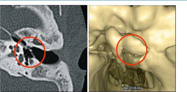
Minor malformations can be present when there is a normal outer ear canal and a normal or nearly normal tympanic membrane. Patients can have had a conductive unilateral or bilateral hearing loss since birth. High-resolution computerized tomography scans should be scrutinized for abnormalities of the internal auditory canal, with the possibility of a gusher, which is seen in 2 % of our material, for the course of the facial nerve and for abnormalities of the ossicles (Fig. 27.1a). Three-dimensional reconstruction can be helpful in selected cases (Fig. 27.1b; courtesy of PD Dr. Randolf Klingbiel, Department of Neuroradiology, Charit´e, Berlin). Stapes fixation is the most frequent finding (41 %) and can be treated as in otosclerosis surgery. A malformed stapes or a stapes remnant may be found at the site of the oval window which is ossified. However, malformations of the other ossicles can be found in different combinations. Behind an intact tympanic membrane, which may be smaller than normal, abnormalities can be seen: of the incus and stapes in 31 % of cases and of the complete chain in 11 % of cases. In other malformations, the malleus, the malleus and incus, malleus and stapes or malleus alone may be affected. Reconstruction procedures depend on the extent of the malformation and the necessity for chain reconstruction using techniques as described in the tympanoplasty chapters.
A persisting stapedial artery is found in 4 % of malformations. The artery can be coagulated.
27 Malformation Surgery 141
Fig. 27.1a |
b |
The occlusion of the round window can be opened by drilling; however, the opening generally recloses.
For surgery an endaural approach is chosen. Sometimes the outer ear canal is narrow and has to be widened after elevation of the skin. The skin of the posterior canal wall can either be worked towards the tympanic membrane as a tympanomeatal flap or partially removed and reimplanted as a free flap. For the anterior wall the same procedures are used as for exostosis surgery or for surgery of a protruding anterior meatal wall in tympanoplasty. The skin should be treated gently to avoid postoperative meatal stenosis. Diamond burrs should be used since they are less traumatic to the skin. Cutting burrs cause more turbulence in the water used for irrigation and may draw the skin flap into the revolving burr.
Small Tympanic Membrane
The tympanic membrane may be smaller than normal. In this situation an underlay graft is used after widening the outer ear canal and the posterior rim of the bony frame of the tympanic membrane. If the ossicular chain has to be reconstructed, a cartilage graft may be necessary.
Chain Reconstruction
A fixed stapes is often accompanied by an obliterated footplate. If the inner ear is normal and the round window is open, an opening at the site of the oval window is drilled. The chain is reconstructed according to the techniques used in

142 27 Malformation Surgery
the surgery of otosclerosis. If possible a prosthesis is crimped to the long process of the incus as in surgery of otosclerosis. If this is impossible, a connection to the handle of the malleus can be made. A total reconstruction with a cartilage footplate in the new oval window is possible but difficult. The hearing results are less reliable.
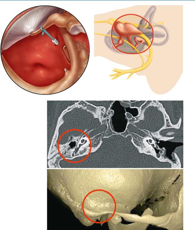
The facial nerve may have an abnormal course. It should therefore be identified before opening the vestibule. Abnormalities of the course of the facial nerve are found in 10 % of cases. The nerve may cover the region of the oval window and make a normal reconstruction impossible. An opening of the basal turn of the scala vestibuli (promontorial window, Plester) is an elegant solution (Fig. 27.2). A prosthesis connects the inner ear to the handle of the malleus.
Major Malformations
Major malformations with complete atresia or stenosis of the ear canal can be associated with absence or dysplasia of the outer ear, downwards dislocation of the auricle and facial malformations. Atresia or stenosis is due to the absence or incomplete development of the tympanic bone. Therefore the facial nerve leaves the temporal bone in a different position and turns forwards into the parotid gland higher than normal. With the lack or incomplete development of the tympanic ring, the temporomandibular joint may be positioned more posteriorly and force the surgeon to drill the ear canal more posteriorly than normal (Fig. 27.3). This results in a different approach to the middle ear cavity, which is situated more posteriorly, with the centre of the new canal in the region of the lateral semicircular canal and the upper mastoidal portion of the facial nerve.
Timing of Diagnosis
Diagnosis for hearing impaiment should be done immediately after birth. In cases of complete atresia the patient has a conductive hearing loss of 60 dB. Generally the inner ear function is normal.
The child is generally fitted with bone conduction hearing aids as early as possible. Bone anchored hearing aids cannot be fitted at that early stage due to the thinness of the cortical bone.
The parents must be counselled. Hearing and speech development is monitored until the age of 7 years.
The preconditions for surgery are: sufficient inner ear function, a favourable anatomy as seen on computerized tomography and family support with acceptance of the possibility of failure. A reduction of the 60 dB hearing loss to 20 dB is a good result. In cases with malformations of the face or the auricle, planning should be done in cooperation with the plastic and maxillofacial surgeon. Incisions should be made avoiding scars in the regions necessary for reconstruction of the auricle. As Jahrsdorfer indicated in his grading system,
27 Malformation Surgery 143
Fig. 27.2 |
Fig. 27.3 |
Fig. 27.4a, b

144 27 Malformation Surgery
computerized tomography is the basic investigation that determines suitability for surgical reconstruction if the inner ear is normal. The mastoid should be sufficiently large to allow the drilling of a new ear canal. The middle ear must be aerated and have a normal or near normal size. If the stapes is present and the facial nerve is in its normal position, the chances for successful surgery are good. Patients with an unfavourable anatomy are not candidates for surgery. They should be fitted with bone anchored hearing aids.
Computerized tomography on the right (Fig. 27.1) shows a favourable anatomical situation with a normally wide middle ear and a wide mastoid with sufficient space for a new external canal. The narrow middle ear and the lack of space in the mastoid are predictive of there being little chance of successful surgery (Fig. 27.4a, b; courtesy of PD Dr. Randolf Klingbiel, Department of Neuroradiology, Charit´e, Berlin). A bone anchored hearing aid is the better solution.
Surgical Steps and Landmarks
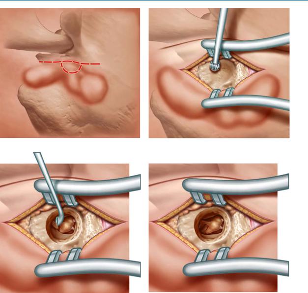
To start with the landmarks are the temporomandibular joint and the tip of the mastoid. The skin is incised behind the temporomandibular joint (Fig. 27.5a). If the auricle is present, an endaural incision is done. It should be noted that the auricle may be placed downward in malformations. A circular skin incision in the area of the future auditory canal should ensure a sufficiently wide entrance at the end of the operation (Fig. 27.5b). A circular ear canal of 1.5 cm is drilled as close as possible to the temporomandibular joint, leaving a bony partition preventing protrusion of connective tissue. The drill hole finds the antrum with the lateral semicircular canal in the centre. The aditus can be identified and worked forward towards the incus. It is often synostotic with the head of the malleus and varies in shape. The end of the malleus handle is malformed and is fixed to the anterior side of the middle ear cavity (Fig. 27.5c). In favourable cases selected as described above the stapes is often intact and mobile. If it is in contact with the incus and the handle of the malleus, the chain can be mobilized and the handle of the malleus detached from the anterior bone of the cavity. The total chain can be used for sound transmission. To prevent noise trauma the separation of the handle is done with a small curette (Fig. 27.5d). If the incus and malleus cannot be preserved, reconstruction is more difficult. Prostheses should be used analogous to tympanoplasty techniques. The results are less favourable. The middle ear is closed with palisades or a thin slice of cartilage of the auricle or the ear remnant. This gives a plain tympanic membrane and avoids niches behind the lateral semicircular canal. The ear canal is covered with a split-thickness skin graft as proposed by Jahrsdorfer. The outer skin is sutured to the bone of the entrance of the canal to avoid narrowing and granulations. The graft is covered with silicone and packed.
27 Malformation Surgery 145
Fig. 27.5a |
b |
c |
d |