

Учебники / Middle Ear Surgery
.pdf126 23 Laser Stapedotomy
Special Cases
Obliterative Otosclerosis
The incidence of obliterative otosclerosis is between 2 % and 10 % of all cases [6, 16, 58, 61]. It was 5 % in the present author’s series. Drilling through a thick footplate obliterating the oval window niche can cause significant vibrationinduced inner ear trauma. The CO2 laser, on the other hand, can vaporize a fenestra in the stapes footplate, regardless of its thickness or degree of fixation, without mechanical trauma to the inner ear.
The settings on the SurgiTouch scanner are the same as for a laser stapedotomy. After the suprastructure is removed, the otosclerotic foci obliterating the oval window niche are uniformly removed over a broad front by laser application with the SurgiTouch scanner. This is continued until the lateral margins of the oval window can be clearly identified. Lower power may have to be used at the periphery of the window niche to avoid accidentally entering the inner ear. Large amounts of char are produced as the bony material is vaporized. Since crystalline char reflects the CO2 laser energy and reduces its ablative effect, it must be removed with a suitable instrument. The vestibule in the posterior part of the oval window niche is opened with the scanner using the same laser parameters as for a one-shot stapedotomy in a footplate without obliterative changes (see Table 23.1). If the diameter of the fenestra is too small to accommodate the prosthesis, the opening can be enlarged either by re-treat- ing the same site with the scanner or by applying a concentric pattern of laser flashes without a scanner. The prosthesis is placed in a routine fashion.
Overhanging Facial Nerve
An overhanging tympanic facial nerve segment, whether covered by bone or occasionally exposed, can be a serious obstacle to surgical access. If the facial nerve is covered by bone, the CO2 laser beam can be carefully applied tangentially at low power (1 – 2 W), using short pulse durations of 0.05 s, to remove the bone. Scanner settings of 4 – 5 W, 0.03 – 0.04 s pulse duration, and 0.3 – 0.4 mm scan diameter are safe and effective. Occasionally this measure is sufficient to obtain a clearer view of the footplate. It is best to avoid completely freeing the facial nerve rom its bony covering to protect it from a direct laser strike and prevent nerve prolapse through the resulting bone defect, which can hamper visibility.
In cases where the facial canal completely obstructs access to the oval window niche and removal of the frequently very thin bone will not significantly improve access, or if the tympanic facial nerve segment is not covered by bone, laser use should be suspended in favour of, e.g. a conventional stapedotomy with a curved perforator. Another option for difficult access is to redirect the CO2 laser beam with a mirror. This may enable the surgeon to perforate a footplate that is not directly accessible to the laser beam.

23 Laser Stapedotomy 127
Overhanging Promontory
Narrowing of the oval window niche by an overhanging promontory wall projecting into the niche generally poses only a minor surgical problem. Using the precautionary measures described earlier (covering the footplate with saline solution or moist gelatin sponge), the bony overhang can be ablated with a tangential beam using the parameters given above to provide a clearer view of the oval window niche.
During removal of the overhanging promontory bone, care is taken to avoid opening the scala tympani. The risk of opening the scalar tympani and damaging the inner ear with the CO2 laser beam, however, is far less than with a conventional instrument such as a diamond bur owing to complete absorption of the laser energy by the perilymph and the very low penetration depth of 0.01 mm. Thus, the inner ear structures are well protected from a direct CO2 laser strike and are safe over a relatively large range of energies.
Inaccessible Footplate
If the footplate is not accessible, for example, due to an abnormal course of the facial nerve or a vascular anomaly, restoration of the sound conduction apparatus may require fenestration of the promontory using the technique described by Plester et al. [56]. Apart from using the CO2 laser to make the fenestra, the surgery is done according to conventional technique. The experimentally determined laser parameters are the same as those recommended for a stapedotomy. The present author has no personal experience with CO2 laser fenestration of the promontory, as he has been able to define the oval window niche in all cases.
Floating Footplate
In a conventional stapedotomy, it is not uncommon for manipulations of the stapes to accidentally mobilize the smallest of the ossicles and create a floating footplate, especially if the stapes is partially fixed. Often it is no longer possible to perforate the footplate in these cases, necessitating a stapedectomy. The CO2 laser, on the other hand, enables the otologic surgeon to create a fenestra of the desired diameter even in a floating footplate. A platinum-Teflon piston can then be placed into the fenestra. The incidence of a floating footplate in laserassisted surgery is very low, however, compared with a conventional stapedotomy. In the author’s series the incidence was 0.5 %, and none of the cases required a stapedectomy.

128 23 Laser Stapedotomy
Problems in Revision Procedures
Successful restoration of hearing in revision stapedotomies involves precise identification and correction of the causative abnormality without traumatizing the inner ear.
The otosurgeon faces a dilemma when exploring the middle ear of a patient after a failed stapedectomy. To determine the reasons for the conductive hearing loss, the surgeon must test the mobility and integrity of the entire ossicular chain and accurately evaluate the status of the oval window and the position of the prosthesis at the entrance to the vestibule. Often one is unable to determine the depth and lateral margins of the oval window or see the connective tissue covering the structures behind the oval window niche. When palpation of these structures is minimized to avoid inner ear trauma, the surgeon may be unable to identify the exact cause(s) of the conductive hearing loss and therefore cannot provide adequate treatment.
Surgical Technique of CO2 Laser Revision Stapedotomy
The tympanomeatal flap is outlined and elevated, and the middle ear is inspected. The malleus and incus are probed with a needle to assess their integrity and mobility. Adhesions are frequently present and are vaporized with the CO2 laser using the safe and effective laser parameters determined experimentally (see Table 23.2). With a beam diameter of 0.18 mm, it is sufficient to use a low power setting of 1 – 2 W with a pulse duration of 0.05 s. When the SurgiTouch scanner is used, settings of 4 – 8 W, pulse duration 0.03 – 0.05 s, and scan diameter 0.3 – 0.7 mm are adequate for soft-tissue ablation. These parameters are used to expose the prosthesis by vaporizing the soft tissue surrounding it.
In patients with a wire prosthesis (e.g. platinum) attached to a connective tissue graft over the oval window, it is not dangerous to strike the wire directly with the laser beam. If the prosthesis is a piston with a fluoroplastic component (e.g. a platinum-fluoroplastic piston), following stapedotomy it should not be struck directly with the beam because the fluoroplastic cannot withstand high temperatures (> 300 °C) and its surface will swell into a “mushroom” shape without disintegrating or combusting.
The prosthesis is exposed by non-contact vaporization of the fibrous attachments. This technique avoids mechanical trauma to the inner ear. Next, the soft tissue covering the oval window niche is uniformly vaporized on a broad front until the lateral margins of the oval window are clearly visualized. If the prosthesis is still embedded in connective tissue, the vaporization is continued until it has been completely freed. Once the distal end of the prosthesis has been cleared of all fibrous attachments, it is detached from the incus and extracted with a 90° hook 2 mm long. If dizziness occurs (under local anaesthesia), the surgeon should stop all manipulations at once and reinspect the distal end of the prosthesis for any remaining fibrous attachments that might be pulling on the inner ear.

23 Laser Stapedotomy 129
The tissue at the centre of the oval window is then uniformly vaporized to create a fenestra 0.5 mm or 0.7 mm in diameter. Vestibular perilymph should be visible through the opening. Depending on what is found in the oval window (a fibrous neomembrane and/or bony footplate), a 4 – 8 W or 20 – 22 W laser application beam with a pulse duration of 0.04 s is applied with a scanner system using one-shot technique, or a 1 – 2 W or 6 – 8 W laser application (pulse duration 0.05 s) is applied in a slightly overlapping pattern of 6 to 12 shots without a scanner.
The length of the revision prosthesis is determined by measuring the distance from the lower surface of the incus to the vestibule and adding 0.2 mm. (The most common is 4.5 – 4.75 mm.) The prosthesis should extend 0.1 – 0.2 mm into the fenestra to help prevent recurrent migration. The plati- num-Teflon piston is inserted into the fenestra and, if the incus is intact, attached to the neck of the incus. If the incus is badly eroded, a malleovestibulopexy will re-establish sound conduction. Finally, the oval window niche is sealed with connective tissue.
Conclusions
Before a new technique can become established, its success rates must be compared with those of traditional techniques. This comparison is difficult to make, however, due to differences in recruitment and data analysis. For example, while the average air-bone gap in older studies was determined for frequencies of 0.5, 1, and 2 kHz, it has additionally been determined for 3 kHz in more recent studies.
Nevertheless, a comparison of the results in major publications shows that the postoperative hearing gain after primary laser stapedotomy [1, 3, 4, 15, 45, 46, 51, 52, 62, 71] does not differ from the good results of conventional surgery [2, 10, 12, 13, 48, 53, 54, 59, 60, 63, 65, 66]. The results published in the literature clearly demonstrate, however, that complications after CO2 laser stapedotomy are less frequent and less severe than after conventional operations [4, 41, 60, 62]. The present author’s results are consistent with these findings.
Laser use in revision surgery offers significant advantages over conventional technique. The principal advantages are improved diagnostic and therapeutic precision, the ability to better stabilize the new prosthesis at the centre of the oval window niche, and the reduction of inner ear trauma. Based on an improvement of the air-bone gap to 20 dB or less, the success rates with laser revision surgery were 70 – 92 % compared with 49 – 85 % with conventional surgery. The higher success rates and lower complication rates are statistically significant and do not depend on the type of laser system used [9, 11, 14, 41, 46, 47, 50, 64, 76].
The CO2 laser appears to be suitable for use in stapes surgery. With advances in laser technology, one-shot stapedotomy can be done in most patients. With strict adherence to recommended settings, the laser helps to optimize this very

130 23 Laser Stapedotomy
exacting procedure and should reduce the incidence of inner ear damage. It is superior to conventional techniques, particularly in the surgery of obliterative otosclerosis and in revision procedures.
Although the relatively new technological advance of laser use in middle ear surgery is gaining acceptance, it must be emphasized that the laser is only a tool, though a highly developed one, and is no substitute for the knowledge, experience, judgment, and manual skills of the surgeon.
Chapter 24 |
131 |
|
|
24 Stapes Revision Surgery
Stefan Dazert, Henning Hildmann
Early Revisions
Immediate reoperation during the first postoperative days is indicated if the patient complains of sensorineural hearing loss (i.e. lateralization of the tuning fork into the contralateral ear) and severe vertigo that do not respond to conservative treatment (i.e. steroids, antibiotics, rheological and antivertiginous drugs).
These symptoms may be caused by granulation tissue inside the oval niche and the tympanic cavity probably due to foreign bodies sucked in from the air during primary operation. Prosthesis and granulations should be carefully removed and the opening of the footplate needs to be covered with connective tissue. Reoperation for hearing improvement is performed after about 6 months.
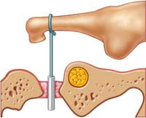
Furthermore, the prosthesis might have slipped into the vestibule if it is not tightly clipped to the incus or if it is too long (Fig. 24.1). In this case, the prosthesis should be replaced and properly clipped to the long process of the incus. Similar symptoms may be found with too long a prosthesis that needs to be replaced with an appropriate length.
Fig. 24.1
132 24 Stapes Revision Surgery
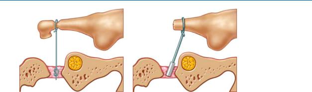
Fig. 24.2 |
Fig. 24.3 |
Late Revisions
Displacement of the Prosthesis
Incus erosion with loosening or displacement of the prosthesis is the most common finding in stapes revision surgery after several years (Fig. 24.2). Patients complain of deterioration of hearing, which often improves shortly after Valsalva’s maneuver. The prosthesis may be refixed by clipping (Fig. 24.3). In cases of severe destruction of the long process of the incus, the prosthesis can be clipped to the proximal part of the long process. Sometimes a groove must be drilled to prevent the prosthesis from slipping. The new prosthesis may have to be bent to adapt to the new anatomical situation. Alternatively, the prosthesis may be fixed using bone cement.
Complete Destruction of the Incus
If refixation of the prosthesis to the incus is impossible, a new slightly longer prosthesis may be attached to the handle of the malleus (malleus to oval window technique = malleovestibulopexy). After creating a tunnel between the fibrous tissue of the tympanic membrane and the bony handle, the wire of the prosthesis is positioned. The surgeon must be careful not to perforate the tympanic membrane because the tissue does not close over the metal ring of the prosthesis. If perforation accidentally occurs, a small thin piece of cartilage should be placed between the metal ring and tympanic membrane.
Reclosure of the Oval Window
The oval window may reclose under a short prosthesis. The reopening of the footplate is more dangerous than the primary operation because scar adhesions may have developed between the inner ear structures, and the footplate and the utricule or the saccule could be torn. Therefore we identify the promontorial side of the footplate and remove some adjacent bone until the vestibulum can be seen. A bony reclosure can now be perforated. Connective tissue can just be pushed cranially without removing tissue.

24 Stapes Revision Surgery 133
Fixation of Incus or Malleus
Malleus or incus fixation should not be overlooked during the first operation. It may also occur later after surgery due to inflammatory reaction after surgical trauma. The malleus to oval window technique as described above is the surgical solution. Drilling away the bony fixations is possible but requires much more extensive surgery.
Schuknecht’s Prosthesis
If a Schuknecht’s wire prosthesis is encountered during revision surgery, the wire must not be removed but cut and left in place. A new opening to the vestibulum is created next to the wire to place an additional new prosthesis.
Perilymphatic Fistula
Perilymphatic fistulas (PLFs) can develop in the region of the oval window inside the connective tissue around the prosthesis due to head injuries or rapid pressure shifts, etc. Patients usually complain of inconsistent vertigo, which might appear a long time after surgery. PLFs can be accompanied by a fluctuating or continuous sensory hearing loss, and in revision surgery they cannot be identified in any case. However, the fistula or the area of the oval window is covered with connective tissue after the tissue in the oval window especially around the fistula is scratched to bleeding.
Vertigo
Slight vertigo after stapes surgery is not uncommon and generally subsides. Disabling vertigo is extremely rare. Hearing can be normal or impaired. If vertigo cannot be controlled by conservative methods, the middle ear must be treated surgically. The prosthesis may be too long, it may have slipped from an eroded incus or a perilymphatic fistula may be seen; extremely seldomly a neurectomy may be indicated, depending on the hearing, using either a translabyrinthine or a transtemporal approach.

134 Chapter 25
25 Middle Ear Trauma
Henning Hildmann, Holger Sudhoff
Traumatic Tympanic Membrane Perforation
Ninety percent of traumatic perforations of the tympanic membrane will heal spontaneously. Myringoplasty must be performed in ears that fail to heal after 3 months, with a typical success rate of 95 %. No surgery should be performed before 3 months unless more serious destruction or foreign bodies are seen or suspected or inner ear symptoms indicate additional inner ear trauma. Subtotal perforations should be revised as soon as the patient is in a stable condition. Otherwise the patient should be instructed to keep water out of the ear and to return for regular checkups and treatment of any infection. Small perforations can be reconstructed by everting the edges with a hook the reconstruction is covered with silicone or cigarette paper. If the situation is unstable, Gelfoam or similar material soaked with an antibiotic solution can be placed on the promontory for support. Larger perforations require an underlay of fascia, performing a type I tympanoplasty using an endaural approach as described below. The middle ear should be checked if ossicular chain or inner ear damage is suspected or foreign bodies may have been introduced into the middle ear. The surgeon, particularly the less experienced, should not hesitate to perform the regular type I tympanoplasty he or she was trained to do instead of trying to reposition large defects. The latter carries the risk of chain subluxation in the office under semisurgical conditions.
Ossicular Chain Trauma
Injuries of the ossicular chain have various aetiologies. Skull traumas from blows to the temporal, parietal or occipital region with or without fracture of the temporal bone are the main causes; direct trauma, explosions, or thermal injuries are other possible causes. The type of injury that results depends upon the type of trauma. Mainly subluxations, luxations and fractures are seen. The incus is most frequently affected. Reconstruction follows the general rules of tympanoplasty. The surgeon should check for foreign bodies during surgery. Wooden splinters, moulding mass for ear moulds, cotton and other materials have been reported.
The stapes may be subluxed. In this case the oval window should be sealed. In complete luxations the stapes is replaced by a prosthesis as in otosclerosis surgery. Subluxations may be treated with repositioning of the footplate and surrounding it with connective tissue.

25 Middle Ear Trauma 135
Perilymph Fistula
The perilymphatic fistula is an opening between the perilymphatic space and the middle ear. They occur rarely in the setting of trauma and are most likely following barotraumas. Patients complain of varying intermittent vertigo. The symptoms may not be immediately apparent. Most frequently a stapes luxation is found, and more seldomly fractures involving other parts of the middle ear can be seen and have to be closed. After previous middle ear surgery a total ossicular prosthesis may perforate the footplate.
The fistulas in areas other than the round and oval window are covered with bone pat´e and a fascia graft or connective tissue grafts.
As a rare cause of vertigo, fistulas of the superior canal to the middle fossa have been described. The aetiology is unknown. After careful imaging and exclusion of the causes, the superior canal can be approached by a transtemporal approach.
Rupture of the Round Window
Ruptures of the round window are rare, occurring after head trauma and barotrauma. Bilateral ruptures are more likely after head trauma. Patients complain of vertigo tinnitus and a sensorineural hearing loss. The symptoms might be incomplete and fluctuating. The diagnosis is difficult since the fistula can only be visualized when a perilymph leak is seen after opening of the middle ear. The absence of perilymph does not exclude a fistula.
If a rupture is suspected the middle ear is opened as in stapes surgery. The round window niche is identified.
To expose the round window the bony ridge over the round window is removed with a small curette or a small drill. The round window membrane is almost perpendicular to the line of inspection. Sometimes an oozing of perilymph can be seen. The round window niche is covered by a small piece of connective tissue and the tympanomeatal flap is reflected to its normal position.
Traumatic Stenosis or Atresia
See surgery of the external ear canal.
Fractures of the Temporal Bone
Fractures of the temporal bone may be caused by direct or indirect impact. Extensive destructions of the lateral skull base exceed the scope of this book. The reconstruction should be adapted to the individual situation often in cooperation with the neighbouring specialities. The reconstruction of the tympanic membrane and the ossicular chain follows the principles of tympanoplasty and ear canal reconstruction as described in this book. Closure of the tympanic membrane prevents later infections of the middle ear and ascending infections of the intracranial space from outside. Reconstruction procedures of the ossicular chain are only sensible if the inner ear is not or is slightly