

Учебники / Middle Ear Surgery
.pdf86 15 Mastoid Cavity
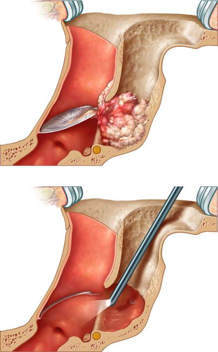
Fig. 15.18
Some cholesteatomas are not invasive and can easily be worked forwards through the antrum without extensive opening of the facial recess (Fig. 15.18). If the sac is worked forwards totally, its removal is safe. If it tears, the interior bony rim of the posterior wall must be controlled with an endoscope and the bone is cleaned with a large diamond burr. For larger cholesteatomas (Fig. 15.19), a wider access to the middle ear through the facial recess is necessary. A final check with an endoscope is helpful (Fig. 15.20).
The anterior epitympanum, the sinus tympani and the hypotympanum cannot be checked by this approach. If in cholesteatoma surgery the epithelium cannot be removed safely, the procedure must be converted to open surgery.
15 Mastoid Cavity |
87 |
|
|
Fig. 15.19
Fig. 15.20

88 15 Mastoid Cavity
Fig. 15.21
Obliteration
Complete obliteration (abdominal fat) with closure of the outer ear canal is seldom performed: especially after tumour removal or in very rare cases in deaf ears with large cholesteatomas and excessive extension of the pneumatic system. In these situations a blind sac closure of the outer ear canal without obliteration is another rare option if there is a possibility of residual cholesteatoma.
After removal of large cholesteatomas a large cavity requires a partial obliteration to facilitate the postoperative treatment and the comfort of the patient. Especially niches and recesses should be levelled to prevent the accumulation of debris. The critical areas are indicated in Fig. 15.21: anterior epitympanum, perilabyrinthine cells, subarcuate tract, retrofacial area, sinus-dura angle, and tip of the mastoid.
For obliterations different materials are described. We use bone pat´e, cartilage and soft tissue flaps. The bone pat´e is collected, and during drilling a col-

15 Mastoid Cavity |
89 |
|
|
Fig. 15.22
Fig. 15.23
lector may be used (Fig. 15.22). It can easily be pressed into niches to produce a smooth cavity. The pat´e must always be covered with connective tissue flaps or cartilage; otherwise it will not remain in place and cannot induce new bone formation.
If cartilage is used it can be placed as large plates or as palisades. Steps and irregularities must be avoided because the covering connective tissue flaps atrophy over time and the irregularities are seen and prevent self-cleansing.
Soft tissue flaps are harvested from the postauricular region. Besides larger free fascia grafts our standard flap is the Palva flap (Fig. 15.23). It is based retroauricularly and covers the outer part of the cavity well. However, it is seldom long enough to reach the lateral semicircular canal. This area should be covered with a free fascia graft (Fig. 15.24).
If a larger flap is needed, especially in revision surgery, the vascularized temporalis fascia flap supplied by a posterior branch of the temporal artery is used in combination with obliteration material. Autologous rib may be used for instance in brain hernias. These procedures require more extensive skin incisions.

90 15 Mastoid Cavity
Fig. 15.24
Fig. 15.25

15 Mastoid Cavity |
91 |
|
|
Reconstruction
The posterior wall can only be reconstructed if the middle ear is aerated and the airway to the mastoid is secured. As described above in small cholesteatomas, the disease is followed with the drill until the sac is removed. The middle ear is checked. There might be a block of aeration between the promontory and handle of the malleus. The chain is reconstructed. Finally the defect is closed with cartilage. Often the cartilage from the cymba fits well due to its slightly rounded shape. Generally one piece with overlapping perichondrium can be fitted (Fig. 15.25). Sometimes the perichondrium is used as additional covering separately. If the cartilage cannot be placed well, insertion as palisades (stripes) may be easier. No edges should be seen on the posterior wall since they might slow down epithelial migration after healing.
Total Reconstruction
For total reconstruction, in our experience cartilage has proven to be the easiest material to work with (Fig. 15.26). The fixation of larger plates is difficult
Fig. 15.26

92 15 Mastoid Cavity
because the cartilage plates tend to be unstable. Fibrin glue resorbs too early. For fixation notches are drilled into the remaining bony walls. Additional cartilage pieces may be necessary to cover spaces at the medial end of the inserted wall, preventing epithelial ingrowth.
Titanium Stabilization
Large defects of the posterior external auditory canal wall resulting from canal wall down technique or which are present in revision surgery have been eliminated by reconstruction using a titanium mesh in selected cases.
The titanium mesh is bent by manual manipulation and cut using special wire scissors, correcting the size to cover the defect of the posterior ear canal wall. It is placed slightly anterior to the facial ridge and reaching in depth the former bony tympanic annulus. The correct size is judged by intraoperative microscopic control of fitting and occasionally supports a 30°-angled endoscope. It is helpful to drill small ridges into the bone to facilitate the stabilization of the titanium mesh. The finally shaped titanium mesh is then removed from its future position. The mesh is subsequently covered with cartilage harvested generally in sufficient amounts from the cavum conchae. In cases of revision it may be helpful to split the cartilage in half to obtain ample material. The perichondrium is left attached to the cartilage. The conchal cartilage is secured with clamps and fixed with two resorbable sutures, such as Vicryl 4.0, on the mesh in order to keep it from moving. The knots are positioned towards the mastoidal segment of the titanium mesh. Uncovered titanium areas must be strictly avoided and access material resected. The composite titanium mesh is then attached to the remaining parts of the adjacent superior and inferior cortical bone and secured with two 3-mm titanium screws (Fig. 15.27). Subsequently it is covered with temporalis fascia and retroauricular split-thickness skin grafts. The canal is packed with Curaspon with an antibiotic ointment. The postauricular incision is closed in the usual manner and the packing removed 21 days after surgery.
Fistulas of the Semicircular Canals
Cholesteatoma tissue eroding a semicircular canal should be left in place until the completion of the procedure. Fistulas of the lateral semicircular canal are more frequent than those of the posterior or superior lateral canal. Fistulas should be expected in about 5 % – 7 % of cholesteatoma cases. Ninety percent of these fistulas are found in the lateral semicircular canal; in 6 % almost the complete labyrinthine portion was destroyed. A positive fistula sign was only present in about 60 % of the affected patients. Therefore the lack of a fistula sign does not exclude its existence. The removal of cholesteatoma matrix adjacent to the semicircular canals needs extra attention. Especially the facial nerve may be dehiscent and the matrix should be gently removed. The cholesteatoma matrix and granulation tissue are cautiously detached. The fistula is

15 Mastoid Cavity |
93 |
|
|
Fig. 15.27
closed with bone pat´e and fascia. The pat´e in over 75 % of patients will lead to osteoneogenesis, achieving its closure in 2 – 3 months. If there are heavily inflamed granulations, the matrix will be removed during second look surgery. The ingrowth of matrix into the lumen of the semicircular canals is rare.
Packing
The mastoid bowl is covered with silicone foil and packed with Curaspon with antibiotic ointment. The package should be left in place for at least 3 weeks.

94 Chapter 16
16 Specific Infections
Holger Sudhoff, Henning Hildmann
Specific infections are generally not expected before surgery but are diagnosed during the histological evaluation of the specimen. The surgical outcome does not differ in comparison to cases of unspecific infection if sufficient medical treatment is started. Even though tissue removal for histology is not routinely done in most centres, surgeons should be encouraged to take specimens more frequently at least in suspected cases.
Middle ear tuberculosis often has a typical whitish appearance and its granulations have a little tendency to bleed. The multiple perforations of the tympanic membrane described in some textbooks as being typical for middle ear tuberculosis have not been seen in our material. Biopsies should be sent for histology where typical Langerhans’ cells are found. As cultures may but often do not produce tubercle bacilli, identification by polymerase chain reaction has become of high diagnostic value. Postoperative general examination of the patient mostly shows other manifestations of the tuberculosis. The treatment is generally done by a pulmonologist according to the localizations and the extent of the disease.
Sarcoidosis may also be seen in patients with pulmonary affection. It is extremely rare and can be diagnosed by histological examination. Sarcoidosis should be considered in patients with lung disease.
Actinomycosis of the middle ear has been described in the literature and can also be identified by histological examination.
Wegener’s granulomatosis is a systemic inflammatory condition affecting the nose, kidneys, lungs and other organs.
Chapter 17 |
95 |
|
|
17 Complications
Jürgen Lautermann, Holger Sudhoff
Complications of infected and draining ears may arise from acute otitis media, cholesteatoma, and rarely from chronic suppurative otitis media. Bacteria and their toxins may spread by direct infective bone erosion, by preformed pathways such as the oval and round windows and by passing along vascular channels (osteo-thrombophlebitis). Anatomically, infective complications of the ear may be roughly divided into intratemporal, extradural and intradural complications. A high-resolution CT scan should be performed in every suspected complication supplemented by MRI if necessary. Rare causes are tuberculosis, Wegener’s granulomatosis and malignant external otitis.
Acute mastoiditis is the most common complication of an otitis media. It presents with retroauricular painful swelling, reddening, general sickness and an elevated erythrocyte sedimentation rate. The early stage of mastoiditis without signs of bone erosion in the CT scan may be treated with systemic antibiotics for a limited period of time. If symptoms progress or do not resolve within 24 h, a mastoidectomy in children simultaneously with myringotomy and/or grommet insertion and adenotomy should be performed to improve the tubal drainage to the epipharynx and to remove a source of inflammation. If uncontrolled the infection can spread into the surrounding tissues. Cholesteatoma can cause the similar complications.
Labyrinthitis and Facial Palsy
Labyrinthitis and facial palsy are rare intratemporal complications.
1.If facial paralysis originates from a cholesteatoma or mastoiditis, an immediate operation is obligatory. The cholesteatoma must be removed, the facial nerve must be identified and decompressed and the neural sheath must be incised if necessary.
2.Sensorineural hearing loss, vertigo and tinnitus indicate inner ear involvement and require revision of the mastoid as well as antibiotic treatment. Purulent labyrinthitis requires opening of the labyrinth.