

Учебники / Micro- and Nanotechnology for Neurotology Zeng 2006
.pdfOssicular Displacement
Silica-coated SNP, average diameter of 16 nm, with a zeta potential of –15 to –20 mV were internalized into epithelia of the tympanic membrane or that covering the incus. The density of SNP at the incus implant site was visible without magnification 8 days following implantation and the FITC-labeled SNP were visible under scanning confocal laser microscopy [fig. 6, 7 in Dormer et al., 2005]. Histopathology revealed no inflammatory response, no giant cells or evidence of apoptosis following 2–15 days of implantation. In this preliminary study, the displacements of the (intact) ossicular chain and tympanic membrane, as measured using single point interferometry, were comparable to 90 dB SPL displacements of the human middle ear [table 1, Dormer et al., 2005]. Frequency doubling occurred as the SNP responded to both polarities of the reversing electromagnetic field. This confirmed the superparamagnetic property of the magnetite SNP.
Discussion
The earliest use of external magnetic field to deliver clinical agents was in 1951, involving catheters for selective angiography. Magnetic microspheres were mostly studied until nanotechnology emerged in the late 1980s. Long-term deposition of iron in vivo is not a toxicity concern, as assessed epidemiologically in miners of hematite whose lung concentrations over lifetimes were 100–1000 times above those produced by drug targeting [Ranney, 1987]. Neither is adverse immunogenetic response a concern as iron is one of the most regulated cellular elements. Today, surface modifications can stabilize SNP in physiological solutions, protect against oxidation, provide functional groups for further derivitization and, in the case of polymeric encapsulation, carry and protect payloads en route to target tissues whereupon biodegradation will release payloads [Neuberger et al., 2005]. Future successes of SNP applications in nanomedicine, like other biomaterials, will be related to the extent of complete physicochemical characterizations (e.g. zeta potential) since surface chemistry dictates cell differentiation [Gupta and Gupta, 2005; Keselowsky and Garcia, 2005].
Middle Ear Biomechanics
For the first time it was shown that SNP, chronically implanted in a tissue, could be used to generate force, although performance data are lacking in this feasibility study [Dormer et al., 2005]. Current implantable middle
ear hearing devices (IMEHD) under development or in clinical trials employ active electronic actuators consisting of motors, solenoid type drivers, piezoelectric crystals or magnets driven by an external magnetic field [Huttenbrink, 1999]. Reduced surgical and device risk, lower cost and direct drive benefit may be an advantage of tissueindwelling, biocompatible SPN. Magnetite nanoparticles caused ossicular displacements in guinea pigs that were comparable to those in human temporal bones in response to a 90-dB SPL sound source. Nevertheless, the mass of the guinea pig ossicular chain is substantially less than in the human and was relatively easy to displace using an external magnetic field. Others have used magnetic particles to exert piconewton forces influencing (bone) cell differentiation [Cartmell et al., 2004]. RGDcoated microparticles bound to integrin receptors on primary human osteoblasts and an external magnetic field oscillated the cells in 2-D monolayer culture or 3-D constructs. Varying load-bearing matrices resulted.
Inner Ear Targeted Delivery
Of the prospective applications in nanomedicine, targeted delivery of therapeutics and enhanced MRI imaging, both utilizing nanoparticulate Fe3O4, may have the greatest clinical impact [Shinkai and Ito, 2004]. Delivering therapeutics only to target tissues may reduce both side effects and cost while improving treatment. Targeting of SNP by an external magnetic field had initially been explored for intravascular delivery. However, traversing the RWM provides a unique nanomedicine application where delivery particles will not be removed by reticuloendothelial organs. Like vascular targeting across the endothelium, inner ear delivery is independent of the membrane status and highly dependent on homogeneity of the magnetic field gradient in the target volume.
Substances with limited access to the inner ear may traverse (permeabilize) the RWM carrying a payload of drugs or genes to the inner ear. We have explored this targeted delivery for the first time utilizing SPN [Lee et al., 2004]. Our in vitro RWM model was used to initially identify candidate SNP for optimal targeted delivery to the inner ear (fig. 1a–c). In vivo testing in rat and guinea pig subsequently validated the RWM model, and we are currently refining the payload release from the biodegradable PLGA in perilymph. The model served to emulate the human RWM, penetrable to SNP, using external magnetic forces. Our results showed that cluster type aggregates (130 nm) containing 10 nm SPN were biocompatible and might be considered as carriers for therapeutic substances or as nonviral vectors for gene therapy. There
Magnetic Nanoparticles and Ear |
Audiol Neurotol 2006;11:123–133 |
131 |

was no observable effect of the SPN on growth and proliferation of human epithelial cells in culture. The SPN crossed the tripartite RWM model much more rapidly than diffusion because of the forces from an external magnetic field. It is not surprising that a small number of particles were detected in perilymph of a guinea pig not exposed to magnetic forces since these particles are small enough to diffuse through the RWM or to be transported through active processes. However, the particles were much more evident after exposure to a magnetic gradient. Future studies are aimed at gathering additional quantitative information.
PLGA nanospheres have been used previously as nonviral vectors of DNA and other biologically active compounds. Labhasetwar and others tested biodegradable nanoparticles ( 200 nm) consisting of PLGA and PVA [Panyam et al., 2002, 2004; Sahoo et al., 2004]. Particles have been loaded with wt-p53 plasmid DNA that transfected a breast cancer cell line [Prabha and Labhasetwar, 2004]. Sustained gene expression resulted from the slow intracellular release of the encapsulated DNA. Cellular uptake of PLGA particles 10–800 nm in diameter was confirmed by fluorescence of 6-coumarin and confocal microscopy [Qaddoumi et al., 2004]. Endocytosis appears to be involved in internalization and cationic surface treatment is facilitatory. PLGA nanoparticles (!200 nm) coated with a PVA-chitosan blend produce a cationic shell with the ability to electrostatically bind DNA to a nanosphere [Kumar et al., 2004a, b].
References
Therapeutic Perspectives
Inner ear medicine represents an expanding field that will benefit from improved targeted delivery strategies for a wide range of therapeutic small molecules, peptides, proteins, oligonucleotides and larger molecules containing genetic information. With the growing understanding of the molecular basis for traumatic, toxic, ischemic, inflammatory, infectious, and degenerative pathologies of the inner ear, specific efficient targeted delivery of mechanismbased therapeutics appears promising. In addition, plasmid gene delivery through an efficient, minimally invasive, safe method may increase the possibility of clinical auditory HC replacement with restoration of hearing. Here, in preliminary studies using materials comparable to those FDA approved and used clinically [Shinkai and Ito, 2004], we have demonstrated that readily achievable magnetic gradients can be created to enhance the delivery of paramagnetic nanoparticles with a biodegradable polymer payload into the mammalian inner ear.
Acknowledgements
Gratitude is expressed to Eric Howard, Associate Professor of Cell Biology, OUHSC, for his invaluable assistance in these ongoing studies.
This study was partially funded by The Shulsky Fund for Medicine and Research, New York City (via the Oklahoma City Community Foundation), the National Institute for Deafness and other Communicative Disorders (SBIR DC05528-01), the Office of Naval Research and INTEGRIS Baptist Medical Center, Oklahoma City.
K.J.D. and D.G. are stockholders in NanoBioMagnetics Inc.
 Adams JC: Clinical implications of inflammatory cytokines in the cochlea: a technical note. Otol Neurotol 2002;23:316–322.
 Aubert-Pouessel A, Venier-Julienne MC, Saulnier P, Sergent M, Benoit JP: Preparation of PLGA microparticles by an emulsion-extraction process using glycofurol as polymer solvent. Pharm Res 2004;21:2384–2391.
 Bala I, Hariharan S, Kumar M: PLGA nanoparticles in drug delivery: the state of the art. Crit Rev Ther Drug Carrier Syst 2004;21:387–422.
Cartmell S, Magnay J, El Haj A, Dobson J: Use of magnetic particles to apply mechanical forces for bone tissue engineering purposes. Int Conf Fine Particle Magnetism, London 2004, pp 41–42.
Dormer KJ: Implantable electronic otologic devices for hearing restoration; in Finn WE, Lopresti PG (ed): Handbook of Neuroprosthetic Research Methods. Boca Raton, CRC Press, 2003, pp 237–261.
 Dormer K, Seeney C, Lewelling K, Lian GD, Gibson D, Johnson M: Epithelial internalization of superparamagnetic nanoparticles and response to external magnetic field. Biomaterials 2005;26:2061–2072.
 Funhoff AM, van Nostrum CF, Lok MC, Fretz MM, Crommelin DJA, Hennink WE: Poly(3- guanidinopropyl methacrylate): a novel cationic polymer for gene delivery. Bioconjug Chem 2004;15:1212–1220.
 Gupta AK, Gupta M: Synthesis and surface engineering of iron oxide nanoparticles for biomedical applications. Biomaterials 2005;26:3995– 4021.
 Hoffer ME, Allen K, Kopke RD, Weisskopf P, Gottshall K, Wester D: Transtympanic versus sustained-release administration of gentamicin: kinetics, morphology, and function. Laryngoscope 2001;111:1343–1357.
 Huang T, Cheng AG, Stupak H, Liu W, Kim A, Staecker H, Lefebvre PP, Malgrange B, Kopke R, Moonen G, Van de Water TR: Oxidative stress-induced apoptosis of cochlear sensory cells: otoprotective strategies. Int J Dev Neurosci 2000;18:259–270.
 Huttenbrink KB: Current status and critical reflections on implantable hearing aids. Am J Otol 1999;20:409–415.
Izumikawa M, Minoda R, Kawamoto K, Abrashkin KA, Swiderski DL, Dolan DF, Brough DE, Raphael Y: Auditory hair cell replacement and hearing improvement by Atoh1 gene therapy in deaf mammals. Nat Med 2005, advanced online publication.
 Jeong JR, Lee SJ, Kim JD, Shin SC: Magnetic properties of Fe3O4 nanoparticles encapsulated with poly(D,L-lactide-co-glycolide). IEEE T Magn 2004;40:3015–3017.
132 |
Audiol Neurotol 2006;11:123–133 |
Kopke/Wassel/Mondalek/Grady/Chen/ |
|
|
Liu/Gibson/Dormer |

 Jiang HL, Jin JF, Hu YQ, Zhu KJ: Improvement of protein loading and modulation of protein release from poly(lactide-co-glycolide) microspheres by complexation of proteins with polyanions. J Microencapsul 2004;21:615–624.
Jones GE, Jackson RL, Liu J, Costello M, Ge X, Coleman JKM, Boasen JF, Harper EA, Kuzdak NE, Kopke RD: Three methods of inner ear trophic factor delivery to recover vestibular function and morphology from gentamicin-in- duced vestibular toxicity in the guinea pig. Abstracts 27th Annu Midwinter Res Meet, Assoc Res Otolaryngol, Daytona Beach, 2004.
 Juhn SK, Hamaguchi Y, Goycoolea M: Review of round window membrane permeability. Acta Otolaryngol Suppl 1988;457:43–48.
 Keselowsky BG, Garcia AJ: Quantitative methods for analysis of integrin binding and focal adhesion formation on biomaterial surfaces. Biomaterials 2005;26:413–418.
 Kopke RD, Coleman JK, Liu J, Campbell KC, Riffenburgh RH: Candidate’s thesis: enhancing intrinsic cochlear stress defenses to reduce noise-induced hearing loss. Laryngoscope 2002;112:1515–1532.
 Kopke RD, Hoffer ME, Wester D, O’Leary MJ, Jackson RL: Targeted topical steroid therapy in sudden sensorineural hearing loss. Otol Neurotol 2001a;22:475–479.
 Kopke RD, Jackson RL, Li GM, Rasmussen MD, Hoffer ME, Frenz DA, Costello M, Schultheiss P, Van de Water TR: Growth factor treatment enhances vestibular hair cell renewal and results in improved vestibular function. Proc Natl Acad Sci USA 2001b;98:5886–5891.
 Kopke RD, Liu W, Gabaizadeh R, Jacono A, Feghali J, Spray D, Garcia P, Steinman H, Malgrange B, Ruben RJ, Rybak L, Van de Water TR: Use of organotypic cultures of Corti’s organ to study the protective effects of antioxidant molecules on cisplatin-induced damage of auditory hair cells. Am J Otol 1997;18:559– 571.
 Korver KD, Rybak LP, Whitworth C, Campbell KM: Round window application of D-methio- nine provides complete cisplatin otoprotection. Otolaryngol Head Neck Surg 2002;126: 683–689.
 Kumar M, Bakowsky U, Lehr CM: Preparation and characterization of cationic PLGA nanospheres as DNA carriers. Biomaterials 2004a;25:1771–1777.
 Kumar M, Mohapatra SS, Kong X, Jena PK, Bakowsky U, Lehr CM: Cationic poly(lactide-co- glycolide) nanoparticles as efficient in vivo gene transfection agents. J Nanosci Nanotechnol 2004b;4:990–994.
Lee SJ, Jeong JR, Shin SC, Kim JC, Chang YH, Chang YM, Kim JD: Nanoparticles of magnetic ferric oxides encapsulated with poly(D,L lactide-co-glycolide) and their applications to magnetic resonance imaging contrast agent. JMMM 2004;272–76:2432–2433.
Lian G, Lewelling K, Johnson M, Gibson D, Dormer K, Seeney C: Fe3O4 magnetite nanoparticles coated by silica for biomedical applications. Microsc Microanal 2004;10(suppl 2):3.
Mamedova N, Kopke R, Liu J, Jackson R, Costello M, Gibson D, Dormer K: Feasibility of superparamagnetic nanoparticles for drug delivery to the inner ear. Abstracts 28th Annu Midwinter Res Meet – Assoc Res Otolaryngol, New Orleans 2005.
 Massart R: Preparation of aqueous magnetic liquids in alkaline and acidic media. IEEE T Magn 1981;17:1247–1248.
Neuberger T, Schopf B, Hofmann H, Hofmann M, von Rechenberg B: Superparamagnetic nanoparticles for biomedical applications: possibilities and limitations of a new drug delivery system. JMMM 2005;1:483–496.
 Nicotera TM, Ding D, McFadden SL, Salvemini D, Salvi R: Paraquat-induced hair cell damage and protection with the superoxide dismutase mimetic M40403. Audiol Neurootol 2004;9: 353–362.
 Pankhurst QA, Connolly J, Jones SK, Dobson J: Applications of magnetic nanopaticles in Biomedicine. J Phys D Appl Phys 2003;36:R161– R181.
 Panyam J, Dali MA, Sahoo SK, Ma WX, Chakravarthi SS, Amidon GL, Levy RJ, Labhasetwar V: Polymer degradation and in vitro release of a model protein from poly(D,L-lactide-co-gly- colide) nanoand microparticles. J Control Release 2003;92:173–187.
 Panyam J, Williams D, Dash A, Leslie-Pelecky D, Labhasetwar V: Solid-state solubility influences encapsulation and release of hydrophobic drugs from PLGA/PLA nanoparticles. J Pharm Sci 2004;93:1804–1814.
 Panyam J, Zhou WZ, Prabha S, Sahoo SK, Labhasetwar V: Rapid endo-lysosomal escape of poly(DL-lactide-co-glycolide) nanoparticles: implications for drug and gene delivery. FASEB J 2002;16:1217–1226.
 Prabha S, Labhasetwar V: Nanoparticle-mediated wild-type p53 gene delivery results in sustained antiproliferative activity in breast cancer cells. Mol Pharm 2004;1:211–219.
 Prabha S, Zhou WZ, Panyam J, Labhasetwar V: Size-dependency of nanoparticle-mediated gene transfection: studies with fractionated nanoparticles. Int J Pharm 2002;244:105– 115.
 Puig-De-Morales M, Grabulosa M, Alcaraz M, Mullol J, Maksym GN, Fredberg JJ, Navajas D: Measurement of cell microrheology by magnetic twisting cytometry with frequency domain demodulation. J Appl Physiol 2001;91: 1152–1159.
 Qaddoumi MG, Ueda H, Yang J, Davda J, Labhasetwar V, Lee VHL: The characteristics and mechanisms of uptake of PLGA nanoparticles in rabbit conjunctival epithelial cell layers. Pharm Res 2004;21:641–648.
Ranney DF: Magnetically controlled devices and biomodulation; in Tyle P (ed): Drug Delivery Devices. New York, NY, Marcel Dekker, 1988, pp 325–368.
 Ravi R, Somani SM, Rybak LP: Mechanism of cisplatin ototoxicity – antioxidant system. Pharmacol Toxicol 1995;76:386–394.
 Ryan AF, Harris JP, Keithley EM: Immune-medi- ated hearing loss: basic mechanisms and options for therapy. Acta Otolaryngol 2002;122: 38–43.
 Sahoo SK, Ma W, Labhasetwar V: Efficacy of transferrin-conjugated paclitaxel-loaded nanoparticles in a murine model of prostate cancer. Int J Cancer 2004;112:335–340.
 Seidman M, Van de Water T: Pharmacologic manipulation of the labyrinth with novel and traditional agents delivered to the inner ear. Ear Nose Throat J 2003;82:276–280, 282–283, 287–288.
 Shinkai M, Ito A: Functional magnetic particles for medical application. Adv Biochem Eng Biotechnol 2004;91:191–220.
 Song BB, Anderson DJ, Schacht J: Protection from gentamicin ototoxicity by iron chelators in guinea pig in vivo. J Pharmacol Exp Ther 1997; 282:369–377.
 Suzuki M, Yamasoba T, Suzukawa K, Kaga K: Adenoviral vector gene delivery via the round window membrane in guinea pigs. Neuroreport 2003;14:1951–1955.
 Wang J, Van de Water TR, Bonny C, de Ribaupierre F, Puel JL, Zine A: A peptide inhibitor of c-Jun N-terminal kinase protects against both aminoglycoside and acoustic trauma-in- duced auditory hair cell death and hearing loss. J Neurosci 2003;23:8596–8607.
 Witte MC, Kasperbauer JL: Round window membrane permeability to transforming growth fac- tor-alpha: an in vitro study. Otolaryngol Head Neck Surg 2000;123:91–96.
 Yamamoto H, Kuno Y, Sugimoto S, Takeuchi H, Kawashima Y: Surface-modified PLGA nanosphere with chitosan improved pulmonary delivery of calcitonin by mucoadhesion and opening of the intercellular tight junctions. J Control Release 2005;102:373–381.
 Zhang YY, Kropp BP, Moore P, Cowan R, Furness PD, Kolligian ME, Frey P, Cheng EY: Coculture of bladder urothelial and smooth muscle cells on small intestinal submucosa: potential applications for tissue engineering technology. J Urol 2000;164:928–934.
Magnetic Nanoparticles and Ear |
Audiol Neurotol 2006;11:123–133 |
133 |


Yamamoto et al., 2002]. These include soluble tropic factors such as growth factors, which are released into the extracellular fluid. Neurites can respond to gradients of these soluble factors with directional growth [e.g. Cao and Shoichet, 2003; Gillespie, 2003; Yoshikawa and Thomas, 2004]. Patterns of extracellular matrix (ECM) molecules such as laminin, fibronectin or collagen, which may be components of extracellular structures or be bound to their surfaces, can provide additional cues [e.g. Gillespie, 2003; Gallo and Letourneau, 2004; Hari et al., 2004; Oster et al., 2004; Yoshikawa and Thomas, 2004]. A wide variety of molecules that reside on the surfaces of cells, such as components of the ephrin/Eph bidirectional signaling system [Marquardt et al., 2005], can provide directional signals at the cellular and the subcellular level.
Many known neurite guidance molecules have been localized to regions or cells of the developing cochlea that are appropriate for a role in regulating neurite growth, providing the substrates for guidance of afferent and efferent projections. Neurotropic growth factors present in the developing organ of Corti include neutrotrophin-3 (NT-3), brain-derived neurotrophic factor and acidic fi- broblast growth factors (FGF-1), all of which are produced by hair cells [Luo et al., 1993; Pirvola et al., 1992, 1994; Ernfors et al., 1994]. Many ECM molecules including fibronectin, laminin, tenascin and various collagens occur in the organ [e.g. Woolf et al., 1992; Tsuprun and Santi, 1999, 2001; Whitlon et al., 1999]. Several ephrins and Eph receptors have also been identified in the developing inner ear [Bianchi and Liu, 1999; van Heumen et al., 2000; Pickles et al., 2002].
Identifying the role played by these individual molecules in spiral ganglion (SG) neurite guidance has proven to be more difficult. Gene deletion studies have provided information, but can result in the death of neurons, e.g. tropic molecules that also provide trophic support [Ernfors et al., 1994; Farinas et al., 1994; Fritzsch et al., 1997a, 1998], or even the organism, e.g. many ECM molecules [George et al., 1993]. While in vitro techniques clearly have their limitations, they can be used to probe different developmental periods independently, in a way that gene deletion studies often cannot. Studies of neurons in culture have the additional advantage that the environment in which neurites are extending can be precisely controlled. In particular, they allow potential guidance factors to be presented in controlled patterns.
This is critical since, in order for a substance to provide directional information to advancing neurites, it must be differentially distributed. Variation in distribution can take many forms, from abrupt gradients in concentration
that are essentially step-like at the dimension of the sampling growth cone, to gradual gradients that extend over many times the sensing radius of the neuron. For example, recent evidence indicates that gradients of soluble neurotropic factors can contribute to neurite guidance over distances of more than 10 mm [Cao and Shoichet, 2003]. Distribution of insoluble substances may be punctate, or can occur in patterns that relate to the underlying structure of the tissue. For example, in rodents, fibronectin appears to occur in tracts that run beneath the hair cells, during the first postnatal week [Woolf et al., 1992]. Cell surface molecules can be confined to individual cells or to cellular regions, or may be broadly distributed across tissues.
We have taken advantage of methods for micropatterning guidance molecules to directly assess SG neurite guidance in culture. Insoluble molecules have been patterned as edges, stripes and puncta. Soluble factors have been represented as discrete fluid gradients or localized cellular sources. Taken together, our studies suggest that SG neurites can respond directionally to differentially distributed guidance factors.
Methods
Culture of SG Neurons
Surgical procedures were approved by the local animal subject committee in accordance with the guidelines laid down by the NIH regarding the care and use of animals for experimental procedures. Threeto five-day-old (P3–P5) rat pups were decapitated and the skulls opened midsagitally under sterile conditions. The membranous labyrinth was exposed by peeling off the cartilaginous cochlear capsule under a dissecting microscope. The stria vascularis and the organ of Corti were removed in order to expose the spiral ganglia. The SG was excised from the entire length of the cochlea and divided into explants that were approximately 300 ! 300 m.
Culture procedures were based on those developed by Van De Water and Ruben [1971] and have been described in detail elsewhere [e.g. Brors et al., 2003]. Briefly, individual explants were cultured on surfaces coated with poly-L-lysine (PLL). Additional molecules were applied to this surface in various patterns as described below. The tissue was initially incubated in attachment media consisting of DMEM, 10% FCS, 5% HEPES and 30 units/ml penicillin for 24 h at 37°C, 5% CO2. After 24 h, the culture medium was changed to maintenance media consisting of DMEM supplemented with 1 ! N2 and 5 g/l glucose. Cultures were maintained for 72–96 h in maintenance media. Various soluble factors were included as described below.
Fixation and Immunohistochemistry
After culture in maintenance media, explants were fixed with 4% paraformaldehyde for 20 min and then washed with PBS. The samples were blocked with 1% donkey serum for 10 min at room temperature to reduce nonspecific binding. Specimens were incubated with rabbit polyclonal anti-200 kDa neurofilament antibody
Micropatterning Neurite Guidance Cues |
Audiol Neurotol 2006;11:134–143 |
135 |
After exposure, the photoresist was developed, and the mold master was cleaned, baked and dried. It was then attached to an aluminum plate prior to silicon wafer removal. The pattern was then molded in polydimethylsiloxane (Sylgard 184, Dow Corning) and cured to the desired level of elasticity.
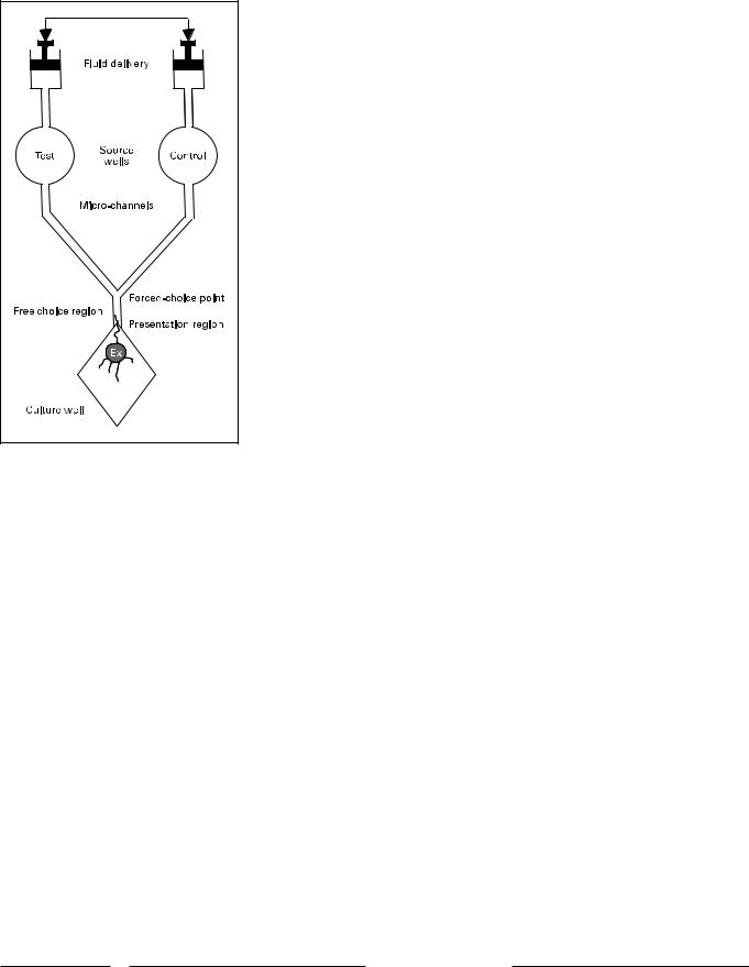
Fig. 1. Schematic diagram of a microfluidic device for creation of a fluid gradient and choice point for growing neurites. The device is generated by photolithography and molding. Test and control fluids are forced through a microchannel network at equal rates. They meet at the forced-choice point of a ‘Y’, and then flow in parallel through a free-choice presentation region to the tissue culture well.
(Sigma-Aldrich) diluted 1:500 at 4°C overnight. Explants were then incubated in donkey antirabbit secondary antibody (Jackson ImmunoResearch) diluted 1:100 in PBS. Either fluorescent or DAB labeling was used to visualize the secondary antibody. Immunolabeling controls in which rabbit serum was substituted for the primary antibody exhibited no labeling. Neurites were digitally imaged on a fluorescence inverted microscope (Olympus).
Microfabrication
Photolithographic techniques used to generate molds for microfluidic devices and for masks used in surface patterning have been described in detail elsewhere [Wittig et al., 2005]. Briefly, mold patterns were generated as negative images in Canvas 5.0 (Deneba Systems, Inc.) and printed as high-resolution transparencies with multiple patterns. The transparencies were used to expose the desired areas of positive photoresist (SU-8 100 or SU-8 1000, Microchem Corp.) spun into a uniform layer of the desired thickness on a 4-inch silicon wafer to ultraviolet light. When a second layer and transparency were required, they were aligned to the initial exposed pattern, which had become translucent.
Results
Soluble Guidance Cues
A microfluidic device for the evaluation of soluble guidance cues in fluid was developed by Wittig et al. [2005]. The device was fabricated and tested for production of fluid gradients with a fluorescent dye. A schematic of the device is illustrated in figure 1. Fluid is delivered at equal rates from two source wells by identical, physically ganged syringes. The resulting fluid gradients are illustrated in figure 2. As the two fluid streams unite at the union of the microchannel ‘Y’ a steep gradient is formed across the 100- m width of the stem of the Y. Dye concentration transitions from 195% of peak concentration to !5% of peak concentration in 20–35 m. This gradient is maintained until the fluid streams exit the narrow stem of the ‘Y’ into an expanding culture well, when the gradient becomes increasingly diffuse. Thus in an experiment using a biologically active diffusible mediator on one side of the device, neurites extending from an SG explant toward the microchannel encounter at first a diffuse and then, as they enter the microchannel, a sharp fluid gradient of the factor.
SG explants presented with a choice between NT-3 and control media showed a strong preference for growth in NT-3. This resulted in changes in neurite direction within the diffuse gradient at the exit from the ‘Y’, in preferential growth of neurites on the NT-3 side of the stem of the ‘Y’ and growth into the NT-3 side of the ‘Y’ bifurcation of a majority of neurites, as illustrated in figure 3. When NT-3 was presented from both delivery wells, no preference for side was observed (data not shown).
Insoluble Guidance Cues
A variety of strategies were employed to produce restricted distributions of substrate-bound guidance cues. Initially, silicone masks were fabricated with a single longitudinal gap. When fitted into a tissue culture well, the mask blocked all but a single stripe of culture surface, to which substances could be attached from solution by incubation. This method produced an abrupt edge of the attached guidance cue over 20 m at the edge of the stripe. SG explants could be placed outside of or on the
136 |
Audiol Neurotol 2006;11:134–143 |
Ryan/Wittig/Evans/Dazert/Mullen |

Pixel intensity (%)
100 |
|
|
|
|
A |
50 |
|
|
|
|
|
0 |
|
|
|
|
|
0 |
20 |
40 |
60 |
80 |
100 |
100 |
|
|
|
|
B |
|
|
50 |
|
|
|
|
|
|
|
0 |
|
|
|
|
|
|
|
0 |
20 |
40 |
60 |
80 |
100 |
|
|
100 |
|
|
|
|
|
|
C |
|
|
|
|
|
|
|
|
50 |
|
|
|
|
|
|
|
0 |
|
|
|
|
|
|
|
0 |
20 |
40 |
60 |
80 |
100 |
120 |
140 |
|
|
|
Distance ( m) |
|
|
|
|
Fig. 2. Test of the fluid gradients produced by delivery of a fluorescent dye versus clear fluid to the forced-choice (A), free-choice (B) and presentation (C) regions of the device. A sharp gradient is created in the forced-choice and free-choice regions. A diffuse gradient is created when the microchannel exits into the presentation region, in both the horizontal and vertical dimensions. Adapted from Wittig et al. [2005].
Fig. 3. Neonatal rat SG explants grown in the microchannel device for 72 h, with delivery of 10 ng/ml NT-3 in neuronal media
(+) versus media alone (–). The explants were subsequently fixed and stained with antineurofilament antibody to visualize neurites. A A sharp inflection in neurite projection toward the microchannel can be observed. B Neurites and associated cells are preferentially observed on the side of the free-choice region that contains NT-3, and in the NT-3-containing microchannel beyond the forced-choice point. Adapted from Wittig et al. [2005].
Micropatterning Neurite Guidance Cues |
Audiol Neurotol 2006;11:134–143 |
137 |

Fig. 4. Neonatal rat SG neurites (green) at the edge of a stripe of EphA4 (dashed line) exhibit growth cone collapse (arrows), termination of extension, and reversal of growth trajectory. From Brors et al. [2003].
Fig. 5. Neurites from a neonatal SG explant (red), grown on a pattern of laminin stripes (green). The neurites demonstrate a strong preference to grow off of laminin. Adapted from Evans et al. [2005].
stripe, to evaluate their response to positive or negative edges, respectively.
This method was used to evaluate the responses of SG neurites to boundaries of the ECM molecule fibronectin [Aletsee et al., 2000], and the Eph receptor EphA4 [Brors et al., 2003]. Fibronectin boundaries proved to inhibit neurite growth, producing a strong tendency for neurites to stop and/or turn in response to a positive edge (data
not shown). EphA4 was actively repulsive, invariably causing neurites to exhibit growth cone collapse and reversal of growth trajectory (fig. 4). No response to a neutral control protein (bovine serum albumin, BSA) was observed (data not shown).
To simulate linear tracts of ECM molecules in the organ of Corti, photolithographic microfabrication techniques were used to manufacture a mask with repeating
138 |
Audiol Neurotol 2006;11:134–143 |
Ryan/Wittig/Evans/Dazert/Mullen |

Fig. 6. Interaction of neurites from an SG explant with point sources of FGF-1. A The neurites cultured with control beads (open arrows) follow a simple trajectory. B The neurites encounter a cluster of FGF-1-coat- ed beads (solid arrows). They exhibit extensive branching. Analysis of numerous such contacts revealed a significant increase in branching (p ! 0.01). From Aletsee et al. [2003].
Fig. 7. Response of SG neurites to a cellular source of FGF-1. In the left panel, neurites grow over control mouse 3T3 fibroblasts. In the right panel, neurites encountering 3T3 cells that have been stably engineered to express human FGF-1 on the cell surface branch and form bouton-like terminations (arrows) on the cells. From Dazert et al. [1998].
microchannels that, after placement on a culture surface, could be filled with fluid containing an ECM molecule, for incubation [Evans et al., 2005]. After removal of the mask and application of PLL to coat the regions between the ECM stripes, this resulted in a pattern of narrow, repeating stripes of the cue.
When SG explants were grown on patterns of laminin, they showed a highly significant (p ! 0.001), dose-depen- dent tendency for preferential growth. When stripes were produced by incubation with low concentrations of laminin, the neurites were preferentially found on laminin. At higher concentrations, the reverse was seen. In the example illustrated in figure 5, neurites exhibit a strong pref-
Micropatterning Neurite Guidance Cues |
Audiol Neurotol 2006;11:134–143 |
139 |

erence to grow off of stripes produced by incubation with 40 g/ml of laminin. In the figure it can be noted that especially neurites approaching the laminin stripes at angles below about 45° tended to grow off of laminin onto PLL. In contrast, neurites preferred PLL to fibronectin at all concentrations, while control stripes of BSA had no effect (data not shown).
Cells that display guidance cues on their surfaces can act as punctate sources. For example, during the first postnatal week, rat hair cells produce high levels of FGF-1. Aletsee et al. [2003] modeled this phenomenon using 2- to 3- m Sepharose microbeads coated with FGF-1, a growth factor that often binds to ECM molecules. SG neurites that encountered beads coated with BSA showed no response. Neurites that encountered FGF-1-coated beads exhibited extensive branching, although they did not terminate on the beads (fig. 6).
Dazert et al. [1998] used an alternative method to model FGF-1 production by hair cells. They co-cultured SG explants with 3T3 fibroblasts that were stably transfected to produce human FGF-1. The cells were placed peripheral to the explants, and were sufficiently different in morphology (larger and more spread out) from cells migrating out of the explants that they could easily be distinguished. In the construct used for transfection, the FGF-1 signal peptide was replaced with that for FGF-3, so that the FGF-1 remained bound to the cell surface rather than being secreted into the surrounding medium. While SG neurites showed a tendency to grow over control fibroblasts, they exhibited branching in response to FGF-1-producing cells. Moreover, they also formed bou- ton-like contacts on the cells (fig. 7).
Discussion
The results presented in this review demonstrate that patterning of neuronal guidance cues, on a microscale that is relevant to sampling by growth cone filopodia, provides a means by which to investigate SG neurite guidance in culture. Using a variety of methods it was possible to produce spatial patterns of cues, as experimental models of distributions that occur in the developing cochlea. In particular, patterning methods based upon photolithographic techniques allowed precise micropatterning. These methods were derived from many previous studies in other systems.
Stripe assays have a long history of use in the evaluation of neurite pathfinding. For example, Walter et al. [1987] created stripes using the cell membranes from ho-
mogenates of different target tissues. They employed a silicon mask with parallel channels similar to that used in the present study. The mask was placed to selectively block (in a striped pattern) the insertion of a membrane suspension into a capillary pore filter when suction was applied beneath the filter. Chick retinal ganglion explants were then cultured on this surface after the matrix was removed and a different membrane suspension was used to fill the empty stripes on the filter. More biochemically specific studies have used purified proteins as substrata. Employing methods similar to those of the present study, based on photolithographic techniques, Vielmetter and Stuermer [1989], Nguyen-Ba-Charvet et al. [2001], Weinl et al. [2003], and Jain et al. [2004] evaluated the responses of primarily retinal neurites to stripes of various substrates.
Our use of photolithography to generate microfluidic networks was also based upon a rich prior history. Methods for the rapid production of microfluidic systems using a positive relief microchannel structure on silicon molded with polydimethylsiloxane were outline by Duffy et al. [1998]. This technique formed the basis for a network used to create complex concentration gradients [Dertinger et al., 2001, 2002] with laminar fluid flow through microchannels. Previous microfluidic designs permanently bonded fluidic delivery tubes, microchannel layers, and surface layers such that each unit could be used only once [Duffy et al., 1998; Dertinger et al., 2001]. We used a compression plate to make each unit reusable across experiments. The compression plate incorporates a fluidic delivery system, simplifying connection of hydraulic pumps (syringe pumps) to the cell culture plate. An additional innovation was to apply the microchannel device to extending neurites.
Using micropatterning, we have found that neonatal SG neurites are sensitive to spatial patterns of a number of potential guidance cues. In particular, SG neurites appeared to display directional growth in response to both diffuse and sharp spatial differences in the concentration of NT-3. Diffuse differences appeared to induce turning, while a near-step difference in concentration produced a tendency for growth restricted to the NT-3 side. While these behaviors can be interpreted to reflect directional responses of SG neurites, an alternate possibility is preferential survival. It is possible that when neurites grew into media without NT-3, the associated neuron died, leaving only neurites in the NT-3 region. We feel that this explanation is less likely than directional behavior, since we and others have found that the requirement for NT-3 is not complete for postnatal P3–P5 neurites extending
140 |
Audiol Neurotol 2006;11:134–143 |
Ryan/Wittig/Evans/Dazert/Mullen |