

Учебники / Rhinosinusitis - A Guide for Diagnosis and Management 2008
.pdf3 Diagnosis and Management of Acute Rhinosinusitis |
31 |
bradykinin, and various cytokines. A downstream effect of this inflammation is the suppression of macrophage and lymphocyte function, which creates a milieu susceptible to bacterial infection and overgrowth [4,5].
Bacteria subsequently superinfect the sinonasal mucosa, as shown by repeated sinus aspiration studies demonstrating that 60% of adults with URI symptoms for 10 days or more have significant bacterial growth in sinus cultures. Isolates from maxillary sinus aspirates show that the most common pathogens are Streptococcus pneumoniae and Haemophilus influenzae, together comprising more than half of the bacterial isolates. Figure 3.1 displays the incidence of bacterial pathogens in acute maxillary rhinosinusitis in an adult population [6].
Epidemiology
Acute rhinosinusitis, either viral or bacterial, carries a significant health burden in the United States even with the most conservative estimates. The most recent data from the National Health Interview Study showed that rhinosinusitis is the most common respiratory disease among Americans, with 13% having been told by a doctor that they have rhinosinusitis in the last year. Female respondents were nearly twice as likely to have the diagnosis; and it was also more common in the South than in other regions of the country [7]. There is a significant cost associated with acute rhinosinusitis, both in health care as well as in the workforce. Rhinosinusitis also ranks in the top 10 most costly physical health conditions affecting U.S. employers [8].
Given that acute rhinosinusitis has such a high prevalence, it is imperative that health care providers are able to accurately diagnose the condition and appropriately prescribe antibiotics. As one recent study illustrated, 81% of patients diagnosed with acute rhinosinusitis have received antibiotics [9]. Appropriate use of antibiotics is vital to avoid a further increase in antibiotic resistance [10]. Prescribing patterns can change the prevalence of specific drug resistance. In Finland, macrolide-resistant group A streptococcal isolates decreased by 50% (from 16.5% to 8.6% of isolates) when new practice guidelines recommended a decrease in macrolide consumption [11].
Presenting Symptoms and Signs
Fever, Nasal Obstruction, Pain, Headache, Purulent Rhinorrhea
Because a diversity of health care professionals are diagnosing and treating acute rhinosinusitis, the bulk of the diagnosis rests on an individual patient’s symptoms and findings on physical examination: purulent nasal drainage, nasal obstruction, and facial pain/pressure/fullness. Although early consensus reports used the major and minor criteria discussed, the more recent reports have strayed from this concept and focused instead on only major symptoms [1–3].
32 |
K.A. Kolln,¨ B.A. Senior |
The time-course of the symptoms then becomes the next most important factor in distinguishing VRS from ABRS. VRS tends to be self-limited, with symptoms peaking at day 2 to 3 and then waning with resolution of symptoms between 10 and 14 days after onset. If symptoms initially improve and then subsequently worsen, or if symptoms persist beyond 10 days, the probability of bacterial infection is increased and the diagnosis of ABRS can be made [2,6,12].
On physical examination, anterior rhinoscopy reveals hyperemia of the nasal mucosa and nasal congestion. If purulence is visualized the diagnosis is secured; pain on palpation over the individual sinuses may aid in the diagnosis. Pharyngeal irritation or purulence in the posterior or lateral pharynx can also be used to aid in the proper diagnosis.
Associated Factors
During evaluation for acute rhinosinusitis, associated factors must be considered. The European Academy of Allergology and Clinical Immunology (EAACI) recommends a query for allergic symptoms such as sneezing, watery rhinorrhea, nasal itching, and itchy watery eyes [3]. Although there are limited data, it does appear that individuals with baseline allergic symptoms may be at increased risk for bacterial rhinosinusitis. Alho and colleagues examined 48 individuals during the first days of a viral upper respiratory infection [13]. Evaluation included a paranasal sinus computed tomography (CT) scan at the initial evaluation and then again after 21 days. The individuals with allergic rhinitis (19%) had significantly poorer CT scan results when compared with nonallergic subjects both initially and at follow-up. These results may indicate impairment in mucociliary clearance with a subsequent predisposition to the development of ABRS [13].
Other factors that must be considered include unilateral symptoms (foreign body, tumor), history of trauma or prior surgery, presence of immunosuppression or systemic disease (Wegener’s granulomatosis, sarcoidosis), or impairments in mucociliary clearance (cystic fibrosis, primary ciliary dyskinesia).
Sinonasal Endoscopy
Examination with a 0◦ endoscope reveals hyperemia, congestion, crusting, and purulence emanating from the sinuses. The location of the purulence can help in localizing the infection, as purulence in the middle meatus streaming anterior to the eustachian tube orifice originates from the maxillary, anterior ethmoid, and frontal sinuses whereas purulence seen in the sphenoethmoid recess and above the eustachian tube orifice comes from the sphenoid and posterior ethmoid sinuses. Endoscopy yielded a sensitivity of 80% and specificity of 94% in one retrospective review, which was an improvement from the standard used in this study, which was standard X-ray [14]. Although sinonasal endoscopy is considered standard in an otolaryngological practice, this is not the case for most practitioners who are confronted with the challenge of diagnosing ARS. Therefore, sinonasal endoscopy
3 Diagnosis and Management of Acute Rhinosinusitis |
33 |
is not considered necessary for an accurate diagnosis, although it should be utilized if available as this enables the practitioner to culture any purulence that is visualized.
Sinonasal Culture
The gold standard for sinus culture has been the maxillary sinus tap via a trocar through the canine fossa or with a needle through the inferior meatus [15]. Although this method is considered the standard for pharmaceutical trials, it is impractical in the clinical realm. Patients are unlikely to agree to the procedure secondary to real or perceived discomfort, and most practitioners treating acute bacterial rhinosinusitis (ABRS) are not skilled in performing the procedure. Nasal cavity swabs, although easily performed, have not been shown to reliably identify a causative organism without endoscopic guidance, as these cultures have only a 65% concordance rate with maxillary antral cultures [16].
With the advent of sinonasal endoscopy, a more directed and less invasive culture technique was introduced. One of the early studies evaluating endoscopic middle meatal cultures (EMMC) was performed by Vogan and colleagues in 2000 [17]. EMMC and antral puncture were performed on 16 individuals presenting with symptoms of acute rhinosinusitis and maxillary sinus air-fluid levels on CT. This group reported a concordance rate of 93.8% for aerobic culture and 87.4% for anaerobic culture [17]. Further support for the validity of EMMC was demonstrated by two meta-analyses, the first in 2005 by Dubin et al., that revealed accuracy of 82% per isolate when compared with maxillary sinus aspirates [18]. Benninger and colleagues then described a sensitivity of 80.9% and specificity of 90.5%, a positive predictive value of 82.6%, a negative predictive value of 89.4%, and an overall accuracy of 87% of EMMC [19].
Culture-directed therapy, although ideal, has remained elusive in the majority of cases of acute rhinosinusitis. The majority of practitioners treating ABRS do not have endoscopic tools at their disposal and, therefore, cultures are reserved for complex and recalcitrant cases necessitating specialty care.
Laboratory Data
Although not routine, serologic markers for inflammation may be helpful in the diagnosis of ABRS. In 1995, Hansen et al. reported on a cohort of 174 patients with physician suspected rhinosinusitis, and the diagnosis was confirmed with maxillary antral puncture in 92 (53%) [20]. The diagnosis was then correlated with CT imaging, physical signs and symptoms, and erythrocyte sedimentation rate (ESR) and C-reactive protein (CRP). Only an ESR more than 20 mm/h in females and more than 10 mm/h in males, as well as a CRP more than 10 mg/l, were significantly associated with the correct diagnosis [20]. Other groups have also supported the use of ESR for increasing the positive predictive value of the diagnosis, but this has not been supported as being cost-effective by others [3,21].
34 |
K.A. Kolln,¨ B.A. Senior |
Imaging
Ultrasound
As a cost-effective means for aiding in the diagnosis of acute maxillary rhinosinusitis, ultrasound has been recommended. Although rapid and noninvasive, the technology is operator dependent and, therefore, has not gained support for standard diagnosis. In one study of 197 adults with symptoms of the common cold, ultrasonography and Waters’ view were performed, and magnetic resonance imaging (MRI) was performed randomly on 40 participants on day 7 of the study [22]. The calculated sensitivity of ultrasonography for the diagnosis of maxillary rhinosinusitis was 64% with a specificity of 95% in this study,[22] which would indicate that a positive ultrasound could be used to diagnose acute rhinosinusitis. However, a negative exam would have little value.
X-Ray
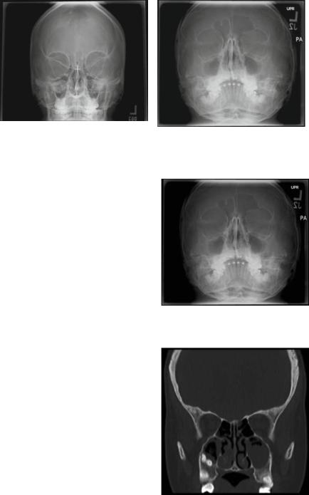
Plain X-ray has been used to evaluate the presence of air-fluid levels or mucosal thickening in the paranasal sinuses. Waters’ (occipitomental) view, where the X-ray beam is oriented through the chin, is used to obtain views of the maxillary and frontal sinuses. In the Caldwell view, the X-ray beam is oriented directly through the forehead and is used to evaluate the frontal sinus. In one report, the sensitivity and specificity are somewhat modest at 76% and 79%, respectively [23]. When evaluating the efficacy of using a single Waters’ view for diagnosing acute maxillary versus frontal rhinosinusitis, the accuracy has been shown to be even worse. When evaluating the maxillary sinus, a single Waters’ view has a false-negative rate of 32% and a mean negative predictive value of 76.9%; and the sensitivity for evaluating the frontal sinus is only 14.6% when compared to CT. Radiologists in this study also could not commit to a diagnosis when evaluating the ethmoid and sphenoid sinuses, indicating that this modality is not adequate for the evaluation of these sinuses [24,25]. Figures 3.2 and 3.3 are plain X-rays of Caldwell and Waters views demonstrating mucosal thickening. These images reveal how the diagnosis of acute rhinosinusitis from X-ray can be difficult. Structural overlapping can lead to the impression of edematous mucosa, a hypoplastic sinus can be misinterpreted as pathological opacification, and infection can be difficult to distinguish from tumor and polyp.
Computed Tomography
Computed tomography (CT) in acute rhinosinusitis demonstrates partial or complete opacification, air-fluid levels, and air bubbles within fluid levels in the paranasal sinuses (Fig. 3.4). This finding contrasts to chronic rhinosinusitis that may show mucosal thickening in addition to complete opacification (Figs. 3.5, 3.6, 3.7). CT, although more sensitive than plain films, is not specific, as demonstrated by partial opacification noted on up to 42% of head CTs performed for various reasons, and
3 Diagnosis and Management of Acute Rhinosinusitis |
35 |
|
(a) |
(b) |
|
Fig. 3.2 Caldwell view of the sinuses demonstrating well-pneumatized paranasal sinuses (A) versus chronic mucosal thickening (B)
Fig. 3.3 Waters’ view demonstrating chronic mucosal thickening
Fig. 3.4 Coronal computed tomography (CT) demonstrating acute maxillary rhinosinusitis

36 |
K.A. Kolln,¨ B.A. Senior |
Fig. 3.5 Coronal CT demonstrating acute maxillary and ethmoid rhinosinusitis with air bubbles within fluid density, indicating purulence in the right maxillary sinus
Fig. 3.6 Coronal CT demonstrating acute on chronic rhinosinusitis with complete opacification of bilateral ethmoid sinuses, left maxillary sinus mucosal thickening, and air bubbles within the right maxillary sinus indicating purulence
Fig. 3.7 Coronal CT demonstrating changes associated with chronic rhinosinusitis, including mucosal thickening of bilateral maxillary sinuses
unrelated to the paranasal sinuses [26]. In addition, CT cannot distinguish between viral and bacterial rhinosinusitis, as opacification of the infundibulum and paranasal sinuses can be seen on CT scan 48 h after the onset of cold-type symptoms [27]. CT radiography has also shown to have no effect on outcome [28].
3 Diagnosis and Management of Acute Rhinosinusitis |
37 |
Imaging, independent of the modality, is neither sensitive nor specific when striving to make the diagnosis of acute rhinosinusitis. Therefore, imaging is not recommended as a first-line procedure when evaluating a patient. CT should be reserved for the diagnosis of complicated acute rhinosinusitis, which is discussed in detail below.
Diagnosis of Complicated Acute Rhinosinusitis
Most episodes of acute rhinosinusitis are self-limited and resolve without further sequelae. However, complicated acute rhinosinusitis involves intracranial and intraorbital spread of infection and must be accurately diagnosed for immediate intervention. Orbital extension is demonstrated by periorbital edema, erythema, conjunctival injection, chemosis, proptosis, diplopia, ophthalmoplegia, and/or decreased visual acuity. Orbital complications are thought to be secondary to extension of infection from osteitis of the thin lamina papyracea or via thrombophlebitis of communicating veins [29,30]. Diagnosis is best performed by a team including an ophthalmologist and otolaryngologist and should include CT scan of the orbit and sinuses to evaluate the extent of the infection, and complete ophthalmologic examination, as well as endoscopic evaluation.
Intracranial complications of rhinosinusitis include subdural empyema, intracerebral abscess, extradural abscess, and meningitis. Infection spreads most commonly from the frontal sinus through direct spread from osteomyelitis of the skull, by retrograde thrombophlebitis through the small diploic veins of the sinus to the small vessels traversing the dura, or via a defect (surgical or traumatic) that directly connects the sinus to the cranial vault. Adolescent and young males are at highest risk for intracranial complications, which is thought to be secondary to an abundant valveless diploic system providing a good conduit for bacterial infection [31]. Individuals most commonly present with altered mental status, headache, fever, seizure, vomiting, hemiparesis, or a cranial neuropathy; CT and MRI are used to confirm the diagnosis.
Controversy
The subjective nature of symptoms-based criteria for the diagnosis of rhinosinusitis presents many challenges. First, interpretation and standardization in the literature are difficult as there is no true “gold standard” with which to compare the various modalities. This problem has been extensively discussed in regard to chronic rhinosinusitis (CRS), as there has been poor correlation between symptoms and findings on CT imaging. For example, Hwang et al. found that 35% of patients with symptoms of CRS had negative CT imaging [32]. In 2002, Stankiewicz and Chow sought to determine the relationship between symptoms (as defined by the 1997 Rhinology task force), nasal endoscopy, and CT scan [33]. They found that neither endoscopy nor CT scanning correlated with the symptoms-based criteria for CRS, as more than 50% and 68% of patients who met the criteria for CRS had negative CT scans and
38 |
K.A. Kolln,¨ B.A. Senior |
normal endoscopic examinations. However, they did find that if purulence, polyps, or polypoid congested mucosa were present on endoscopy, sinus disease was usually present on the CT scan and that a negative endoscopic exam was a relatively good predictor of a negative CT scan [33].
This observation then brings into question the importance of clinical symptoms, imaging, and nasal endoscopy in acute rhinosinusitis. Given the diversity of health care professionals who are involved with diagnosing ARS, and lack of better noninvasive physical examination techniques, we support the symptoms-based schemes that have been discussed. We also believe that, given the poor sensitivity and specificity of diagnostic imaging, there is little role for this modality in the diagnosis of uncomplicated rhinosinusitis. Endoscopic evaluation on the other hand, if available, should be used as an adjunct in the diagnosis of rhinosinusitis and should be considered before imaging. Endoscopy may aid greatly in the diagnosis of acute rhinosinusitis, especially in two scenarios: when negative, it may help to avoid antibiotic use; and further, for the patient who is not responding to therapy, endoscopically obtained cultures may provide guidance in antibiotic choice.
References
1.Lanza DC, Kennedy DW. Adult rhinosinusitis defined. Otolaryngol Head Neck Surg 1997:117;S1–S7.
2.Rosenfeld RM. Clinical practice guideline on adult sinusitis. Otolaryngol Head Neck Surg 2007;137:365–377.
3.Fokkens W, Lund V, Bachert C, et al. EAACI position paper on rhinosinusitis and nasal polyps executive summary. Allergy 2005;60:583–601.
4.Makela MJ, Puhakka T, Ruuskanen O, et al. Viruses and bacteria in the etiology of the common cold. J Clin Microbiol 1998;36:539–542.
5.Winther B, Gwaltney JM Jr, Mygind N, et al. Viral induced rhinitis. Am J Rhinol 1998;12: 17–20.
6.Gwaltney JM, Scheld WM, Sande MA, et al. The microbial etiology and antimicrobial therapy of adults with acute community-acquired sinusitis: a fifteen year experience at the University of Virginia and review of other selected studies. J Allergy Clin Immunol 1992;90:457–462.
7.Pleis JR, Lethbridge-C¸ ejku M. Summary health statistics for U.S. adults: National health interview survey, 2005. National Center for Health Statistics. Vital Health Stat 2006;10(232).
8.Goetzel RZ, Hawkins K, Ozmikowski RJ, et al. The health and productivity cost burden of the “Top 10” physical and mental health conditions affecting six large U.S. employers in 1999. J Occup Environ Med 2003;45:5–14.
9.Gill JM, Fleischut P, Haas S, et al. Use of antibiotics for adult upper respiratory infections in outpatient settings: a national ambulatory network study. Fam Med 2006;38(5):349–354.
10.Dowell SF, Schwartz B. Resistant pneumococci: protecting patients through judicious use of antibiotics. Am Fam Physician 1997;55:1647–1658.
11.Seppala H, Klaukka T, Vuopio-Varkila J, et al. The effect of changes in the consumption of macrolide antibiotics on erythromycin resistance in group A streptococci in Finland. N Engl J Med 1997;337:441–446.
12.Gwaltney JM, Hendley JO, Simon G, et al. Rhinovirus infections in an industrial population. II. Characteristics of illness and antibody response. JAMA 1967;202:494–500.
13.Alho OP, Karttunen TJ, Karttunen R, et al. Subjects with allergic rhinitis show signs of more severely impaired paranasal sinus functioning during viral colds than nonallergic subjects. Allergy 2003;58:767–771.
3 Diagnosis and Management of Acute Rhinosinusitis |
39 |
14.Berger G, Steinberg DM, Popovtzer A, et al. Endoscopy versus radiography for the diagnosis of acute bacterial rhinosinusitis. Eur Arch Otorhinolaryngol 2005;262:416–422.
15.Carenfelt C, Lundberg C, Nord CE, et al. Bacteriology of maxillary sinusitis in relation to quality of the retained secretion. Acta Otolaryngol 1978;86:298–302.
16.Axelsson A, Brorson JE. The correlation between bacterial findings in the nose and maxillary sinus in acute maxillary sinusitis. Laryngoscope 1973;83:2003–2011.
17.Vogan JC, Bolger WE, Keyes AS. Endoscopically guided sinonasal cultures: a direct comparison with maxillary sinus aspirate cultures. Otolaryngol Head Neck Surg 2000;122:370–373.
18.Dubin MG, Ebert CS, Coffey CS, et al. Concordance of middle meatal swab and maxillary sinus aspirate in acute and chronic sinusitis: a meta-analysis. Am J Rhinol 2005;19:462–470.
19.Benninger MS, Payne SC, Ferguson BJ, et al. Endoscopically directed middle meatal cultures versus maxillary sinus taps in acute bacterial maxillary rhinosinusitis: a meta-analysis. Otolaryngol Head Neck Surg 2006;134:3–9.
20.Hansen JG, Schmidt H, Rosborg J, et al. Predicting acute maxillary sinusitis in a general practice population. BMJ 1995;311:233–236.
21.Lindbaek M, Hjortdahl P. The clinical diagnosis of acute purulent sinusitis in general practice: a review. Br J Gen Pract 2002;52:491–495.
22.Puhakka T, Heikkinen T, Makel¨a¨ MJ, et al. Validity of ultrasonography in diagnosis of acute maxillary sinusitis. Arch Otolaryngol Head Neck Surg 2000;126:1482–1486.
23.Lau J, Zucker D, Engels EA, et al. Diagnosis and treatment of acute bacterial rhinosinusitis. Evidence Report/Technology Assessment No. 9. Agency for Health Care Policy and Research, Rockville, MD. Publication no. 99-E016.
24.Konen E, Faibel M, Kleinbaum Y, et al. The value of the occipitomental (Waters’) view in diagnosis of sinusitis: a comparative study with computed tomography. Clin Radiol 2000;55:856–860.
25.Aalokken TM, Hagtvedt T, Dalen I, et al. Conventional sinus radiography compared with CT in the diagnosis of acute sinusitis. Dentomaxillofac Radiol 2003;32:60–62.
26.Havas TE, Motbey JA, Gullane PJ. Prevalence of incidental abnormalities on computed tomographic scans of the paranasal sinuses. Arch Otolaryngol Head Neck Surg 1998;114:856–859.
27.Gwaltney JM Jr, Phillips CD, Miller RD, et al. Computed tomographic study of the common cold. N Engl J Med 1994;330:25–30.
28.van Buchem FL, Knottnerus JA, Schrijnemaekers VJJ, et al. Primary-care-based randomised placebo-controlled trial of antibiotic treatment in acute maxillary sinusitis. Lancet 1997;349:683–687.
29.Chandler JR, Langenbruner DJ, Stevens ER. The pathogenesis of orbital complications in acute sinusitis. Laryngoscope 1970;80:141–148.
30.Krohel GB, Krauss HR, Winnick J. Orbital abscess: presentation diagnosis, therapy, and sequalae. Ophthalmology 1982;89:492–498.
31.Kaplan RJ. Neurological complications of infections of the head and neck. Otolaryngol Clin N Am 1976;9:729–749.
32.Hwang PH, Irwin SB, Griest SE, et al. Radiologic correlates of symptom-based diagnostic criteria for chronic rhinosinusitis. Otolaryngol Head Neck Surg 2003; 128:489–496.
33.Stankiewicz JA, Chow JM. Nasal endoscopy and the definition and diagnosis of chronic rhinosinusitis. Otolaryngol Head Neck Surg 2002;126:623–627.

Chapter 4
Diagnosis of Chronic Rhinosinusitis
Rodney J. Schlosser and Richard J. Harvey
Chronic mucosal inflammation of the nose and paranasal sinuses is common. The symptoms of chronic rhinosinusitis (CRS) are thought to affect 16%, or 30 million, Americans in population-based studies [1,2]. While these surveys are questionnaire based and may overdiagnose the condition, even physician-diagnosed incidences are reported as 2% to 4% [3,4], which still represents an enormous disease burden. No solid data have been published on the duration of episodes, whether treated or from natural resolution. CRS affects an increasing proportion of the adult population until the sixth decade, then declines [3]. The quality-of-life impact of the disease, as measured by SF-36 scores, is comparable or worse to other chronic conditions such as chronic obstructive pulmonary disease (COPD), congestive heart failure (CHF), and back pain [5].
Identifying CRS patients correctly, from other sinonasal conditions, and providing health care interventions can greatly reduce the burden of disease within this group. We discuss the following concepts for the diagnosis of CRS:
•Defining CRS
•Presenting symptoms and differential diagnoses
•Other significant patient history
•Primary and secondary investigations
•Diagnostic schemes and clinical workup
This chapter discusses the current concepts of CRS with or without nasal polyps and an evidence-based review of the diagnostic methods.
Defining CRS
Defining the condition of chronic rhinosinusitis (CRS) has been one of the greatest challenges for otolaryngology. Patients with chronic nasal symptoms have been collectively diagnosed as CRS without further inclusive features. This situation has
R.J. Schlosser
Department of Otolaryngology, Medical University of South Carolina, Charleston, SC, USA e-mail: schlossr@musc.edu
E.R. Thaler, D.W. Kennedy (eds.), Rhinosinusitis, DOI: 10.1007/978-0-387-73062-2 4, |
41 |
C Springer Science+Business Media, LLC 2008 |
|