REJR_17
.pdf
RUSSIAN ELECTRONIC JOURNAL OF RADIOLOGY
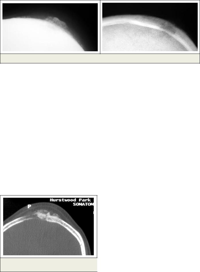
Fig.1. Plain radiographs of the skull showing erosion of the outer skull table with a soft tissue mass.
Scalp masses are seen quite commonly in patients of all age groups. In most cases the lump represents a benign condition and is incidentally noted. In children the common aetiologies for a scalp lump are dermoid cyst, epidermoid cyst, hemangiomas, hematomas and abscesses [1]. In the adult group trichilemmal cyst, inflammatory lesions, hemangiomas and lipomas are the usual causes. However, in a lump which is painful and/or is rapidly increasing in size, the possibility of a metastatic deposit should be considered. This is particularly true in a patient who has a known primary elsewhere.
In a major review of 388 scalp lumps, Carson et al [2] found 30 malignant lesions. 16 of these were metastatic deposits from a known primary in lungs, breast, kidney or thyroid. The rest were leukemia (three), lymphoma (four), melanoma (three), adenocarcinoma with unknown primary (two) and sarcomas (two). Bardales and Stanley [3] in their review of scalp lumps noted 9 lesions which were metastatic. They also showed five patients with metastasis who did not have a known primary elsewhere. Only two patients had a benign lump in their cohort of 16 patients who had been referred for a biopsy of a scalp mass.
Meningioma is a common tumour of the brain and spinal cord. In various studies they
Fig.2. CT of the skull, axial view, bone window shows the mass lesion causing skull vault erosion.
have been shown to represent 13-18% of all the primary intracranial tumors and up to 12% of all the primary intraspinal tumours [4,5]. The presentation of meningioma as a scalp lump is however uncommon.
These benign tumours arise from the arachnoid cells. Ectopic meningiomas are thought to arise from ectopic arachnoid cell rests or from pluripotential mesenchymal cells [6]. The ectopic arachnoid cell rests are formed because of delayed closure of the neural tube, with consequent herniation of the meninges or from premature closure of the neural tube with meningeal tissue being pinched off into the skin [7]. Arachnoid cells have been shown to be present in the diploic space of the skull. This explains the presence of ectopic meningiomas in the scalp, in the skull vault (the intra-osseous meningioma, where they are more commonly present along the sutures) and along the cranial nerves [8,9,10]. Intracranial meningiomas can extend outside the skull and malignant lesions can metastasize extra cranially [11,12]. Formation of ectopic meningiomas after trauma, which results in meningeal herniation outside the skull, has also been reported [7,13].
Pendergrass and Hope [14] described a meningioma in the frontal region which was causing erosion of the outer skull table without any intracranial connection. This was the first such case described in the radiology literature. Since then many reports of similar lesions have been published in the neurology and neurosurgery literature [28-31], but there have been very few reports in radiology journals. However, there have been reports of intradiploic/intraosseous meningiomas [8,16,17,18,19].
Unlike meningiomas related to the central neuraxis, ectopic meningiomas are rare. According to Farr et al [20] out of 405 cases of meningiomas only 3 were confirmed as primary extra cranial and extra spinal meningiomas present in the soft tissues. Wicker [11] and Waga [27] found the incidence of these ectopic meningiomas at 0.9% after reviewing 1768 and 226 cases respectively. Am-
REJR | www.rejr.ru | Том 5 №1 2015. Страница 71
Перейти в содержание

RUSSIAN ELECTRONIC JOURNAL OF RADIOLOGY
Fig.3. A transmitted light microscopic image of an haematoxylin and eosin-stained section showing a cellular lesion in which the cells have poorly defined borders and form the characteristic whorls around vessels or stromal elements. The finely distributed chromatin within the nuclei, with inconspicuous nucleoli, is characteristic of meningiomas.
mirati et al [21] found 4 intra diploic meningiomas in a series of 373 intra cranial lesions.
Apart from the scalp, meningiomas have been reported in the skin [7,23], orbit, paranasal sinuses, neck, nasal cavity and in the parotid glands [7,11,15,20].
Although commoner in adults, ectopic meningiomas occur at all ages and affect females more commonly than males [7,11,15,20].
Depending on their location, the symptoms range from proptosis, cranial nerve palsies, nasal blockage, or scalp/skin swelling.
It is important to be certain that the apparent scalp or sub-cutaneous tumour does not have an intra cranial component. Even when the lesion is extra cranial, there may be a rudimentary track, connecting the lump to the dura and if not careful, intervention can lead to meningitis or oth-
References:
1.Willatt JMG, Quaghebeur G. Calvarial masses of infants and children. A radiological approach. Clin Radiol.2004;59:474-486.
2.Carson HJ, Gattuso P, Castelli MJ,Reddy V. Scalp lesions: A review of histopathologic and FNA biopsy findings. Am J Der- matopathol.1995;17:256-259.
3.Bardales RH, Stanley MW. Subcutaneous masses of the scalp and forehead: Diagnosis by FNA. Diagn Cytopathol. 1995;12:131-134.
4.Buetow MP, Beutow PC, Smirniotopoulous JG. Typical, atypical and misleading features in meningioma. Radiographics 1991; 11:1087-1106.
5.Rohringer M, Sutherland GR, Louw DF, Sima AAF. Incidence and clinicopathological features of meningioma. J Neurosurg 1989;71:665-672.
6.Russell DS, Rubinstein LJ. Pathology of tumors of the nervous
er intracranial complication [7]. Moreover, an intracranial meningioma may have an extra cranial component. Farr et al [20] studied 405 cases of meningiomas and 71 of these tumors (20%) had extra cranial extensions. Fifteen of these patients had an ill-conceived extra cranial surgical intervention due to mis-diagnosis. In a review of the literature by Geoffray et al [15], out of 39 meningiomas of the naso-oral cavity and para nasal sinuses, 15 had documented intra cranial extension. In 84 extra cranial meningiomas 14 were present in the scalp with erosion of the outer skull table. None of these patients had any intra cranial extension.
Intra-diploic meningiomas can have osteolytic, mixed lytic and sclerotic or more rarely completely osteolytic appearance on plain x-rays [9]. Osteoblastic response is said to be secondary to the infiltration of meningeal cells in the bone resulting in osteoblastic activity (19). Sclerosis can affect the diploic space or one or both of the skull tables [8]. In most cases there is inward bulging of the inner skull table. Lesions on the inner or outer aspects of skull can cause erosion of the adjacent skull table. Calcification is better seen on CT [23,24,26]. MR shows a low signal mass on T1W, which is high signal on T2W, and after gadolinium administration, the intraosseous lesion enhances in a spotty manner and the extra cranial component enhances uniformly [23,24,26].
The diagnostic work-up of a scalp lump should include a CT which helps to show the skull erosion and the nature of the soft tissue. MR shows any intracranial extension. Biopsy is needed to establish a tissue diagnosis and is most easily done under ultrasound guidance.
In conclusion an ectopic meningioma should be considered in the differential diagnosis of a scalp lump. It is important to investigate for an intracranial extension of an apparent extra cranial mass lesion to avoid a potentially serious complication.
system.5th ed. Baltimore:Williams and Wilkins,1989;449-483.
7.Lopez DA, Silvers DN, Major MC, Helwig EB. Cutaneous men- ingiomas-a clinico-pathologic study. Cancer 1974; 34:728-744.
8.Kim KS, Rogers LF, Goldblatt D. CT features of hyperostosing meningioma en plaque. AJNR 1987; 8: 853-859.
9.Azar-Kia B, Sarwar M, Mare JA, Schecter MM. Intraosseous meningioma. Neuroradiology 1974; 6: 246-253.
10.Ohaegbulam SC. Ectopic epidural calvarial meningioma. Surg Neurol 1979; 12:33-35.
11.Whicker JH, Devine KD, MacCart CS. Diagnostic and therapeutic problems in extracranial meningiomas. Am J Surg1973; 126: 452-457.
12.Figueroa BE, Quint DJ, McKeever PE, Chandler WF. Extracranial metastatic meningioma. Br J Radiol 1999 ;72:513-516.
13.Walters GA, Ragland RL, Knorr JR,Malhotra R, Gelber
REJR | www.rejr.ru | Том 5 №1 2015. Страница 72
Перейти в содержание
RUSSIAN ELECTRONIC JOURNAL OF RADIOLOGY
ND. Post traumatic cutaneous meningioma of the face. Am J Neuroradiol.1994;15:393-395.
14.Pendergrass EP, Hope JW. An extracranial meningioma with no apparent intracranial source. Am J Roentgenol Radium Ther Nucl Med 1953; 70: 967-970.
15.Geoffray A, Lee Y-Y, Jing B-S, Wallace S. Extracranial meningiomas of the head and neck.AJNR 1984; 5: 599-604.
16.Hsin-Yi Lee , Prager J, Hahn Y, Ramsey R. Intraosseous meningioma: CT and MR appearance. J Comput Assist Tomogr 1992; 16: 1000-1001.
17.Okamoto S, Hisaoka M, Aoki T, Kadoya C, Kobanawa S, Hashimoto. Intraosseous microcystic meningioma. Skeletal Ra- diol.2000;29:354-357.
18.Daffner RH, Yakulis R, Maroon JC. Intraosseous meningioma. Skeletal Radiol 1998; 27:108-111.
19.Van Tassel P, Lee Y, Ayala A, Carrasco HC, Klima T. Case
680.Skeletal Radiol 1991; 20:383-386.
20.Farr HW, Gray GF, Vrana M, Panio M. Extracranial meningioma. Journal of Surgical Oncology 1973; 5: 412-420.
21.Ammirati M, Mirzai Sh, Samii M. Primary intraosseous meningioma of the skull base.Acta Neurochir (Wien) 1990;107:56-60.
23.Christopherson LA, Finelli DA, Wyatt-Ashmead J, Likavec MJ. Ectopic extraspinal meningioma: CT and MR appearance. AJNR 1997;18:1335-1337.
24.Mighell AJ, Stassen LFA, Soames JV. Meningioma-an unusual forehead swelling: a case report. Br J Oral Maxillofac Surg 1994; 32:253-256.
25.Bitoh S, Obashi J, Kobayashi Y. Primary ectopic extracalvarial meningioma. Surg Neurol 1986;25:591-594.
26.Morrison MC, Weiss KL, Moskos MM. Clinical image. CT and MR appearance of a primary intraosseous meningioma. J Comput Assist Tomogr 1988;12:169-170.
27.Waga S, Nishikawa M, Ohtsubo K, Kamijyo Y, Handa H. Extra calvarial meningiomas. Neurology 1970;20:368-372.
28.Oka K, Hirakawa K, Yoshida S, Tomonaga M. Primary calvarial meningioma. Surg Neurol 1989; 32: 304-310.
29.Ghobashy A, Tobler W. Intraosseous calvarial meningioma of the skull presenting as a solitary osteolytic skull lesion. Acta Nerochir(Wein) 1994; 129: 105-108.
30.Spinnato S, Cristofori L, Iuzzolino P, Pinna G, Bricolo A. Intradiploic meningioma of the skull. J Neurosurg Sci 1999; 43: 149-152.
22.Anderson SE, Johnston JO, Zalaudek CJ, Stauffer 31. McWhorter JM, Ghatak NR, Kelly DL. Extracranial meningi-
E,Steinbach LS. Peripheral nerve ectopic meningioma at the el- |
oma presenting as lytic skull lesion. Srg Neurol 1976; 5:223- |
bow joint. Skeletal Radiol 2001;30:639-642. |
224. |
REJR | www.rejr.ru | Том 5 №1 2015. Страница 73
Перейти в содержание

RUSSIAN ELECTRONIC JOURNAL OF RADIOLOGY
МАСТЕР-КЛАСС
ЗНАЧЕНИЕ 3D/4D ЭХОГРАФИИ В I-М ТРИМЕСТРЕ ДЛЯ ПРОФИЛАКТИКИ РОЖДЕНИЯ ДЕТЕЙ С ПОРОКАМИ РАЗВИТИЯ
Воеводин С.М.
Научный центр
Проблема пороков развития у плода является одной из актуальных задач совре- акушерства, гинеко- менного акушерства. Какие задачи стоят перед эхографией на этапе I-го три- логии и перинатоло- местра беременности. В чем заключается диагностическое значение 3D/4D эхо- гии им.
графии и в чем принципиальное отличие от 2D эхографии? Какие возможности и пре- В. И. Кулакова имущества несет в себе данный метод? Почему так важно использование 3D/4D эхогра- Москва, Россия. фии в I-м триместре беременности? Что такое экологическое 3D/4D ультразвуковое исследование? На эти и другие вопросы дает ответ сегодняшний мастер-класс.
Ключевые слова: эхография, 3D/4D эхография, пороки развития, экологическое 3D/4D ультразвуковое исследование.
VALUE OF 3D/4D ULTRASOUND IN THE FIRST TRIMESTER FOR THE PREVENTION OF
CHILDREN’S CONGENITAL ABNORMALITIES
Voevodin S.M.
t the moment the problem of fetus congenital abnormalities retains its relevance. |
V.I. Kulakov Federal |
What are the challenges of the ultrasound during the first trimester? What is the di- |
Research Center for |
А agnostic value of 3D/4D ultrasound and what is the fundamental difference between |
Obstetrics, Gynecology, |
3D/4D and 2D ultrasound? What are the possibilities and advantages of the method? Why is |
and Perinatology, |
it so important to perform 3D/4D ultrasound during the first trimester? What is the ecologi- |
Moscow, Russia. |
cal 3D/4D ultrasound? Current master-class gives answers to all these and other questions. |
|
Keywords: ultrasound, 3D/4D ultrasound, congenital abnormalities, ecological 3D/4D ultrasound.
REJR | www.rejr.ru | Том 5 №1 2015. Страница 74
Перейти в содержание

RUSSIAN ELECTRONIC JOURNAL OF RADIOLOGY
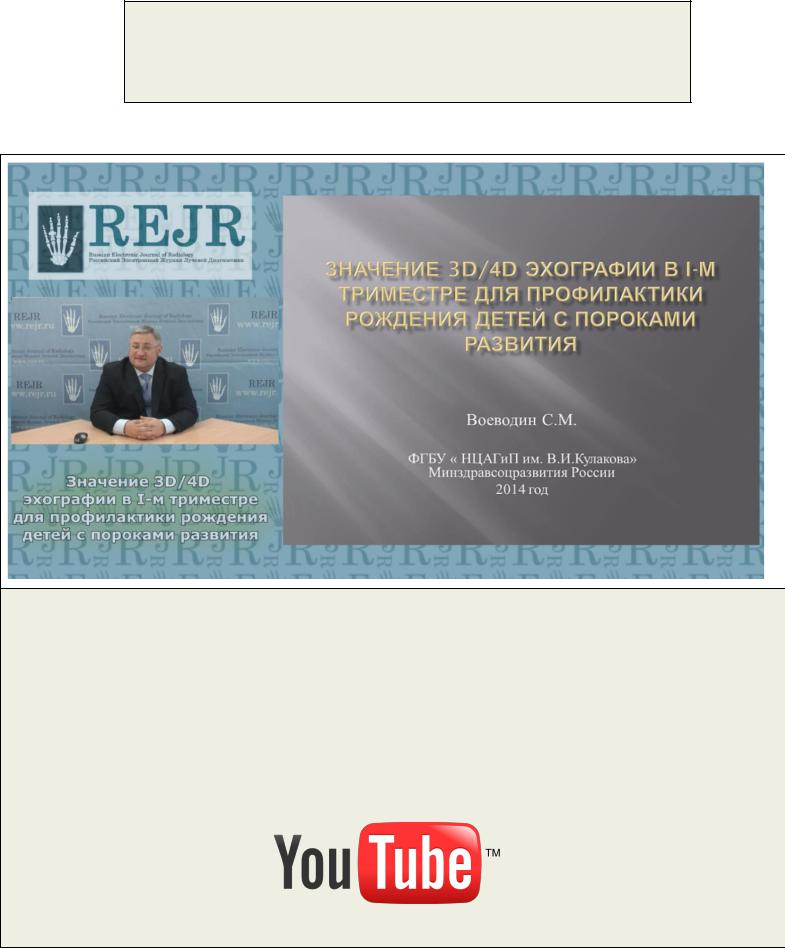
Для просмотра мастер-класса перейдите на сайт: https://rejr.ru/seventeenth_nomer/master-class.html
Руководитель отдела визуальной диагностики, д.м.н. Воеводин С.М.
Мастер-класс. ЗНАЧЕНИЕ 3D/4D ЭХОГРАФИИ В I-М ТРИМЕСТРЕ ДЛЯ ПРОФИЛАКТИКИ РОЖДЕНИЯ ДЕТЕЙ С ПОРОКАМИ РАЗВИТИЯ
Для запуска презентации нажмите на любое место в области презентации, чтобы она загрузилась (если Вы просматриваете журнал в окне браузера, то вначале сохраните журнал к себе на компьютер и откройте его с локального диска, иначе презентация не пойдет).
1)Используйте кнопки влево и вправо в левом нижнем углу страницы для перемещения по слайдам.
2)Каждая презентация сопровождается текстовым или звуковым комментарием автора. Включите в верхнем левом углу третью вкладку – ЗАМЕТКИ. Следите за текстом автора при переключении презентации на новый слайд. Если презентация сопровождается звуком, то отрегулируйте уровень звука, нажав на иконку динамика.
3)Чтобы включить полноэкранный просмотр презентации достаточно нажать левой кнопкой мыши на правую нижнюю клавишу перехода в полноэкранный режим.
Если у Вас не отображается мастер-класс – установите Adobe Flash Player: http://get.adobe.com/ru/flashplayer/
Внимание! Презентация защищена авторскими правами. Полное или частичное копирование материала запрещено, без предварительного согласия авторов.
REJR | www.rejr.ru | Том 5 №1 2015. Страница 75
Перейти в содержание

RUSSIAN ELECTRONIC JOURNAL OF RADIOLOGY
ПРЕПОДАВАНИЕ СПЕЦИАЛЬНОСТИ
Вэтом году мы открываем в нашем журнале новую рубрику, посвященную струк-
туре и преподаванию лучевой диагностики – «Преподавание специальности».
Вэтом номере мы разместили материалы заседания учебно-методической комиссии (УМК) по лучевой диагностике и терапии УМО по медицинскому и фармацевтическому образованию вузов России, которое состоялось 20 января 2015г. На этом заседа-
нии были рассмотрены вопросы целесообразности проведения коротких программ про-
фессиональной переподготовки (ПП) и объединения в составе научной специальности «лучевая диагностика, лучевая терапия» специальности «функциональная диагностика».
Вследующих номерах мы разместим материалы и по другим направлениям подго-
товки специалистов, в том числе о роли компаний-производителей диагностического
оборудования в повышении квалификации врачей-рентгенологов, радиологов и специалистов по УЗ диагностике, о вкладе профессиональных объединений, национальных и регионарных конгрессов и симпозиумов.
Прошу всех заинтересованных специалистов, руководителей отделений, препода-
вателей и заведующих кафедрами и циклами присылать нам ваши материалы, посвя-
щенные этой теме.
Широкое обсуждение данной проблемы поможет нам оценить плюсы и минусы тех или иных форм преподавания. В конечном счёте это, безусловно, пойдёт на пользу каче-
ству преподавания и уровню подготовки специалистов по лучевой диагностике.
Главный редактор REJR,
председатель УМК по лучевой диагностике и терапии, профессор С.К. Терновой.
REJR | www.rejr.ru | Том 5 №1 2015. Страница 76
Перейти в содержание

RUSSIAN ELECTRONIC JOURNAL OF RADIOLOGY
ЗАСЕДАНИЕ УЧЕБНО-МЕТОДИЧЕСКОЙ КОМИССИИ ПО ЛУЧЕВОЙ ДИАГНОСТИКЕ И ЛУЧЕВОЙ ТЕРАПИИ УЧЕБНО-МЕТОДИЧЕСКОГО ОЪЕДИНЕНИЯ ПО МЕДИЦИНСКОМУ И ФАРМАЦЕВТИЧЕСКОМУ ОБРАЗОВАНИЮ ВУЗОВ РОССИИ (20.01.2015)
ДОКЛАД ЧЛЕНА УМК ПО ЛУЧЕВОЙ ДИАГНОСТИКЕ, ЗАВ. КАФЕДРОЙ ЛУЧЕВОЙ ДИАГНОСТИКИ РОСТОВСКОГО ГОСУДАРСТВЕННОГО МЕДИЦИНСКОГО УНИВЕРСИТЕТА, ПРОФЕССОРА В.И. ДОМБРОВСКОГО
REJR | www.rejr.ru | Том 5 №1 2015. Страница 77
Перейти в содержание

RUSSIAN ELECTRONIC JOURNAL OF RADIOLOGY
REJR | www.rejr.ru | Том 5 №1 2015. Страница 78
Перейти в содержание

RUSSIAN ELECTRONIC JOURNAL OF RADIOLOGY
REJR | www.rejr.ru | Том 5 №1 2015. Страница 79
Перейти в содержание

RUSSIAN ELECTRONIC JOURNAL OF RADIOLOGY
REJR | www.rejr.ru | Том 5 №1 2015. Страница 80
Перейти в содержание
