

Книги фарма 2 / Bertram G. Katzung-Basic & Clinical Pharmacology(9th Edition)
.pdfthe evidence for some of these compounds follows.
Table 21–2. Summary of Neurotransmitter Pharmacology in the Central Nervous System.
|
Transmitter |
|
Anatomy |
|
Receptor |
|
|
Receptor |
|
|
Mechanisms |
|
||
|
|
|
|
|
Subtypes and |
|
|
Antagonists |
|
|
|
|
|
|
|
|
|
|
|
Preferred |
|
|
|
|
|
|
|
|
|
|
|
|
|
|
Agonists |
|
|
|
|
|
|
|
|
|
|
Acetylcholine |
|
Cell bodies at |
|
Muscarinic (M1): |
|
|
Pirenzepine, |
|
|
Excitatory: in K+ |
|
||
|
|
|
all levels; long |
|
muscarine, McN- |
|
|
atropine |
|
|
conductance; |
|
||
|
|
|
and short |
|
A-343 |
|
|
|
|
|
IP3, DAG |
|
||
|
|
|
connections |
|
|
|
|
|
|
|
|
|
|
|
|
|
|
|
|
|
|
|
|
|
|
|
|
||
|
|
|
|
|
Muscarinic (M2): |
|
|
Atropine, |
|
|
Inhibitory: K+ |
|
||
|
|
|
|
|
muscarine, |
|
|
methoctramine |
|
|
conductance; |
|
||
|
|
|
|
|
bethanechol |
|
|
|
|
|
cAMP |
|
||
|
|
|
|
|
|
|
|
|
|
|
|
|
|
|
|
|
|
|
|
|
|
|
|
|
|
|
|
||
|
|
|
Motoneuron- |
|
Nicotinic: |
|
|
Dihydro- - |
|
|
Excitatory: |
|
||
|
|
|
Renshaw cell |
|
nicotine |
|
|
erythroidine, - |
|
|
cation |
|
||
|
|
|
synapse |
|
|
|
|
bungarotoxin |
|
|
conductance |
|
||
|
Dopamine |
|
Cell bodies at |
|
D1: SKF 38393 |
|
|
Phenothiazines, |
|
|
Inhibitory (?): |
|
||
|
|
|
all levels; short, |
|
|
|
|
SCH 23390 |
|
|
cAMP |
|
||
|
|
|
medium, and |
|
|
|
|
|
|
|
|
|
|
|
|
|
|
|
D2: quinpirole, |
|
|
Phenothiazines, |
|
|
Inhibitory |
|
|||
|
|
|
long |
|
|
|
|
|
|
|||||
|
|
|
|
bromocriptine |
|
|
butyrophenones |
|
|
(presynaptic): |
|
|||
|
|
|
connections |
|
|
|
|
|
|
|||||
|
|
|
|
|
|
|
|
|
|
Ca |
2+ |
; Inhibitory |
|
|
|
|
|
|
|
|
|
|
|
|
|
|
|
||
|
|
|
|
|
|
|
|
|
|
|
(postsynaptic): |
|
||
|
|
|
|
|
|
|
|
|
|
|
in K+ |
|
||
|
|
|
|
|
|
|
|
|
|
|
conductance, |
|
||
|
|
|
|
|
|
|
|
|
|
|
cAMP |
|
||
|
|
|
|
|
|
|
|
|
||||||
|
GABA |
|
Supraspinal |
|
GABAA: |
|
|
Bicuculline, |
|
|
Inhibitory: Cl- |
|
||
|
|
|
|
|
|
|
|
|||||||
|
|
|
interneurons |
|
muscimol |
|
|
picrotoxin |
|
|
conductance |
|
||
|
|
|
involved in pre- |
|
|
|
|
|
|
|
|
|
|
|
|
|
|
and |
|
|
|
|
|
|
|
|
|
|
|
|
|
|
|
GABAB: |
|
|
2-OH saclofen, |
|
|
Inhibitory |
|
|||
|
|
|
postsynaptic |
|
|
|
|
|
|
|||||
|
|
|
|
baclofen |
|
|
CGP 35348, |
|
|
(presynaptic): |
|
|||
|
|
|
inhibition |
|
|
|
|
|
|
|||||
|
|
|
|
|
|
|
CGP55845 |
|
|
Ca |
2+ |
|
|
|
|
|
|
|
|
|
|
|
|
|
|
|
|
||
|
|
|
|
|
|
|
|
|
|
|
conductance; |
|
||
|
|
|
|
|
|
|
|
|
|
|
Inhibitory |
|
||
|
|
|
|
|
|
|
|
|
|
|
(postsynaptic): |
|
||
|
|
|
|
|
|
|
|
|
|
|
K+ conductance |
|
||
|
|
|
|
|
|
|
|
|
||||||
|
Glutamate |
|
Relay neurons |
|
N-Methyl-D- |
|
|
2-Amino-5- |
|
|
Excitatory: |
|
||
|
|
|
|
|
|
|
|
|||||||
|
|
|
at all levels and |
|
aspartate |
|
|
phosphonovalerate, |
|
|
cation |
|
||
|
|
|
some |
|
(NMDA): |
|
|
CPP, MK-801 |
|
|
conductance, |
|
||
|
|
|
interneurons |
|
NMDA |
|
|
|
|
|
particularly Ca2+ |
|
||
|
|
|
|
|
|
|
|
|
|
|
|
|
|
|
|
|
|
|
|
|
|
|
|
|
|
|
|
||
|
|
|
|
|
AMPA: AMPA |
|
|
CNQX, |
|
|
Excitatory: |
|
||
|
|
|
|
|
|
|
|
|
|
|
|
|
|
|

|
|
|
|
|
|
|
|
|
GYKI52466 |
|
cation |
|
|
|
|
|
|
|
|
|
|
|
|
|
|
conductance |
|
|
|
|
|
|
|
Kainate: kainate, |
|
|
CNQX |
|
|||
|
|
|
|
|
|
|
|
|
|
|
|
||
|
|
|
|
|
|
domoic acid |
|
|
|
|
|
|
|
|
|
|
|
|
|
|
|
|
|
|
|
|
|
|
|
|
|
|
|
Metabotropic: |
|
|
MCPG |
|
|
Inhibitory |
|
|
|
|
|
|
|
ACPD, |
|
|
|
|
|
(presynaptic): |
|
|
|
|
|
|
|
quisqualate |
|
|
|
|
|
Ca2+ |
|
|
|
|
|
|
|
|
|
|
|
|
|
conductance; |
|
|
|
|
|
|
|
|
|
|
|
|
|
cAMP; |
|
|
|
|
|
|
|
|
|
|
|
|
|
Excitatory: K+ |
|
|
|
|
|
|
|
|
|
|
|
|
|
conductance, |
|
|
|
|
|
|
|
|
|
|
|
|
|
IP3, DAG |
|
|
|
|
|
|
|
|
|
|
|
|
|
|
|
|
|
|
|
|
|
|
|
|
|
|
|
|
|
|
Glycine |
|
|
Spinal |
|
Taurine, - |
|
|
Strychnine |
|
|
Inhibitory: Cl- |
|
|
|
|
|
interneurons |
|
alanine |
|
|
|
|
|
conductance |
|
|
|
|
|
and some brain |
|
|
|
|
|
|
|
|
|
|
|
|
|
stem |
|
|
|
|
|
|
|
|
|
|
|
|
|
interneurons |
|
|
|
|
|
|
|
|
|
|
5- |
|
|
Cell bodies in |
|
5-HT1A: LSD, 8- |
|
|
Metergoline, |
|
|
Inhibitory: K+ |
|
|
Hydroxytryptamine |
|
|
midbrain and |
|
OH-DPAT |
|
|
spiperone |
|
|
conductance, |
|
|
(serotonin) |
|
|
pons project to |
|
|
|
|
|
|
|
cAMP |
|
|
|
|
|
all levels |
|
|
|
|
|
|
|
|
|
|
|
|
|
|
|
|
|
|
|
|
|
|
|
|
|
|
|
|
|
5-HT2A: LSD, |
|
|
Ketanserin |
|
|
Excitatory: K+ |
|
|
|
|
|
|
|
DOB |
|
|
|
|
|
conductance, |
|
|
|
|
|
|
|
|
|
|
|
|
|
IP3, DAG |
|
|
|
|
|
|
|
|
|
|
|
|
|
|
|
|
|
|
|
|
|
|
|
|
|
|
|
|
|
|
|
|
|
|
|
5-HT3: 2-methyl- |
|
|
ICS 205930, |
|
|
Excitatory: |
|
|
|
|
|
|
|
5-HT, |
|
|
ondansetron |
|
|
cation |
|
|
|
|
|
|
|
phenylbiguanide |
|
|
|
|
|
conductance |
|
|
|
|
|
|
|
|
|
|
|
|
|
|
|
|
|
|
|
|
|
|
|
|
|
|
|
|
|
|
|
|
|
|
|
5-HT4: BIMU8 |
|
|
GR 1138089 |
|
|
Excitatory: K+ |
|
|
|
|
|
|
|
|
|
|
|
|
|
conductance |
|
|
|
|
|
|
|
|
|
|
|
|
|
|
|
|
Norepinephrine |
|
|
Cell bodies in |
|
1: phenylephrine |
|
|
Prazosin |
|
|
Excitatory: K+ |
|
|
|
|
|
|
|
|
|
|
|||||
|
|
|
|
pons and brain |
|
|
|
|
|
|
|
conductance, |
|
|
|
|
|
stem project to |
|
|
|
|
|
|
|
IP3, DAG |
|
|
|
|
|
all levels |
|
|
|
|
|
|
|
|
|
|
|
|
|
|
|
|
|
|
|
|
|
|
|
|
|
|
|
|
|
2: clonidine |
|
|
Yohimbine |
|
|
Inhibitory |
|
|
|
|
|
|
|
|
|
|
|
|
|
(presynaptic): |
|
|
|
|
|
|
|
|
|
|
|
|
|
Ca2+ |
|
|
|
|
|
|
|
|
|
|
|
|
|
conductance: |
|
|
|
|
|
|
|
|
|
|
|
|
|
Inhibitory: K+ |
|
|
|
|
|
|
|
|
|
|
|
|
|
conductance, |
|
|
|
|
|
|
|
|
|
|
|
|
|
cAMP |
|
|
|
|
|
|
|
|
|
|
|
|
|
|
|
|
|
|
|
|
|
|
|
|
|
|
|
|
|
|
|
|
|
|
|
1: isoproterenol, |
|
|
Atenolol, practolol |
|
|
Excitatory: K+ |
|
|
|
|
|
|
|
dobutamine |
|
|
|
|
|
conductance, |
|
|
|
|
|
|
|
|
|
|
|
|
|
cAMP |
|
|
|
|
|
|
|
|
|
|
|
|
|
|
|

|
|
|
|
|
|
|
|
|
|
|
|
|
|
|
|
|
|
|
|
|
|
|
|
|
|
|
|
|
|
|
|
|
|
|
|
2: salbutamol |
|
|
Butoxamine |
|
|
Inhibitory: may |
|
|
|
|
|
|
|
|
|
|
|
|
|
|
|
involve in |
|
|
|
|
|
|
|
|
|
|
|
|
|
|
electrogenic |
|
|
|
|
|
|
|
|
|
|
|
|
|
|
sodium pump; |
|
|
|
|
|
|
|
|
|
|
|
|
|
|
cAMP |
|
|
Histamine |
|
|
Cells in ventral |
|
H1: 2(m- |
|
|
|
Mepyramine |
|
|
Excitatory: K+ |
|
|
|
|
|
|
|
|
|
|
|
|||||
|
|
|
|
posterior |
|
fluorophenyl)- |
|
|
|
|
|
conductance, |
|
|
|
|
|
|
hypothalamus |
|
histamine phenyl- |
|
|
|
|
|
IP3, DAG |
|
|
|
|
|
|
|
|
histamine |
|
|
|
|
|
|
|
|
|
|
|
|
|
|
|
|
|
|
|
|
|
|
|
|
|
|
|
|
|
|
|
|
|
|
|
|
|
|
|
|
|
|
|
|
H2: dimaprit |
|
|
|
Ranitidine |
|
|
Excitatory: K+ |
|
|
|
|
|
|
|
|
|
|
|
|
|
|
conductance, |
|
|
|
|
|
|
|
|
|
|
|
|
|
|
cAMP |
|
|
|
|
|
|
|
|
|
|
|
|
|
|||
|
Opioid peptides |
|
|
Cell bodies at |
|
Mu: bendorphin, |
|
|
Naloxone, CTOP |
|
|
Inhibitory |
|
|
|
|
|
|
all levels; long |
|
DAMGO |
|
|
|
|
|
|
(presynaptic): |
|
|
|
|
|
and short |
|
|
|
|
|
|
|
|
Ca2+ |
|
|
|
|
|
connections |
|
|
|
|
|
|
|
|
conductance, |
|
|
|
|
|
|
|
|
|
|
|
|
|
|
cAMP |
|
|
|
|
|
|
|
|
|
|
|
|
|
|
|
|
|
|
|
|
|
|
Delta: |
|
|
|
Naloxone |
|
|
Inhibitory |
|
|
|
|
|
|
|
|
|
|
|
|
|
|||
|
|
|
|
|
|
enkephalin, |
|
|
|
|
|
|
(postsynaptic): |
|
|
|
|
|
|
|
DPDPE |
|
|
|
|
|
|
K+ conductance, |
|
|
|
|
|
|
|
|
|
|
|
|
|
|
cAMP |
|
|
|
|
|
|
|
Kappa: |
|
|
|
Naloxone, nor-BNI |
|
|
|
|
|
|
|
|
|
|
|
|
|
|
|
|
|
||
|
|
|
|
|
|
dynorphin, U- |
|
|
|
|
|
|
|
|
|
|
|
|
|
|
69593 |
|
|
|
|
|
|
|
|
|
Tachykinins |
|
|
Primary sensory |
|
NK1: Substance |
|
|
CP99994 |
|
|
Excitatory: K+ |
|
|
|
|
|
|
|
|
|
|
|
||||||
|
|
|
|
neurons, cell |
|
P methylester |
|
|
|
|
|
conductance, |
|
|
|
|
|
|
bodies at all |
|
|
|
|
|
|
|
|
IP3, DAG |
|
|
|
|
|
8 |
] |
|
|
SR48968 |
|
|
||||
|
|
|
|
levels; long and |
|
NK2: -[Ala |
|
|
|
|
|
|
||
|
|
|
|
|
NKA4–10 |
|
|
|
|
|
|
|
|
|
|
|
|
|
short |
|
|
|
|
|
|
|
|
|
|
|
|
|
|
connections |
|
|
|
|
|
|
|
|
|
|
|
|
|
|
|
|
|
|
|
|
|
|
|
|
|
|
|
|
|
|
NK3: GR138676 |
|
|
[Pro7]NKB |
|
|
|
|
||
|
|
|
|
|
|
|
|
|
|
|
|
|||
|
|
|
|
|
|
|
|
|
|
|
|
|
|
|
|
|
|
|
|
|
|
|
|
|
|
|
|
|
|
|
Endocannabinoids |
|
|
Widely |
|
CB1: WIN55212- |
|
|
SR141716 |
|
|
Inhibitory |
|
|
|
(anandamide, 2- |
|
|
distributed |
|
2 methylester |
|
|
|
|
|
|
(presynaptic): |
|
|
arachidonylglyerol |
|
|
|
|
|
|
|
|
|
|
|
Ca2+ |
|
|
|
|
|
|
|
|
|
|
|
|
|
|
conductance, |
|
|
|
|
|
|
|
|
|
|
|
|
|
|
cAMP |
|
|
|
|
|
|
|
|
|
|
|
|
|
|
|
|
|
|
|
|
|
|
|
|
|
|
|
|
|
|
|
8-OH DPAT, 8-hydroxy-2(di-n-propylamino)tetralin; ACPD, trans-1-amino-cyclopentyl-1,3- dicarboxylate; AMPA, DL- -amino-3-hydroxy-5-methylisoxazole-4-propionate; BIMU8, [endo-N-8- methyl-8-azabicyclo(3.2.1)oct-3-yl]-2,3-dihydro-3-isopropyl-2-oxo-1H-benzimidazol-1- carboxamide hydrochloride; CGP 35348, 3-aminopropyl(diethoxymethyl)phosphinic acid; CNQX, 6-cyano-7-nitroquinoxaline-2,3-dione; CP 99994, (+)-(2S, 3S)-3-(2-methoxybenzylamino)-2-

phenylpiperidine; CPP, 3-(2-carboxypiperazin-4-yI)propyl-1-phosphonic acid; CTOP, D-Phe-Cys- Tyr-D-Trp-Orn-Thr-Pen-Thr-NH2; DAG, diacylglycerol; DAMGO, D-ala-2,Me-Phe4,Gly1- enkephalin; DOB, 5-bromo-2,5-dimethoxyamphetamine; DPDPE, D-pen2D-pen5(-enkephalin); GR 113808, (1-{2-[(methylsulfonyl)amino]ethyl}-4-piperidinyl)methyl-1-methyl-1H-indole-3- carboxylate; GYKI 52466, 1-(4-aminophenyl)-4-methyl-7,8-methylenedioxy-5H-2,3- benzodiazepine; IP3, inositol trisphosphate; MCPG, 
 -methyl-4-carboxyphenylglycine; MK-801, (dizocilpine), 10,11-dihydro-5-methyl-5H-dibenzo(a,d)cyclohepten-5,10-imine; NK1,2,3, neurokinin and derivatives; nor-BNI, nor-binaltorphimine; SR 141716, N-(piperidine-1yl)-5-(4- chlorophenyl)-1-(2,4- dichlorophenyl)-4-methyl-1H-pyrazole-3-carboxamide; SR 48968, (S)-N- methyl-N-(4-acetylamino-4-phenylpiperidino)-2-(3,4-dichlorophenyl)butylbenzamide; U-69593,
(+)-(5
 ,7
,7
 ,8
,8
 )-N-methyl-N-[7-(1-pyrrolidinyl)]-1-oxaspiro(4,5)dec-8-yl-benzenacetamide; WIN 55212-2, (R)-(+)-[2,3-dihydro-5-methyl-3-[(morpholino)methyl]pyrrolo-[1,2,3-de]-1,4-benzoxazin- 6-yl] (1-naphthyl)methanone
Amino Acids
The amino acids of primary interest to the pharmacologist fall into two categories: the neutral amino acids glycine and GABA and the acidic amino acid glutamate. All of these compounds are present in high concentrations in the CNS and are extremely potent modifiers of neuronal excitability.
Neutral Amino Acids
The neutral amino acids are inhibitory and increase membrane permeability to chloride ions, thus mimicking the IPSP. Glycine concentrations are particularly high in the gray matter of the spinal cord, and strychnine, which is a potent spinal cord convulsant and has been used in some rat poisons, selectively antagonizes both the action of glycine and the IPSPs recorded in spinal cord neurons. Thus, it is generally agreed that glycine is released from spinal cord inhibitory local circuit neurons involved in postsynaptic inhibition.
GABA receptors are divided into two types: GABAA and GABAB. GABAA receptors open chloride channels and are antagonized by picrotoxin and bicuculline, which both cause generalized convulsions. GABAB receptors, which can be selectively activated by the antispastic drug baclofen, are coupled to G proteins that either inhibit calcium channels or activate potassium channels. In most regions of the brain, IPSPs have a fast and slow component mediated by GABAA and GABAB receptors, respectively. Immunohistochemical studies indicate that a large majority of the local circuit neurons synthesize GABA. A special class of local circuit neuron localized in the dorsal horn of the spinal cord also synthesizes GABA. These neurons form axoaxonic synapses with primary sensory nerve terminals and are responsible for presynaptic inhibition (Figure 21–5 B).
Acidic Amino Acids
Glutamate is present in very high concentrations in the CNS. Virtually all neurons that have been tested are strongly excited by this amino acid. This excitation is caused by the activation of both ionotropic and metabotropic receptors, which have been extensively characterized by molecular cloning. The ionotropic receptors can be further divided into three subtypes based on the action of the selective agonists: kainate (KA), 
 -amino-3-hydroxy-5-methylisoxazole-4-propionate (AMPA), and N-methyl-D-aspartate (NMDA). The AMPAand KA-activated channels are permeable to sodium and potassium ions and, for certain subtypes, calcium as well. They are often grouped together and referred to as non-NMDA channels. The NMDA-activated channel is highly permeable to sodium, potassium, and calcium ions.
The metabotropic glutamate receptors act indirectly on ion channels via G proteins. They are selectively activated by trans-1-amino-cyclopentyl-1,3-dicarboxylate (ACPD). These G proteincoupled receptors are either positively coupled to (ie, stimulate) phospholipase C or negatively coupled to adenylyl cyclase. Depending on the type of synapse, metabotropic glutamate receptors can initiate a slow postsynaptic excitation or a presynaptic inhibition. Although the presence of metabotropic receptors at excitatory synapses varies, most excitatory synapses contain both NMDA receptors and non-NMDA receptors in the postsynaptic membrane.
The role of NMDA receptors has received considerable attention. These receptors play a critical role in synaptic plasticity, which is thought to underlie certain forms of learning and memory. They are selectively blocked by the dissociative anesthetic ketamine and the hallucinogenic drug phencyclidine. These drugs exert their effects by entering and blocking the open channel. Some drugs that block this receptor channel have potent antiepileptic activity in animal models, though these drugs have yet to be tested clinically. Considerable evidence exists that the release of glutamate during neuronal injury can, by activating the NMDA receptor, cause further cell injury and death. Thus, a particularly exciting finding is that blocking the NMDA receptor can attenuate the neuronal damage caused by anoxia in experimental animals. The potential therapeutic benefits of this action are considerable, although clinical trials to date have been disappointing.
Acetylcholine
Acetylcholine was the first compound to be identified pharmacologically as a transmitter in the CNS. Eccles showed in the early 1950s that excitation of Renshaw cells by motor axon collaterals was blocked by nicotinic antagonists. Furthermore, Renshaw cells were extremely sensitive to nicotinic agonists. These experiments were remarkable for two reasons. First, this early success at identifying a transmitter for a central synapse was followed by disappointment, because it remained the sole central synapse for which the transmitter was known until the late 1960s, when comparable data became available for the neutral amino acids. Second, the motor axon collateral synapse remains one of the best-documented examples of a cholinergic nicotinic synapse in the mammalian CNS, despite the rather widespread distribution of nicotinic receptors as defined by in situ hybridization studies. Most CNS responses to acetylcholine are mediated by a large family of G protein-coupled muscarinic receptors. At a few sites, acetylcholine causes slow inhibition of the neuron by activating the M2 subtype of receptor, which opens potassium channels. A far more widespread muscarinic action in response to acetylcholine is a slow excitation that in some cases is mediated by M1 receptors. These muscarinic effects are much slower than either nicotinic effects on Renshaw cells or the effect of amino acids. Furthermore, this muscarinic excitation is unusual in that acetylcholine produces it by decreasing the membrane permeability to potassium, ie, the opposite of conventional transmitter action.
A number of pathways contain acetylcholine, including neurons in the neostriatum, the medial septal nucleus, and the reticular formation. Cholinergic pathways appear to play an important role in cognitive functions, especially memory. Presenile dementia of the Alzheimer type is reportedly associated with a profound loss of cholinergic neurons. However, the specificity of this loss has been questioned since the levels of other putative transmitters, eg, somatostatin, are also decreased.
Monoamines
Monoamines include the catecholamines (dopamine and norepinephrine) and 5-hydroxytryptamine. Although these compounds are present in very small amounts in the CNS, they can be localized using extremely sensitive histochemical methods. These pathways are the site of action of many drugs; for example, the CNS stimulants cocaine and amphetamine are believed to act primarily at

catecholamine synapses. Cocaine blocks the reuptake of dopamine and norepinephrine, while amphetamines cause presynaptic terminals to release these transmitters.
Dopamine
The major pathways containing dopamine are the projection linking the substantia nigra to the neostriatum and the projection linking the ventral tegmental region to limbic structures, particularly the limbic cortex. The therapeutic action of the antiparkinsonism drug levodopa is associated with the former area, whereas the therapeutic action of the antipsychotic drugs is thought to be associated with the latter area. Dopamine-containing neurons in the tuberobasal ventral hypothalamus play an important role in regulating hypothalamohypophysial function. A number of dopamine receptors have been identified, and they fall into two categories: D1-like and D2-like. All dopamine receptors are metabotropic. Dopamine generally exerts a slow inhibitory action on CNS neurons. This action has been best characterized on dopamine-containing substantia nigra neurons, where D2 receptor activation opens potassium channels.
Norepinephrine
This system has already been discussed. Most noradrenergic neurons are located in the locus ceruleus or the lateral tegmental area of the reticular formation. Although the density of fibers innervating various sites differs considerably, most regions of the central nervous system receive diffuse noradrenergic input. All noradrenergic receptor subtypes are metabotropic. When applied to neurons, norepinephrine can hyperpolarize them by increasing potassium conductance. This effect is mediated by  2 receptors and has been characterized most thoroughly on locus ceruleus neurons. In many regions of the CNS, norepinephrine actually enhances excitatory inputs by both indirect and direct mechanisms. The indirect mechanism involves disinhibition, ie, inhibitory local circuit neurons are inhibited. The direct mechanism is blockade of potassium conductances that slow neuronal discharge. Depending on the type of neuron, this effect is mediated by either
 1 or
1 or  receptors. Facilitation of excitatory synaptic transmission is in accordance with many of the behavioral processes thought to involve noradrenergic pathways, eg, attention and arousal.
5-Hydroxytryptamine
Most 5-hydroxytryptamine (5-HT, serotonin) pathways originate from neurons in the raphe or midline regions of the pons and upper brain stem. 5-HT is contained in unmyelinated fibers that diffusely innervate most regions of the CNS, but the density of the innervation varies. 5-HT acts on more than a dozen receptor subtypes. Except for the 5-HT3 receptor, all of these receptors are metabotropic. The ionotropic 5-HT3 receptor exerts a rapid excitatory action at a very limited number of sites in the CNS. In most areas of the central nervous system, 5-HT has a strong inhibitory action. This action is mediated by 5-HT1A receptors and is associated with membrane hyperpolarization caused by an increase in potassium conductance. It has been found that 5-HT1A receptors and GABAB receptors share the same potassium channels. Some cell types are slowly excited by 5-HT owing to its blockade of potassium channels via 5-HT2 or 5-HT4 receptors. Both excitatory and inhibitory actions can occur on the same neurons. It has often been speculated that 5- HT pathways may be involved in the hallucinations induced by LSD, since this compound can antagonize the peripheral actions of 5-HT. However, LSD does not appear to be a 5-HT antagonist in the central nervous system, and typical LSD-induced behavior is still seen in animals after raphe nuclei are destroyed. Other proposed regulatory functions of 5-HT-containing neurons include sleep, temperature, appetite, and neuroendocrine control.
Peptides

A great many CNS peptides have been discovered that produce dramatic effects both on animal behavior and on the activity of individual neurons. Many of the peptides have been mapped with immunohistochemical techniques and include opioid peptides (enkephalins, endorphins, etc), neurotensin, substance P, somatostatin, cholecystokinin, vasoactive intestinal polypeptide, neuropeptide Y, and thyrotropin-releasing hormone. As in the peripheral autonomic nervous system, peptides often coexist with a conventional nonpeptide transmitter in the same neuron. A good example of the approaches used to define the role of these peptides in the central nervous system comes from studies on substance P and its association with sensory fibers. Substance P is contained in and released from small unmyelinated primary sensory neurons of the spinal cord and brain stem and causes a slow EPSP in target neurons. These sensory fibers are known to transmit noxious stimuli, and it is therefore surprising that—while substance P receptor antagonists can modify responses to certain types of pain—they do not block the response. Glutamate, which is released with substance P from these synapses, presumably plays an important role in transmitting pain stimuli. Substance P is certainly involved in many other functions, since it is found in many areas of the central nervous system that are unrelated to pain pathways.
Many of these peptides are also found in peripheral structures, including peripheral synapses. They are described in Chapter 6: Introduction to Autonomic Pharmacology and Chapter 17: Vasoactive Peptides.
Nitric Oxide
The CNS contains a substantial amount of nitric oxide synthase (NOS), which is found within certain classes of neurons. This neuronal NOS is an enzyme activated by calcium-calmodulin, and activation of NMDA receptors, which increases intracellular calcium, results in the generation of nitric oxide. While a physiologic role for nitric oxide has been clearly established for vascular smooth muscle, its role in synaptic transmission and synaptic plasticity remains controversial.
Endocannabinoids
The primary psychoactive ingredient in cannabis, 

 9-tetrahydrocannabinol (

 9-THC), affects the brain mainly by activating a specific cannabinoid receptor, CB1. CB1 is expressed at high levels in many brain regions, and several endogenous brain lipids, including anandamide and 2- arachidonylglycerol, have been identified as CB1 ligands. These ligands are not stored, as are classic neurotransmitters, but instead are rapidly synthesized by neurons in response to depolarization and consequent calcium influx. In further contradistinction to classic neurotransmitters, endogenous cannabinoids can function as retrograde synaptic messengers: they are released from postsynaptic neurons and travel backward across synapses, activating CB1 receptors on presynaptic neurons and suppressing transmitter release. Cannabinoids may affect memory, cognition, and pain perception by this mechanism.
Katzung PHARMACOLOGY, 9e > Section V. Drugs That Act in the Central Nervous System > Chapter 21. Introduction to the Pharmacology of CNS Drugs >
Chapter 22. Sedative-Hypnotic Drugs
Sedative-Hypnotic Drugs: Introduction
Assignment of a drug to the sedative-hypnotic class indicates that its major therapeutic use is to cause sedation (with concomitant relief of anxiety) or to encourage sleep. Because there is considerable chemical variation within this group, this drug classification is based on clinical uses
rather than on similarities in chemical structure. Anxiety states and sleep disorders are common problems, and sedative-hypnotics are among the most widely prescribed drugs worldwide. Katzung PHARMACOLOGY, 9e > Section V. Drugs That Act in the Central Nervous System > Chapter 22. Sedative-Hypnotic Drugs >
Basic Pharmacology of Sedative-Hypnotics
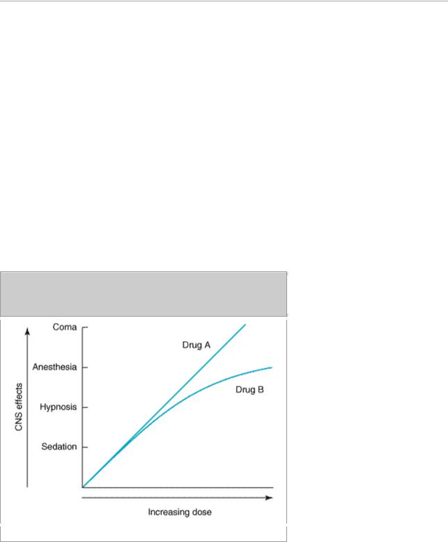
An effective sedative (anxiolytic) agent should reduce anxiety and exert a calming effect. The degree of central nervous system depression caused by a sedative should be the minimum consistent with therapeutic efficacy. A hypnotic drug should produce drowsiness and encourage the onset and maintenance of a state of sleep. Hypnotic effects involve more pronounced depression of the central nervous system than sedation, and this can be achieved with most drugs in this class simply by increasing the dose. Graded dose-dependent depression of central nervous system function is a characteristic of sedative-hypnotics. However, individual drugs differ in the relationship between the dose and the degree of central nervous system depression. Two examples of such dose-response relationships are shown in Figure 22–1. The linear slope for drug A is typical of many of the older sedative-hypnotics, including the barbiturates and alcohols. With such drugs, an increase in dose above that needed for hypnosis may lead to a state of general anesthesia. At still higher doses, sedative-hypnotics may depress respiratory and vasomotor centers in the medulla, leading to coma and death. Deviations from a linear dose-response relationship, as shown for drug B, will require proportionately greater dosage increments in order to achieve central nervous system depression more profound than hypnosis. This appears to be the case for benzodiazepines and certain newer hypnotics; the greater margin of safety this offers is an important reason for their widespread use to treat anxiety states and sleep disorders.
Figure 22–1.
Dose-response curves for two hypothetical sedative-hypnotics.
Chemical Classification
The benzodiazepines (Figure 22–2) are the most widely used sedative-hypnotics. All of the structures shown are 1,4-benzodiazepines, and most contain a carboxamide group in the 7-
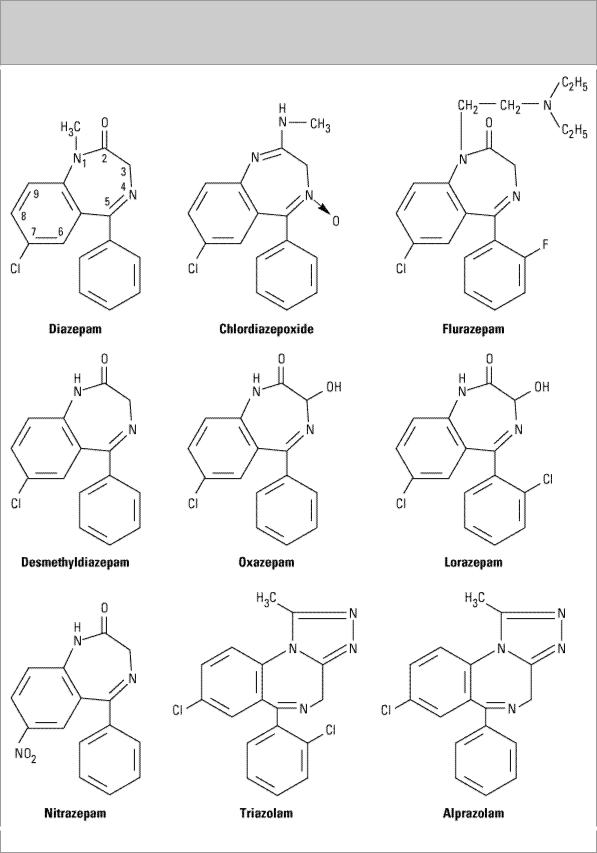
membered heterocyclic ring structure. A substituent in the 7 position, such as a halogen or a nitro group, is required for sedative-hypnotic activity. The structures of triazolam and alprazolam include the addition of a triazole ring at the 1,2-position, and such drugs are sometimes referred to as triazolobenzodiazepines.
Figure 22–2.
Chemical structures of benzodiazepines.
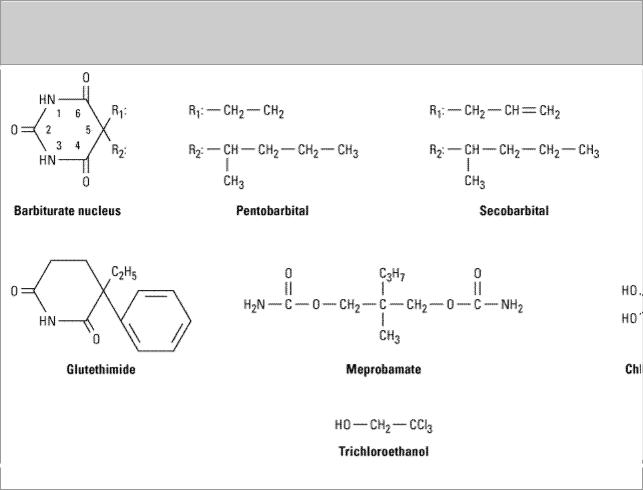
The chemical structures of some older and less commonly used sedative-hypnotics, including several barbiturates, are shown in Figure 22–3. Glutethimide (a piperidinedione) and meprobamate (a carbamate) are of distinctive chemical structure but are practically equivalent to barbiturates in their pharmacologic effects, and their clinical use is rapidly declining. The sedative-hypnotic class also includes compounds of simple chemical structure, including ethanol (see Chapter 23: The Alcohols), chloral hydrate, trichloroethanol, and paraldehyde (not shown).
Figure 22–3.
Chemical structures of barbiturates and other sedative-hypnotics.
Several drugs with novel chemical structures have been introduced recently. Buspirone is an anxiolytic agent that has actions different from those of conventional sedative-hypnotic drugs. Zolpidem and zaleplon, while structurally unrelated to benzodiazepines, share a similar mechanism of action.