

Книги фарма 2 / Bertram G. Katzung-Basic & Clinical Pharmacology(9th Edition)
.pdfacetylcholine receptor (AChR), which allows Na+ to flow down its concentration gradient into cells, producing a localized excitatory postsynaptic potential—a depolarization.
The AChR (Figure 2–9) is one of the best-characterized of all cell-surface receptors for hormones or neurotransmitters. One form of this receptor is a pentamer made up of five polypeptide subunits (eg, two 
 chains plus one
  , one
, one 
 , and one

 chain, all with molecular weights ranging from 43,000 to 50,000). These polypeptides, each of which crosses the lipid bilayer four times, form a cylindric structure 8 nm in diameter. When acetylcholine binds to sites on the
 subunits, a conformational change occurs that results in the transient opening of a central aqueous channel through which sodium ions penetrate from the extracellular fluid into the cell.
Figure 2–9.
The nicotinic acetylcholine receptor, a ligand-gated ion channel. The receptor molecule is depicted as embedded in a rectangular piece of plasma membrane, with extracellular fluid above and cytoplasm below. Composed of five subunits (two 
 , one
, one  , one
, one  , and one
 ), the receptor opens a central transmembrane ion channel when acetylcholine (ACh) binds to sites on the extracellular domain of its
 subunits.
The time elapsed between the binding of the agonist to a ligand-gated channel and the cellular response can often be measured in milliseconds. The rapidity of this signaling mechanism is crucially important for moment-to-moment transfer of information across synapses. Ligand-gated ion channels can be regulated by multiple mechanisms, including phosphorylation and internalization. In the central nervous system, these mechanisms contribute to synaptic plasticity involved in learning and memory.
G Proteins & Second Messengers
Many extracellular ligands act by increasing the intracellular concentrations of second messengers such as cyclic adenosine-3',5'-monophosphate (cAMP), calcium ion, or the phosphoinositides

(described below). In most cases they use a transmembrane signaling system with three separate components. First, the extracellular ligand is specifically detected by a cell-surface receptor. The receptor in turn triggers the activation of a G protein located on the cytoplasmic face of the plasma membrane. The activated G protein then changes the activity of an effector element, usually an enzyme or ion channel. This element then changes the concentration of the intracellular second messenger. For cAMP, the effector enzyme is adenylyl cyclase, a transmembrane protein that converts intracellular adenosine triphosphate (ATP) to cAMP. The corresponding G protein, Gs, stimulates adenylyl cyclase after being activated by hormones and neurotransmitters that act via a specific receptor (Table 2–1).
Table 2–1. A Partial List of Endogenous Ligands and Their Associated Second Messengers.
Ligand |
Second Messenger |
|
|
Adrenocorticotropic hormone |
cAMP |
|
|
Acetylcholine (muscarinic receptors) |
Ca2+, phosphoinositides |
|
|
|
|
Angiotensin |
Ca2+, phosphoinositides |
|
|
|
|
Catecholamines ( 1-adrenoceptors) |
Ca2+, phosphoinositides |
|
|
|
|
Catecholamines ( -adrenoceptors) |
cAMP |
|
|
Chorionic gonadotropin |
cAMP |
|
|
Follicle-stimulating hormone |
cAMP |
|
|
Glucagon |
cAMP |
|
|
Histamine (H2 receptors) |
cAMP |
|
|
|
|
Luteinizing hormone |
cAMP |
|
|
Melanocyte-stimulating hormone |
cAMP |
|
|
Parathyroid hormone |
cAMP |
|
|
Platelet-activating factor |
Ca2+, phosphoinositides |
|
|
|
|
Prostacyclin, prostaglandin E2 |
cAMP |
|
|
Serotonin (5-HT4 receptors) |
cAMP |
|
|
|
|
Serotonin (5-HT1C and 5-HT2 receptors) |
Ca2+, phosphoinositides |
|
|
|
|
Thyrotropin |
cAMP |
|
|
Thyrotropin-releasing hormone |
Ca2+, phosphoinositides |
|
|
|
|
Vasopressin (V1 receptors) |
Ca2+, phosphoinositides |
|
|
|
|
Vasopressin (V2 receptors) |
cAMP |
|
|

Key: cAMP = cyclic adenosine monophosphate.
Gs and other G proteins use a molecular mechanism that involves binding and hydrolysis of GTP (Figure 2–10). This mechanism allows the transduced signal to be amplified. For example, a neurotransmitter such as norepinephrine may encounter its membrane receptor for only a few milliseconds. When the encounter generates a GTP-bound Gs molecule, however, the duration of activation of adenylyl cyclase depends on the longevity of GTP binding to Gs rather than on the receptor's affinity for norepinephrine. Indeed, like other G proteins, GTP-bound Gs may remain active for tens of seconds, enormously amplifying the original signal. This mechanism explains how signaling by G proteins produces the phenomenon of spare receptors (described above). At low concentrations of agonist the proportion of agonist-bound receptors may be much less than the proportion of G proteins in the active (GTP-bound) state; if the proportion of active G proteins correlates with pharmacologic response, receptors will appear to be spare (ie, a small fraction of receptors occupied by agonist at any given time will appear to produce a proportionately larger response).
Figure 2–10.
The guanine nucleotide-dependent activation-inactivation cycle of G proteins. The agonist activates the receptor (R), which promotes release of GDP from the G protein (G), allowing entry of GTP into the nucleotide binding site. In its GTP-bound state (G-GTP), the G protein regulates activity of an effector enzyme or ion channel (E). The signal is terminated by hydrolysis of GTP, followed by return of the system to the basal unstimulated state. Open arrows denote regulatory effects. (Pi, inorganic phosphate.)
The family of G proteins contains several functionally diverse subfamilies (Table 2–2), each of which mediates effects of a particular set of receptors to a distinctive group of effectors. Receptors coupled to G proteins comprise a family of "seven-transmembrane" or "serpentine" receptors, so called because the receptor polypeptide chain "snakes" across the plasma membrane seven times (Figure 2–11). Receptors for adrenergic amines, serotonin, acetylcholine (muscarinic but not

nicotinic), many peptide hormones, odorants, and even visual receptors (in retinal rod and cone cells) all belong to the serpentine family. All were derived from a common evolutionary precursor. Some serpentine receptors exist as dimers, but it is thought that dimerization is not usually required for activation.
Table 2–2. G Proteins and Their Receptors and Effectors.
G |
Receptors for: |
|
Effector/Signaling Pathway |
|
|
Protein |
|
|
|
|
|
|
|
|
|
|
|
Gs |
-Adrenergic amines, glucagon, histamine, |
|
Adenylyl cyclase |
cAMP |
|
|
serotonin, and many other hormones |
|
|
|
|
|
|
|
|
|
|
Gi1, Gi2, |
2-Adrenergic amines, acetylcholine |
|
Several, including: |
|
|
Gi3 |
(muscarinic), opioids, serotonin, and many others |
|
Adenylyl cyclase |
cAMP |
|
|
|
|
|
||
|
|
|
Open cardiac K+ channels |
heart |
|
|
|
|
rate |
|
|
|
|
|
|
|
|
Golf |
Odorants (olfactory epithelium) |
|
Adenylyl cyclase |
cAMP |
|
|
|
|
|
|
|
|
|
|
|
|
|
Go |
Neurotransmitters in brain (not yet specifically |
|
Not yet clear |
|
|
|
identified) |
|
|
|
|
|
|
|
|
|
|
Gq |
Acetylcholine (eg, muscarinic), bombesin, |
|
Phospholipase C |
IP3, |
|
|
serotonin (5-HT1C), and many others |
|
diacylglycerol, cytoplasmic Ca2+ |
||
|
|
|
|
|
|
|
|
|
|
|
|
Gt1, Gt2 |
Photons (rhodopsin and color opsins in retinal |
|
cGMP phosphodiesterase |
cGMP |
|
|
rod and cone cells) |
|
(phototransduction) |
|
|
|
|
|
|
|
|
Key: cAMP = cyclic adenosine monophosphate; cGMP = cyclic guanosine monophosphate.
Serpentine receptors transduce signals across the plasma membrane in essentially the same way. Often the agonist ligand—eg, a catecholamine, acetylcholine, or the photon-activated chromophore of retinal photoreceptors—is bound in a pocket enclosed by the transmembrane regions of the receptor (as in Figure 2–11). The resulting change in conformation of these regions is transmitted to cytoplasmic loops of the receptor, which in turn activate the appropriate G protein by promoting replacement of GDP by GTP, as described above. Considerable biochemical evidence indicates that G proteins interact with amino acids in the third cytoplasmic loop of the serpentine receptor polypeptide (shown by arrows in Figure 2–11). The carboxyl terminal tails of these receptors, also located in the cytoplasm, can regulate the receptors' ability to interact with G proteins, as described below.
Figure 2–11.

Transmembrane topology of a typical serpentine receptor. The receptor's amino (N) terminal is extracellular (above the plane of the membrane), and its carboxyl (C) terminal intracellular. The terminals are connected by a polypeptide chain that traverses the plane of the membrane seven times. The hydrophobic transmembrane segments (light color) are designated by roman numerals (I–VII). The agonist (Ag) approaches the receptor from the extracellular fluid and binds to a site surrounded by the transmembrane regions of the receptor protein. G proteins (G) interact with cytoplasmic regions of the receptor, especially with portions of the third cytoplasmic loop between transmembrane regions V and VI. The receptor's cytoplasmic terminal tail contains numerous serine and threonine residues whose hydroxyl (–OH) groups can be phosphorylated. This phosphorylation may be associated with diminished receptor-G protein interaction.
Receptor Regulation
Receptor-mediated responses to drugs and hormonal agonists often desensitize with time (Figure 2– 12, top). After reaching an initial high level, the response (eg, cellular cAMP accumulation, Na+ influx, contractility, etc) gradually diminishes over seconds or minutes, even in the continued presence of the agonist. This desensitization is usually reversible; a second exposure to agonist, if provided a few minutes after termination of the first exposure, results in a response similar to the initial response.
Figure 2–12.

Possible mechanism for desensitization of the 
 -adrenoceptor. The upper part of the figure depicts the response to a
  -adrenoceptor agonist (ordinate) versus time (abscissa). The break in the time axis indicates passage of time in the absence of agonist. Temporal duration of exposure to agonist is indicated by the light-colored bar. The lower part of the figure schematically depicts agonistinduced phosphorylation (P) by
  -adrenoceptor kinase (
 -adrenergic receptor kinase,
  ARK) of carboxyl terminal hydroxyl groups (–OH) of the
 -adrenoceptor. This phosphorylation induces binding of a protein,
 -arrestin (
 -arr), which prevents the receptor from interacting with Gs. Removal of agonist for a short period of time allows dissociation of
 -arr, removal of phosphate (Pi) from the receptor by phosphatases (P'ase), and restoration of the receptor's normal responsiveness to agonist.
Although many kinds of receptors undergo desensitization, the mechanism is in many cases obscure. A molecular mechanism of desensitization has been worked out in some detail, however, in the case of the  -adrenoceptor (Figure 2–12, bottom). The agonist-induced change in conformation of the receptor causes it to bind, activate, and serve as a substrate for a specific kinase,
  -adrenoceptor kinase (also called
  ARK).
ARK).  ARK then phosphorylates serine or threonine

residues in the receptor's carboxyl terminal tail. The presence of phosphoserines increases the receptor's affinity for binding a third protein,  -arrestin. Binding of
  -arrestin to cytoplasmic loops of the receptor diminishes the receptor's ability to interact with Gs, thereby reducing the agonist response (ie, stimulation of adenylyl cyclase). Upon removal of agonist, however, cellular phosphatases remove phosphates from the receptor and
  ARK stops putting them back on, so that the receptor—and consequently the agonist response—return to normal. This mechanism of desensitization, which rapidly and reversibly modulates the ability of the receptor to signal to G protein, turns out to regulate many G protein–coupled receptors. Another important regulatory process is down-regulation. Down-regulation, which decreases the actual number of receptors present in the cell or tissue, occurs more slowly than rapid desensitization and is less readily reversible. This is because down-regulation involves a net degradation of receptors present in the cell, requiring new receptor biosynthesis for recovery, in contrast to rapid desensitization which involves reversible phosphorylation of existing receptors. Many G protein–coupled receptors are down-regulated by undergoing ligand-induced endocytosis and delivery to lysosomes, similar to down-regulation of protein tyrosine kinases such as the EGF receptor. Down-regulation generally occurs only after prolonged or repeated exposure of cells to agonist (over hours to days). Brief periods of agonist exposure (several minutes) can also induce internalization of receptors. In this case, many receptors, including the
 -adrenoceptor, do not down-regulate but instead recycle intact to the plasma membrane. This rapid cycling through endocytic vesicles promotes dephosphorylation of receptors, increasing the rate at which fully functional receptors are replenished in the plasma membrane. Thus, depending on the particular receptor and duration of activation, internalization can mediate quite different effects on receptor signaling and regulation.
Well-Established Second Messengers
Cyclic Adenosine Monophosphate (cAMP)
Acting as an intracellular second messenger, cAMP mediates such hormonal responses as the mobilization of stored energy (the breakdown of carbohydrates in liver or triglycerides in fat cells stimulated by 
 -adrenomimetic catecholamines), conservation of water by the kidney (mediated by vasopressin), Ca2+ homeostasis (regulated by parathyroid hormone), and increased rate and contraction force of heart muscle (
 -adrenomimetic catecholamines). It also regulates the production of adrenal and sex steroids (in response to corticotropin or follicle-stimulating hormone), relaxation of smooth muscle, and many other endocrine and neural processes.
cAMP exerts most of its effects by stimulating cAMP-dependent protein kinases (Figure 2–13). These kinases are composed of a cAMP-binding regulatory (R) dimer and two catalytic (C) chains. When cAMP binds to the R dimer, active C chains are released to diffuse through the cytoplasm and nucleus, where they transfer phosphate from ATP to appropriate substrate proteins, often enzymes. The specificity of cAMP's regulatory effects resides in the distinct protein substrates of the kinases that are expressed in different cells. For example, liver is rich in phosphorylase kinase and glycogen synthase, enzymes whose reciprocal regulation by cAMP-dependent phosphorylation governs carbohydrate storage and release.
Figure 2–13.

The cAMP second messenger pathway. Key proteins include hormone receptors (Rec), a stimulatory G protein (Gs), catalytic adenylyl cyclase (AC), phosphodiesterases (PDE) that hydrolyze cAMP, cAMP-dependent kinases, with regulatory (R) and catalytic (C) subunits, protein substrates (S) of the kinases, and phosphatases (P'ase), which remove phosphates from substrate proteins. Open arrows denote regulatory effects.
When the hormonal stimulus stops, the intracellular actions of cAMP are terminated by an elaborate series of enzymes. cAMP-stimulated phosphorylation of enzyme substrates is rapidly reversed by a diverse group of specific and nonspecific phosphatases. cAMP itself is degraded to 5'-AMP by several cyclic nucleotide phosphodiesterases (PDE, Figure 2–13). Competitive inhibition of cAMP degradation is one way caffeine, theophylline, and other methylxanthines produce their effects (see Chapter 20: Drugs Used in Asthma).
Calcium and Phosphoinositides
Another well-studied second messenger system involves hormonal stimulation of phosphoinositide hydrolysis (Figure 2–14). Some of the hormones, neurotransmitters, and growth factors that trigger this pathway (see Table 2–1) bind to receptors linked to G proteins, while others bind to receptor tyrosine kinases. In all cases, the crucial step is stimulation of a membrane enzyme, phospholipase C (PLC), which splits a minor phospholipid component of the plasma membrane, phosphatidylinositol-4,5-bisphosphate (PIP2), into two second messengers, diacylglycerol and inositol-1,4,5-trisphosphate (IP3 or InsP3). Diacylglycerol is confined to the membrane where it activates a phospholipidand calcium-sensitive protein kinase called protein kinase C. IP3 is watersoluble and diffuses through the cytoplasm to trigger release of Ca2+ from internal storage vesicles. Elevated cytoplasmic Ca2+ concentration promotes the binding of Ca2+ to the calcium-binding
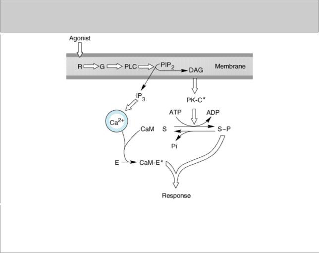
protein calmodulin, which regulates activities of other enzymes, including calcium-dependent protein kinases.
Figure 2–14.
The Ca2+-phosphoinositide signaling pathway. Key proteins include hormone receptors (R), a G protein (G), a phosphoinositide-specific phospholipase C (PLC), protein kinase C substrates of the kinase (S), calmodulin (CaM), and calmodulin-binding enzymes (E), including kinases, phosphodiesterases, etc. (PIP2, phosphatidylinositol-4,5-bisphosphate; DAG, diacylglycerol. Asterisk denotes activated state. Open arrows denote regulatory effects.)
With its multiple second messengers and protein kinases, the phosphoinositide signaling pathway is much more complex than the cAMP pathway. For example, different cell types may contain one or more specialized calciumand calmodulin-dependent kinases with limited substrate specificity (eg, myosin light chain kinase) in addition to a general calciumand calmodulin-dependent kinase that can phosphorylate a wide variety of protein substrates. Furthermore, at least nine structurally distinct types of protein kinase C have been identified.
As in the cAMP system, multiple mechanisms damp or terminate signaling by this pathway. IP3 is inactivated by dephosphorylation; diacylglycerol is either phosphorylated to yield phosphatidic acid, which is then converted back into phospholipids, or it is deacylated to yield arachidonic acid; Ca2+ is actively removed from the cytoplasm by Ca2+ pumps.
These and other nonreceptor elements of the calcium-phosphoinositide signaling pathway are now becoming targets for drug development. For example, the therapeutic effects of lithium ion, an established agent for treating manic-depressive illness, may be mediated by effects on the metabolism of phosphoinositides (see Chapter 29: Antipsychotic Agents & Lithium).
Cyclic Guanosine Monophosphate (cGMP)
Unlike cAMP, the ubiquitous and versatile carrier of diverse messages, cGMP has established signaling roles in only a few cell types. In intestinal mucosa and vascular smooth muscle, the cGMP-based signal transduction mechanism closely parallels the cAMP-mediated signaling mechanism. Ligands detected by cell surface receptors stimulate membrane-bound guanylyl cyclase to produce cGMP, and cGMP acts by stimulating a cGMP-dependent protein kinase. The actions of cGMP in these cells are terminated by enzymatic degradation of the cyclic nucleotide and by dephosphorylation of kinase substrates.
Increased cGMP concentration causes relaxation of vascular smooth muscle by a kinase-mediated mechanism that results in dephosphorylation of myosin light chains (see Figure 12–2). In these smooth muscle cells, cGMP synthesis can be elevated by two different transmembrane signaling mechanisms utilizing two different guanylyl cyclases. ANP, a blood-borne peptide hormone, stimulates a transmembrane receptor by binding to its extracellular domain, thereby activating the guanylyl cyclase activity that resides in the receptor's intracellular domain. The other mechanism mediates responses to NO (see Chapter 19: Nitric Oxide, Donors, & Inhibitors), which is generated in vascular endothelial cells in response to natural vasodilator agents such as acetylcholine and histamine (NO is also called endothelium-derived relaxing factor [EDRF]). After entering the target cell, NO binds to and activates a cytoplasmic guanylyl cyclase. A number of useful vasodilating drugs act by generating or mimicking NO, or by interfering with the metabolic breakdown of cGMP by phosphodiesterase (see Chapter 11: Antihypertensive Agents and Chapter 12: Vasodilators & the Treatment of Angina Pectoris).
Interplay among Signaling Mechanisms
The calcium-phosphoinositide and cAMP signaling pathways oppose one another in some cells and are complementary in others. For example, vasopressor agents that contract smooth muscle act by IP3-mediated mobilization of Ca2+, whereas agents that relax smooth muscle often act by elevation of cAMP. In contrast, cAMP and phosphoinositide second messengers act together to stimulate glucose release from the liver.
Phosphorylation: A Common Theme
Almost all second messenger signaling involves reversible phosphorylation, which performs two principal functions in signaling: amplification and flexible regulation. In amplification, rather like GTP bound to a G protein, the attachment of a phosphoryl group to a serine, threonine, or tyrosine residue powerfully amplifies the initial regulatory signal by recording a molecular memory that the pathway has been activated; dephosphorylation erases the memory, taking a longer time to do so than is required for dissociation of an allosteric ligand. In flexible regulation, differing substrate specificities of the multiple protein kinases regulated by second messengers provide branch points in signaling pathways that may be independently regulated. In this way, cAMP, Ca2+, or other second messengers can use the presence or absence of particular kinases or kinase substrates to produce quite different effects in different cell types. Inhibitors of protein kinases have great potential as therapeutic agents, particularly in neoplastic diseases. Trastuzumab, an antibody that antagonizes growth factor receptor signaling, was discussed earlier as a therapeutic agent for breast cancer. Another example of this general approach is imatinib (Gleevec, STI571), a small molecule inhibitor of the cytoplasmic tyrosine kinase Bcr/Abl, which is activated by growth factor signaling pathways and is overexpressed in chronic myelogenous leukemia (CML). This compound, a promising agent for treating CML, was recently approved by the US Food and Drug Administration (FDA) for clinical use.