

Книги фарма 2 / Bertram G. Katzung-Basic & Clinical Pharmacology(9th Edition)
.pdf
|
|
prazosin |
|
|
|
|
|
|
|
1B |
|
CEC |
|
C8 |
|
|
(irreversible) |
|
|
|
|
|
|
|
1D |
|
WB4101 |
|
|
|
|
|
|
|
|
|
|
|
|
2 type |
|
Rauwolscine, |
cAMP common to |
C20 |
|
|
yohimbine |
all |
|
|
|
|
|
|
2A |
Clonidine, BHT920 |
|
cAMP; K+channels; |
C10 |
|
Oxymetazoline |
|
Ca2+ channels |
|
|
|
|
|
|
|
|
|
|
|
2B |
|
Prazosin |
cAMP; Ca2+ |
C2 |
|
|
|
channels |
|
|
|
|
|
|
|
|
|
|
|
2C |
|
Prazosin |
cAMP |
C4 |
|
|
|
|
|
|
|
|
|
|
type |
Isoproterenol |
Propranolol |
cAMP common to |
|
|
|
|
all |
|
|
|
|
|
|
1 |
Dobutamine |
Betaxolol |
cAMP |
C10 |
|
|
|
|
|
|
|
|
|
|
2 |
Procaterol, |
Butoxamine |
cAMP |
C5 |
|
terbutaline |
|
|
|
|
|
|
|
|
3 |
BRL37344 |
|
cAMP |
C8 |
|
|
|
|
|
|
|
|
|
|
Dopamine |
Dopamine |
|
|
|
type |
|
|
|
|
|
|
|
|
|
D1 |
Fenoldopam |
|
cAMP |
C5 |
|
|
|
|
|
|
|
|
|
|
D2 |
Bromocriptine |
|
cAMP; K+ |
C11 |
|
|
|
channels; Ca2+ |
|
|
|
|
channels |
|
|
|
|
|
|
|
|
|
|
|
D3 |
Quinpirol |
AJ76 |
cAMP; K+ |
C3 |
|
|
|
channels; Ca2+ |
|
|
|
|
channels |
|
|
|
|
|
|
|
|
|
|
|
D4 |
|
Clozapine |
cAMP |
C11 |
|
|
|
|
|
|
|
|
|
|
D5 |
|
|
cAMP |
C4 |
|
|
|
|
|
|
|
|
|
|
Key: BRL37344 = Sodium-4-(2-[2-hydroxy-{3-chlorophenyl}ethylamino]propyl)phenoxyacetate
BHT920 = 6-Allyl-2-amino-5,6,7,8-tetrahydro-4H-thiazolo-[4,5-d]-azepine

CEC = Chloroethylclonidine
DAG = Diacylglycerol
IP3 = Inositol trisphosphate
WB4101 = N-[2-(2,6-dimethoxyphenoxy)ethyl]-2,3-dihydro-1,4-benzodioxan-2-methanamine
Alpha Adrenoceptors
Following the demonstration of the 
 subtypes, it was found that there are two major groups of
 receptors:
 1 and
1 and 
 2. These receptors were originally identified with antagonist drugs that distinguished between
 1 and
1 and 
 2 receptors. For example,
 adrenoceptors were identified in a variety of tissues by measuring the binding of radiolabeled antagonist compounds that are considered to have a high affinity for these receptors, eg, dihydroergocryptine (
 1 and
1 and 
 2), prazosin (
 1), and yohimbine (
 2). These radioligands were used to measure the number of receptors in tissues and to determine the affinity (by displacement of the radiolabeled ligand) of other drugs that interact with the receptors.
The concept of subtypes within the 
 1 group emerged out of pharmacologic experiments that demonstrated complex shapes of agonist dose-response curves of smooth muscle contraction as well as differences in antagonist affinities in inhibiting contractile responses in various tissues. These experiments demonstrated the existence of two subtypes of
 1 receptor that could be distinguished on the basis of their reversible affinities for a variety of drugs and experimental compounds. A third
  1 receptor subtype was subsequently identified by molecular cloning techniques. These
 1 receptors are termed
 1A,
1A, 
 1B, and
 1D receptors. There is evidence that the
 1A receptor has splice variants. A major current area of investigation is determining the importance of each of these various subtypes in mediating
 1 receptor responses in a variety of organs.
The hypothesis that there are subtypes of 
 2 receptors emerged from pharmacologic experiments and molecular cloning. It is now known that there are three subtypes of
 2 receptors, termed
 2A,
2A, 
 2B, and
 2C, that are products of distinct genes.
Dopamine Receptors
The endogenous catecholamine dopamine produces a variety of biologic effects that are mediated by interactions with specific dopamine receptors (Table 9–1). These receptors are distinct from 
 and
and 
 receptors and are particularly important in the brain (see Chapter 21: Introduction to the Pharmacology of CNS Drugs and Chapter 29: Antipsychotic Agents & Lithium) and in the splanchnic and renal vasculature. There is now considerable evidence for the existence of at least five subtypes of dopamine receptors. Pharmacologically distinct dopamine receptor subtypes, termed D1 and D2, have been known for some time. Molecular cloning has identified several distinct genes encoding each of these subtypes. Further complexity occurs because of the presence of introns within the coding region of the D2-like receptor genes, which allows for alternative splicing of the exons in this major subtype. There is extensive polymorphic variation in the D4 human receptor gene. The terminology of the various subtypes is D1, D2, D3, D4, and D5. They comprise two D1-like receptors (D1 and D5) and three D2-like (D2, D3, and D4). These subtypes may have importance for understanding the efficacy and adverse effects of novel antipsychotic drugs (see Chapter 29: Antipsychotic Agents & Lithium).
Receptor Selectivity

Examples of clinically useful sympathomimetic agonists that are relatively selective for 
 1-,
1-, 
 2-, and
 -adrenoceptor subgroups are compared with some nonselective agents in Table 9–2. Selectivity means that a drug may preferentially bind to one subgroup of receptors at concentrations too low to interact extensively with another subgroup. For example, norepinephrine preferentially activates
  1 receptors as compared with
  2 receptors. However, selectivity is not usually absolute (nearly absolute selectivity has been termed "specificity"), and at higher concentrations related classes of receptor may also interact with the drug. As a result, the "numeric" subclassification of adrenoceptors is clinically important mainly for drugs that have relatively marked selectivity. Given interpatient variations in drug kinetics and dynamics, the extent of a drug's selectivity should be kept in mind if this property is viewed as clinically important in the treatment of an individual patient.
Table 9–2. Relative Selectivity of Adrenoceptor Agonists.
|
Relative Receptor Affinities |
|||
|
|
|
|
|
Alpha agonists |
|
|
|
|
|
|
|
|
|
Phenylephrine, methoxamine |
1 |
> |
2 >>>>> |
|
|
|
|
|
|
|
|
|
|
|
Clonidine, methylnorepinephrine |
2 |
> |
1 >>>>> |
|
|
|
|
|
|
|
|
|
|
|
Mixed alpha and beta agonists |
|
|
|
|
|
|
|
|
|
Norepinephrine |
1 |
= |
2; |
1 >> 2 |
|
|
|
|
|
|
|
|
|
|
Epinephrine |
1 |
= |
2; |
1 = 2 |
|
|
|
|
|
|
|
|
|
|
Beta agonists |
|
|
|
|
|
|
|
||
Dobutamine1 |
1 |
> 2 >>>> |
||
|
|
|
|
|
|
|
|
|
|
Isoproterenol |
1 |
= |
2 >>>> |
|
|
|
|
|
|
|
|
|
|
|
Terbutaline, metaproterenol, albuterol, ritodrine |
2 |
>>> |
1 >>>> |
|
|
|
|
|
|
|
|
|
|
|
Dopamine agonists |
|
|
|
|
|
|
|||
Dopamine |
D1 = D2 >>> >>> |
|||
|
|
|
|
|
|
|
|||
Fenoldopam |
D1 >>> D2 |
|||
|
|
|
|
|
|
|
|
|
|
1See text.
The exact number of adrenoceptor subtypes that are actually expressed in human tissues is uncertain, but expression of subtypes has been demonstrated in tissues where the physiologic or

tissue. For example, determining which blood vessels express which subtypes of 
 1 and
1 and 
 2 receptors could lead to design of drugs having selectivity for certain vascular beds such as the splanchnic or coronary vessels. Similarly, there has been extensive investigation into the
 1 receptor subtypes mediating pharmacologic responses in the human prostate.
Molecular Mechanisms of Sympathomimetic Action
The effects of catecholamines are mediated by cell surface receptors. As described in Chapter 2: Drug Receptors & Pharmacodynamics, these receptors are coupled by G proteins to the various effector proteins whose activities are regulated by those receptors. Each G protein is a heterotrimer consisting of 
 ,
,  , and
, and  subunits. G proteins are classified on the basis of their distinctive
 subunits. G proteins of particular importance for adrenoceptor function include Gs, the stimulatory G protein of adenylyl cyclase; Gi, the inhibitory G protein of adenylyl cyclase; and Gq, the protein coupling
  receptors to phospholipase C. The activation of G protein-coupled receptors by catecholamines promotes the dissociation of GDP from the
 subunit of the appropriate G protein. GTP then binds to this G protein, and the
 subunit dissociates from the
  -
-  unit. The activated GTP-bound
 subunit then regulates the activity of its effector. Effectors of adrenoceptor-activated
 subunits include adenylyl cyclase, cGMP phosphodiesterase, phospholipase C, and ion channels. The
 subunit is inactivated by hydrolysis of the bound GTP to GDP and Pi, and the subsequent reassociation of the
  subunit with the
  -
- subunit. The
  -
- subunits have additional independent effects, acting on a variety of effectors such as ion channels and enzymes.
Receptor Types
Alpha Receptors
Alpha1 receptors stimulate polyphosphoinositide hydrolysis, leading to the formation of inositol 1,4,5-trisphosphate (IP3) and diacylglycerol (DAG) (Figure 9–1). G proteins in the Gq family couple 
 1 receptors to phospholipase C. IP3 promotes the release of sequestered Ca2+ from intracellular stores, which increases the cytoplasmic concentration of free Ca2+ and the activation of various calcium-dependent protein kinases. Activation of these receptors may also increase influx of calcium across the cell's plasma membrane. Inositol 1,4,5-trisphosphate is sequentially dephosphorylated, which ultimately leads to the formation of free inositol. DAG activates protein kinase C that modulates activity of many signaling pathways. In addition,
 1 receptors activate signal transduction pathways that were originally described for peptide growth factor receptors which activate tyrosine kinases. For example,
 1 receptors have been found to activate mitogenactivated kinases (MAP kinases) and polyphosphoinositol-3-kinase (PI-3-kinase). These pathways may have importance for the
 1 receptor-mediated stimulation of cell growth and proliferation through the regulation of gene expression. The physiologic significance of this "cross talk" between major signaling pathways remains to be determined.
Figure 9–1.
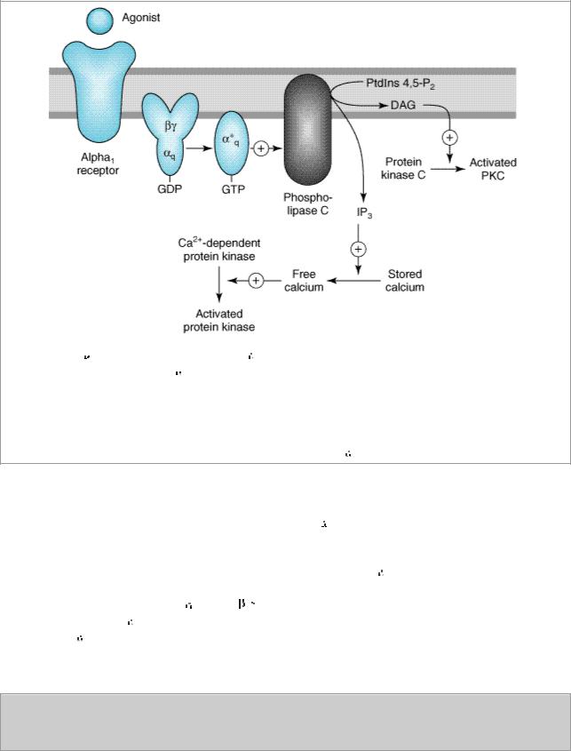
Activation of 
 1 responses. Stimulation of
 1 receptors by catecholamines leads to the activation of a Gq coupling protein. The
 subunit of this G protein activates the effector, phospholipase C, which leads to the release of IP3 (inositol 1,4,5-trisphosphate) and DAG (diacylglycerol) from phosphatidylinositol 4,5-bisphosphate (PtdIns 4,5-P2). IP3 stimulates the release of sequestered stores of calcium, leading to an increased concentration of cytoplasmic Ca2+. Ca2+ may then activate Ca2+-dependent protein kinases, which in turn phosphorylate their substrates. DAG activates protein kinase C. See text for additional effects of
 1 receptor activation.
Alpha2 receptors inhibit adenylyl cyclase activity and cause intracellular cAMP levels to decrease. In addition to this well-documented effect, activation of 
 2-receptors utilize additional signaling pathways, including regulation of ion channel activities and the activities of important enzymes involved in signal transduction. Alpha2-receptor–mediated inhibition of adenylyl cyclase activity is transduced by the inhibitory regulatory protein, Gi, which couples
 2 receptors to the inhibition of adenylyl cyclase (Figure 9–2). How the activation of Gi leads to the inhibition of adenylyl cyclase is unclear, but it is likely that both
 and the
  -
-
 subunits of Gi contribute to this response. In addition, some of the effects of
 2 adrenoceptors are independent of their ability to inhibit adenylyl cyclase; for example,
 2-receptor agonists cause platelet aggregation and a decrease in platelet cAMP levels, but it is not clear that aggregation is the result of the decrease in cAMP or other mechanisms involving Gi-regulated effectors.
Figure 9–2.
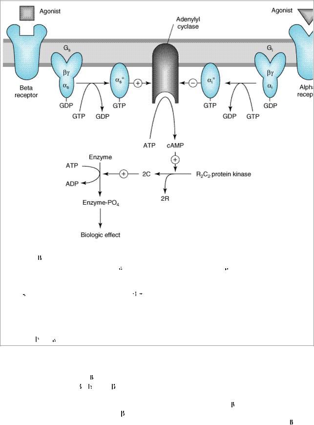
Activation and inhibition of adenylyl cyclase by agonists that bind to catecholamine receptors. Binding to  adrenoceptors stimulates adenylyl cyclase by activating the stimulatory G protein, Gs, which leads to the dissociation of its
 subunit charged with GTP. This
 s subunit directly activates adenylyl cyclase, resulting in an increased rate of synthesis of cAMP. Alpha2 adrenoceptor ligands inhibit adenylyl cyclase by causing dissociation of the inhibitory G protein, Gi, into its subunits; ie, an
 i subunit charged with GTP and a
 -
- unit. The mechanism by which these subunits inhibit adenylyl cyclase is uncertain. cAMP binds to the regulatory subunit (R) of cAMP-dependent protein kinase, leading to the liberation of active catalytic subunits (C) that phosphorylate specific protein substrates and modify their activity. These catalytic units also phosphorylate the cAMP response element binding protein (CREB), which modifies gene expression. See text for other actions of

 and
and 
 2 adrenoceptors.
Beta Receptors
The mechanism of action of  agonists has been studied in considerable detail. Activation of all three receptor subtypes (
 1,
1,  2, and
  3) results in activation of adenylyl cyclase and increased conversion of ATP to cAMP (Figure 9–2). Activation of the cyclase enzyme is mediated by the stimulatory coupling protein Gs. cAMP is the major second messenger of
  -receptor activation. For example, in the liver of many species,
  -receptor activation increases cAMP synthesis, which leads to a cascade of events culminating in the activation of glycogen phosphorylase. In the heart,
  - receptor activation increases the influx of calcium across the cell membrane and its sequestration inside the cell. Beta-receptor activation also promotes the relaxation of smooth muscle. While the mechanism is uncertain, it may involve the phosphorylation of myosin light chain kinase to an inactive form (see Figure 12–1). Beta adrenoceptors may activate voltage-sensitive calcium

channels in the heart via Gs-mediated enhancement independently of changes in cAMP concentration. Under certain circumstances,  2 receptors may couple to Gi proteins. These receptors have been demonstrated to activate additional kinases, such as MAP kinases, by forming multisubunit complexes, found within cells, containing multiple signaling molecules. In addition, recent evidence suggests that formation of dimers of
  receptors themselves (both homodimers and heterodimers of
  1 and
1 and  2 receptors) is importantly involved in their signaling mechanisms.
Dopamine Receptors
The D1 receptor is typically associated with the stimulation of adenylyl cyclase (Table 9–1); for example, D1-receptor-induced smooth muscle relaxation is presumably due to cAMP accumulation in the smooth muscle of those vascular beds where dopamine is a vasodilator. D2 receptors have been found to inhibit adenylyl cyclase activity, open potassium channels, and decrease calcium influx.
Receptor Regulation
Responses mediated by adrenoceptors are not fixed and static. The number and function of adrenoceptors on the cell surface and their responses may be regulated by catecholamines themselves, other hormones and drugs, age, and a number of disease states. These changes may modify the magnitude of a tissue's physiologic response to catecholamines and can be important clinically during the course of treatment. One of the best-studied examples of receptor regulation is the desensitization of adrenoceptors that may occur after exposure to catecholamines and other sympathomimetic drugs. After a cell or tissue has been exposed for a period of time to an agonist, that tissue often becomes less responsive to further stimulation by that agent. Other terms such as tolerance, refractoriness, and tachyphylaxis have also been used to denote desensitization. This process has potential clinical significance because it may limit the therapeutic response to sympathomimetic agents.
Many mechanisms have been found to contribute to desensitization. Operating at transcriptional, translational, and protein levels, some mechanisms function relatively slowly—over the course of hours or days. Other mechanisms of desensitization occur quickly, within minutes. Rapid modulation of receptor function in desensitized cells may involve critical covalent modification of the receptor, especially by phosphorylation on specific amino acid residues, association of these receptors with other proteins, or changes in their subcellular location.
There are two major categories of desensitization of responses mediated by G protein coupled receptors. Homologous desensitization refers to loss of responsiveness exclusively of the receptors that have been exposed to repeated or sustained activation by a drug. Heterologous desensitization refers to loss of responsiveness of some cell surface receptors that have not been directly activated by the drug in question.
A major mechanism of desensitization that occurs rapidly involves phosphorylation of receptors by members of the G protein-coupled receptor kinase (GRK) family, of which there are at least seven members. Specific adrenoreceptors are substrates for these kinases only when they are bound to an agonist. This mechanism is an example of homologous desensitization since it specifically involves only agonist-occupied receptors.
Phosphorylation of these receptors enhances their affinity for  -arrestins; upon binding of a
 - arrestin molecule, the capacity of the receptor to activate G proteins is blunted, presumably due to steric hindrance (see Figure 2–12). Arrestins constitute another large family of widely expressed

proteins. Receptor phosphorylation followed by  -arrestin binding has been linked to subsequent endocytosis of the receptor. This response may be facilitated by the capacity of
 -arrestins to bind to the structural protein clathrin. In addition to blunting responses requiring the presence of the receptor on the cell surface, these regulatory processes may also contribute to novel mechanisms of receptor signaling via intracellular pathways.
Receptor desensitization may also be mediated by second-messenger feedback. For example, 
 adrenoceptors stimulate cAMP accumulation, which leads to activation of protein kinase A; protein kinase A can phosphorylate residues on
 receptors, resulting in inhibition of receptor function. For example, for the
  2 receptor, phosphorylation occurs on serine residues both in the third cytoplasmic loop and carboxyl terminal tail of the receptor. Similarly, activation of protein kinase C by Gq- coupled receptors may lead to phosphorylation of this class of G protein-coupled receptors. This second-messenger feedback mechanism has been termed heterologous desensitization because activated protein kinase A or protein kinase C may phosphorylate any structurally similar receptor with the appropriate consensus sites for phosphorylation by these enzymes.
Adrenoreceptor Polymorphisms
Since elucidation of the sequences of the genes encoding the 
 1,
1, 
 2, and
  subtypes of adrenoceptors, it has become clear that there are relatively common genetic polymorphisms for many of these receptor subtypes in humans. Some of these may lead to changes in critical amino acid sequences that have pharmacologic importance. There is evidence that some of these polymorphisms may change the susceptibility to diseases such as heart failure, alter the propensity of a receptor to desensitize, and alter therapeutic responses to drugs in diseases such as asthma.
Chemistry & Pharmacokinetics of Sympathomimetic Drugs
Phenylethylamine may be considered the parent compound from which sympathomimetic drugs are derived (Figure 9–3). This compound consists of a benzene ring with an ethylamine side chain. Substitutions may be made (1) on the terminal amino group, (2) on the benzene ring, and (3) on the 
 or
or 
 carbons. Substitution by –OH groups at the 3 and 4 positions yields sympathomimetic drugs collectively known as catecholamines. The effects of modification of phenylethylamine are to change the affinity of the drugs for
 and
and  receptors as well as to influence the intrinsic ability to activate the receptors. In addition, chemical structure determines the pharmacokinetic properties of these molecules. Sympathomimetic drugs may activate both
 and
and 
 receptors; however, the relative
  -receptor versus
 -receptor activity spans the range from almost pure
 activity (methoxamine) to almost pure
 activity (isoproterenol).
Figure 9–3.
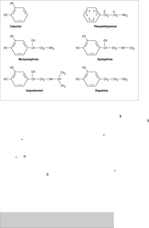
Phenylethylamine and some important catecholamines. Catechol is shown for reference.
Substitution on the Amino Group
Increasing the size of alkyl substituents on the amino group tends to increase  -receptor activity. For example, methyl substitution on norepinephrine, yielding epinephrine, enhances activity at
  2 receptors. Beta activity is further enhanced with isopropyl substitution at the amino nitrogen (isoproterenol). Beta2-selective agonists generally require a large amino substituent group. The larger the substituent on the amino group, the lower the activity at
 receptors; eg, isoproterenol is very weak at
 receptors.
Substitution on the Benzene Ring
Maximal  and
and  activity are found with catecholamines (drugs having –OH groups at the 3 and 4 positions). The absence of one or the other of these groups, particularly the hydroxyl at C3, without other substitutions on the ring may dramatically reduce the potency of the drugs. For example, phenylephrine (Figure 9–4) is much less potent than epinephrine; indeed,
 receptor affinity is decreased about 100-fold and
  activity is almost negligible except at very high concentrations. However, catecholamines are subject to inactivation by catechol-O-methyltransferase (COMT), an enzyme found in gut and liver (see Chapter 6: Introduction to Autonomic Pharmacology). Therefore, absence of one or both –OH groups on the phenyl ring increases the bioavailability after oral administration and prolongs the duration of action. Furthermore, absence of ring –OH groups tends to increase the distribution of the molecule to the central nervous system. For example, ephedrine and amphetamine (Figure 9–4) are orally active, have a prolonged duration of action, and produce central nervous system effects not typically observed with the catecholamines.
Figure 9–4.
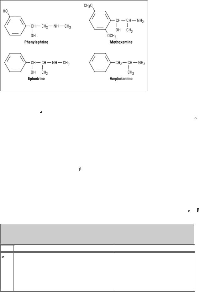
Some examples of noncatecholamine sympathomimetic drugs.
Substitution on the Alpha Carbon
Substitutions at the 
 carbon block oxidation by monoamine oxidase (MAO) and prolong the action of such drugs, particularly the noncatecholamines. Ephedrine and amphetamine are examples of
 - substituted compounds (Figure 9–4). Alpha-methyl compounds are also called phenylisopropylamines. In addition to their resistance to oxidation by MAO, some phenylisopropylamines have an enhanced ability to displace catecholamines from storage sites in noradrenergic nerves. Therefore, a portion of their activity is dependent upon the presence of normal norepinephrine stores in the body; they are indirectly acting sympathomimetics.
Substitution on the Beta Carbon
Direct-acting agonists typically have a 
 -hydroxyl group, though dopamine does not. In addition to activating adrenoceptors, this hydroxyl group may be important for storage of sympathomimetic amines in neural vesicles.
Organ System Effects of Sympathomimetic Drugs
General outlines of the cellular actions of sympathomimetics are presented in Tables 6–3 and 9–3. The net effect of a given drug in the intact organism depends on its relative receptor affinity (
 or
or  ), intrinsic activity, and the compensatory reflexes evoked by its direct actions.
Table 9–3. Distribution of Adrenoceptor Subtypes.
Type Tissue |
Actions |
1 |
Most vascular smooth muscle (innervated) |
Contraction |
|
|
|
|
|
|
Pupillary dilator muscle |
Contraction (dilates pupil) |
|
|
|
|
|
|
Pilomotor smooth muscle |
Erects hair |
|
|
|
|
|
|
Prostate |
Contraction |
|
|
|
|
|
|
Heart |
Increases force of contraction |
|
|
|
|
|