

Книги фарма 2 / Bertram G. Katzung-Basic & Clinical Pharmacology(9th Edition)
.pdfhave recently been found to block farnesyl pyrophosphate synthase, an enzyme in the mevalonate pathway that appears to be critical for osteoclast survival. Statins, which block mevalonate synthesis, stimulate bone formation at least in animal studies. Thus, the mevalonate pathway appears to be important in bone cell function and provides new targets for drug development. These effects vary depending on the bisphosphonate being studied (ie, only amino bisphosphonates have this property) and may account for some of the clinical differences observed in the effects of the various bisphosphonates on bone mineral homeostasis. However, with the exception of the induction of a mineralization defect by higher than approved doses of etidronate and gastric and esophageal irritation by pamidronate and by high doses of alendronate, these drugs have proved to be remarkably free of adverse effects. Esophageal irritation can be minimized by taking the drug with a full glass of water and remaining upright for 30 minutes.
Plicamycin (Mithramycin)
Plicamycin is a cytotoxic antibiotic (see Chapter 55: Cancer Chemotherapy) that has been used clinically for two disorders of bone mineral metabolism: Paget's disease and hypercalcemia. The cytotoxic properties of the drug appear to involve its binding to DNA and interruption of DNAdirected RNA synthesis. The reasons for its usefulness in the treatment of Paget's disease and hypercalcemia are unclear but may relate to the need for protein synthesis to sustain bone resorption. The doses required to treat Paget's disease and hypercalcemia are about one tenth the amounts required to achieve cytotoxic effects.
Thiazides
The chemistry and pharmacology of this family of drugs are covered in Chapter 15: Diuretic Agents. The principal application of thiazides in the treatment of bone mineral disorders is in reducing renal calcium excretion. Thiazides may increase the effectiveness of parathyroid hormone in stimulating reabsorption of calcium by the renal tubules or may act on calcium reabsorption secondarily by increasing sodium reabsorption in the proximal tubule. In the distal tubule, thiazides block sodium reabsorption at the luminal surface, increasing the calcium-sodium exchange at the basolateral membrane and thus enhancing calcium reabsorption into the blood at this site. Thiazides have proved to be quite useful in reducing the hypercalciuria and incidence of stone formation in subjects with idiopathic hypercalciuria. Part of their efficacy in reducing stone formation may lie in their ability to decrease urine oxalate excretion and increase urine magnesium and zinc levels (both of which inhibit calcium oxalate stone formation).
Fluoride
Fluoride is well established as effective for the prophylaxis of dental caries and has been under investigation for the treatment of osteoporosis. Both therapeutic applications originated from epidemiologic observations that subjects living in areas with naturally fluoridated water (1–2 ppm) had less dental caries and fewer vertebral compression fractures than subjects living in nonfluoridated water areas. Fluoride is accumulated by bones and teeth, where it may stabilize the hydroxyapatite crystal. Such a mechanism may explain the effectiveness of fluoride in increasing the resistance of teeth to dental caries, but it does not explain new bone growth.
Fluoride in drinking water appears to be most effective in preventing dental caries if consumed prior to the eruption of the permanent teeth. The optimum concentration in drinking water supplies is 0.5–1 ppm. Topical application is most effective if done just as the teeth erupt. There is little further benefit to giving fluoride after the permanent teeth are fully formed. Excess fluoride in drinking water leads to mottling of the enamel proportionate to the concentration above 1 ppm.

Because of the paucity of agents that stimulate new bone growth in patients with osteoporosis, the use of fluoride for this disorder has been examined (see Osteoporosis). Results of earlier studies indicated that fluoride alone without adequate calcium supplementation produced osteomalacia. More recent studies, in which calcium supplementation has been adequate, have demonstrated an improvement in calcium balance, an increase in bone mineral, and an increase in trabecular bone volume. However, studies of the ability of fluoride to reduce fractures reach opposite conclusions. Adverse effects observed—at the doses used for testing fluoride's effect on bone—include nausea and vomiting, gastrointestinal blood loss, arthralgias, and arthritis in a substantial proportion of patients. Such effects are usually responsive to reduction of the dose or giving fluoride with meals (or both). At present, fluoride is not approved by the Food and Drug Administration for use in osteoporosis.
Katzung PHARMACOLOGY, 9e > Section VII. Endocrine Drugs > Chapter 42. Agents That Affect Bone Mineral Homeostasis >
Clinical Pharmacology
Disorders of bone mineral homeostasis generally present with abnormalities in serum or urine calcium levels (or both), often accompanied by abnormal serum phosphate levels. These abnormal mineral concentrations may themselves cause symptoms requiring immediate treatment (eg, coma in malignant hypercalcemia, tetany in hypocalcemia). More commonly, they serve as clues to an underlying disorder in hormonal regulators (eg, primary hyperparathyroidism), target tissue response (eg, chronic renal failure), or drug misuse (eg, vitamin D intoxication). In such cases, treatment of the underlying disorder is of prime importance.
Since bone and kidney play central roles in bone mineral homeostasis, conditions that alter bone mineral homeostasis usually affect either or both of these tissues secondarily. Effects on bone can result in osteoporosis (abnormal loss of bone; remaining bone histologically normal), osteomalacia (abnormal bone formation due to inadequate mineralization), or osteitis fibrosa (excessive bone resorption with fibrotic replacement of resorption cavities). Biochemical markers of skeletal involvement include changes in serum levels of the skeletal isoenzyme of alkaline phosphatase and osteocalcin (reflecting osteoblastic activity) and urine levels of hydroxyproline and pyridinoline cross-links (reflecting osteoclastic activity). The kidney becomes involved when the calcium-times- phosphate product in serum exceeds the point at which ectopic calcification occurs (nephrocalcinosis) or when the calcium-times-oxalate (or phosphate) product in urine exceeds saturation, leading to nephrolithiasis. Subtle early indicators of such renal involvement include polyuria, nocturia, and hyposthenuria. Radiologic evidence of nephrocalcinosis and stones is not generally observed until later. The degree of the ensuing renal failure is best followed by monitoring the decline in creatinine clearance.
Abnormal Serum Calcium & Phosphate Levels
Hypercalcemia
Hypercalcemia causes central nervous system depression, including coma, and is potentially lethal. Its major causes (other than thiazide therapy) are hyperparathyroidism and cancer with or without bone metastases. Less common causes are hypervitaminosis D, sarcoidosis, thyrotoxicosis, milkalkali syndrome, adrenal insufficiency, and immobilization. With the possible exception of hypervitaminosis D, these latter disorders seldom require emergency lowering of serum calcium. A number of approaches are used to manage the hypercalcemic crisis.

Saline Diuresis
In hypercalcemia of sufficient severity to produce symptoms, rapid reduction of serum calcium is required. The first steps include rehydration with saline and diuresis with furosemide. Most patients presenting with severe hypercalcemia have a substantial component of prerenal azotemia owing to dehydration, which prevents the kidney from compensating for the rise in serum calcium by excreting more calcium in the urine. Therefore, the initial infusion of 500—1000 mL/h of saline to reverse the dehydration and restore urine flow can by itself substantially lower serum calcium. The addition of a loop diuretic such as furosemide not only enhances urine flow but also inhibits calcium reabsorption in the ascending limb of the loop of Henle (see Chapter 15: Diuretic Agents). Monitoring central venous pressure is important to forestall the development of heart failure and pulmonary edema in predisposed subjects. In many subjects, saline diuresis will suffice to reduce serum calcium levels to a point at which more definitive diagnosis and treatment of the underlying condition can be achieved. If this is not the case or if more prolonged medical treatment of hypercalcemia is required, the following agents are available (discussed in order of preference).
Bisphosphonates
Etidronate, 7.5 mg/kg in 250–500 mL saline, infused over several hours each day for 3 days, has proved quite useful in treating hypercalcemia of malignancy. More recently, pamidronate, 60–90 mg, infused over 2–4 hours, and zoledronate, 4 mg, infused over 15 minutes, have been approved for the same indication and appear to be more effective. This form of treatment is remarkably free of toxicity. The effects generally persist for weeks, but treatment can be repeated after a 7-day interval if necessary and if renal function is not impaired.
Calcitonin
Calcitonin has proved useful as ancillary treatment in a large number of patients. Calcitonin by itself seldom restores serum calcium to normal, and refractoriness frequently develops. However, its lack of toxicity permits frequent administration at high doses (200 MRC units or more). An effect on serum calcium is observed within 4–6 hours and lasts for 6–10 hours. Calcimar (salmon calcitonin) is available for parenteral and nasal administration.
Gallium Nitrate
Gallium nitrate is approved by the Food and Drug Administration for the management of hypercalcemia of malignancy and is undergoing trials for the treatment of advanced Paget's disease. This drug acts by inhibiting bone resorption. At a dosage of 200 mg/m2 body surface area per day given as a continuous intravenous infusion in 5% dextrose for 5 days, gallium nitrate proved superior to calcitonin in reducing serum calcium in cancer patients. Because of potential nephrotoxicity, patients should be well-hydrated and have good renal output before starting the infusion.
Plicamycin (Mithramycin)
Because of its toxicity, plicamycin (mithramycin) is not the drug of first choice for the treatment of hypercalcemia. However, when other forms of therapy fail, 25–50  g/kg given intravenously usually lowers serum calcium substantially within 24–48 hours. This effect can last for several days. This dose can be repeated as necessary. The most dangerous toxic effect is sudden thrombocytopenia followed by hemorrhage. Hepatic and renal toxicity can also occur. Hypocalcemia, nausea, and vomiting may limit therapy. Use of this drug must be accompanied by
careful monitoring of platelet counts, liver and kidney function, and serum calcium levels.
Phosphate
Giving intravenous phosphate is probably the fastest and surest way to reduce serum calcium, but it is a hazardous procedure if not done properly. Intravenous phosphate should be used only after other methods of treatment (pamidronate, calcitonin, saline diuresis with furosemide, and plicamycin) have failed to control symptomatic hypercalcemia. Phosphate must be given slowly (50 mmol or 1.5 g elemental phosphorus over 6–8 hours) and the patient switched to oral phosphate (1– 2 g/d elemental phosphorus, as one of the salts indicated below) as soon as symptoms of hypercalcemia have cleared. The risks of intravenous phosphate therapy include sudden hypocalcemia, ectopic calcification, acute renal failure, and hypotension. Oral phosphate can also lead to ectopic calcification and renal failure if serum calcium and phosphate levels are not carefully monitored, but the risk is less and the time of onset much longer. Phosphate is available in oral and intravenous forms as the sodium or potassium salt. Amounts required to provide 1 g of elemental phosphorus are as follows:
Intravenous:
In-Phos: 40 mL
Hyper-Phos-K: 15 mL
Oral:
Fleet Phospho-Soda: 6.2 mL
Neutra-Phos: 300 mL
K-Phos-Neutral: 4 tablets
Glucocorticoids
Glucocorticoids have no clear role in the acute treatment of hypercalcemia. However, the chronic hypercalcemia of sarcoidosis, vitamin D intoxication, and certain cancers may respond within several days to glucocorticoid therapy. Prednisone in oral doses of 30–60 mg daily is generally used, though equivalent doses of other glucocorticoids are effective. The rationale for the use of glucocorticoids in these diseases differs, however. The hypercalcemia of sarcoidosis is secondary to increased production of 1,25(OH)2D, possibly by the sarcoid tissue itself. Glucocorticoid therapy directed at the reduction of sarcoid tissue results in restoration of normal serum calcium and 1,25(OH)2D levels. The treatment of hypervitaminosis D with glucocorticoids probably does not alter vitamin D metabolism significantly but is thought to reduce vitamin D-mediated intestinal calcium transport. An action of glucocorticoids to reduce vitamin D-mediated bone resorption has not been excluded, however. The effect of glucocorticoids on the hypercalcemia of cancer is probably twofold. The malignancies responding best to glucocorticoids (ie, multiple myeloma and related lymphoproliferative diseases) are sensitive to the lytic action of glucocorticoids, so part of the effect may be related to decreased tumor mass and activity. Glucocorticoids have also been shown to inhibit the secretion or effectiveness of cytokines elaborated by multiple myeloma and related cancers that stimulate osteoclastic bone resorption. Other causes of hypercalcemia— particularly primary hyperparathyroidism—do not respond to glucocorticoid therapy.
Hypocalcemia

The main features of hypocalcemia are neuromuscular—tetany, paresthesias, laryngospasm, muscle cramps, and convulsions. The major causes of hypocalcemia in the adult are hypoparathyroidism, vitamin D deficiency, renal failure, and malabsorption. Neonatal hypocalcemia is a common disorder that usually resolves without therapy. The roles of PTH, vitamin D, and calcitonin in the neonatal syndrome are under active investigation. Large infusions of citrated blood can produce hypocalcemia by the formation of citrate-calcium complexes. Calcium and vitamin D (or its metabolites) form the mainstay of treatment of hypocalcemia.
Calcium
A number of calcium preparations are available for intravenous, intramuscular, and oral use. Calcium gluceptate (0.9 meq calcium/mL), calcium gluconate (0.45 meq calcium/mL), and calcium chloride (0.68–1.36 meq calcium/mL) are available for intravenous therapy. Calcium gluconate is the preferred form because it is less irritating to veins. Oral preparations include calcium carbonate (40% calcium), calcium lactate (13% calcium), calcium phosphate (25% calcium), and calcium citrate (21% calcium). Calcium carbonate is often the preparation of choice because of its high percentage of calcium, ready availability (eg, Tums), low cost, and antacid properties. In achlorhydric patients, calcium carbonate should be given with meals to increase absorption or the patient switched to calcium citrate, which is somewhat better absorbed. Combinations of vitamin D and calcium are available, but treatment must be tailored to the individual patient and individual disease, a flexibility lost by fixed-dosage combinations. Treatment of severe symptomatic hypocalcemia can be accomplished with slow infusion of 5–20 mL of 10% calcium gluconate. Rapid infusion can lead to cardiac arrhythmias. Less severe hypocalcemia is best treated with oral forms sufficient to provide approximately 400–800 mg of elemental calcium (1–2 g calcium carbonate) per day. Dosage must be adjusted to avoid hypercalcemia and hypercalciuria.
Vitamin D
When rapidity of action is required, 1,25(OH)2D3 (calcitriol), 0.25–1  g daily, is the vitamin D metabolite of choice, since it is capable of raising serum calcium within 24–48 hours. Calcitriol also raises serum phosphate, though this action is usually not observed early in treatment. The combined effects of calcitriol and all other vitamin D metabolites and analogs on both calcium and phosphate make careful monitoring of these mineral levels especially important to avoid ectopic calcification secondary to an abnormally high serum calcium x phosphate product. Since the choice of the appropriate vitamin D metabolite or analog for long-term treatment of hypocalcemia depends on the nature of the underlying disease, further discussion of vitamin D treatment will be found under the headings of the specific diseases.
Hyperphosphatemia
Hyperphosphatemia is a frequent complication of renal failure but is also found in all types of hypoparathyroidism (idiopathic, surgical, and pseudo), vitamin D intoxication, and the rare syndrome of tumoral calcinosis. Emergency treatment of hyperphosphatemia is seldom necessary but can be achieved by dialysis or glucose and insulin infusions. In general, control of hyperphosphatemia involves restriction of dietary phosphate plus the use of phosphate binding gels such as sevelamer and of calcium supplements. Because of their potential to induce aluminumassociated bone disease, aluminum-containing antacids should be used sparingly and only when other measures fail to control the hyperphosphatemia.
Hypophosphatemia
A variety of conditions are associated with hypophosphatemia, including primary hyperparathyroidism, vitamin D deficiency, idiopathic hypercalciuria, vitamin D-resistant rickets, various other forms of renal phosphate wasting (eg, Fanconi's syndrome), overzealous use of phosphate binders, and parenteral nutrition with inadequate phosphate content. Acute hypophosphatemia may lead to a reduction in the intracellular levels of high-energy organic phosphates (eg, ATP), interfere with normal hemoglobin-to-tissue oxygen transfer by decreasing red cell 2,3-diphosphoglycerate levels, and lead to rhabdomyolysis. However, clinically significant acute effects of hypophosphatemia are seldom seen, and emergency treatment is generally not indicated. The long-term effects of hypophosphatemia include proximal muscle weakness and abnormal bone mineralization (osteomalacia). Therefore, hypophosphatemia should be avoided during other forms of therapy and treated in conditions such as hypophosphatemic rickets of which it is a cardinal feature. Oral forms of phosphate available for use are listed above in the section on hypercalcemia.
Specific Disorders Involving the Bone Mineral-Regulating Hormones
Primary Hyperparathyroidism
This rather common disease, if associated with symptoms and significant hypercalcemia, is best treated surgically. Oral phosphate and bisphosphonates have been tried but cannot be recommended. Asymptomatic patients with mild disease in general do not get worse and may be left untreated. The calcimimetic agents, a new class of drugs that act through the calcium receptor, are in clinical trials. If they prove efficacious, medical management of this disease will need to be reconsidered.
Hypoparathyroidism
In the absence of PTH (idiopathic or surgical hypoparathyroidism) or a normal target tissue response to PTH (pseudohypoparathyroidism), serum calcium falls and serum phosphate rises. In such patients, 1,25(OH)2D levels are usually low, presumably reflecting the lack of stimulation by PTH of 1,25(OH)2D production. The skeletons of patients with idiopathic or surgical hypoparathyroidism are normal except for a slow turnover rate. A number of patients with pseudohypoparathyroidism appear to have osteitis fibrosa, suggesting that the normal or high PTH levels found in such patients are capable of acting on bone but not on the kidney. The distinction between pseudohypoparathyroidism and idiopathic hypoparathyroidism is made on the basis of normal or high PTH levels but deficient renal response (ie, diminished excretion of cAMP or phosphate) in patients with pseudohypoparathyroidism.
The principal therapeutic concern is to restore normocalcemia and normophosphatemia. Under most circumstances, vitamin D (25,000–100,000 units three times per week) and dietary calcium supplements suffice. More rapid increments in serum calcium can be achieved with calcitriol, though it is not clear that this metabolite offers a substantial advantage over vitamin D itself for long-term therapy. Many patients treated with vitamin D develop episodes of hypercalcemia. This complication is more rapidly reversible with cessation of therapy using calcitriol rather than vitamin D. This would be of importance to the patient in whom such hypercalcemic crises are common. Dihydrotachysterol and 25(OH)D have not received much study as therapy for hypoparathyroidism, though both should be effective. Whether they offer advantages over vitamin D sufficient to justify their added expense remains to be seen.
Nutritional Rickets

Vitamin D deficiency, once thought to be rare in this country, is being recognized more often, especially in the pediatric and geriatric populations on vegetarian diets and with reduced sunlight exposure. This problem can be avoided by daily intake of 400–800 units of vitamin D and treated by higher dosages (4000 units per day). No other metabolite is indicated. The diet should also contain adequate amounts of calcium and phosphate.
Chronic Renal Failure
The major problems of chronic renal failure that impact on bone mineral homeostasis are the loss of 1,25(OH)2D and 24,25(OH)2D production, the retention of phosphate that reduces ionized calcium levels, and the secondary hyperparathyroidism that results. With the loss of 1,25(OH)2D production, less calcium is absorbed from the intestine and less bone is resorbed under the influence of PTH. As a result hypocalcemia usually develops, furthering the development of hyperparathyroidism. The bones show a mixture of osteomalacia and osteitis fibrosa.
In contrast to the hypocalcemia that is more often associated with chronic renal failure, some patients may become hypercalcemic from two causes (in addition to overzealous treatment with calcium). The most common cause of hypercalcemia is the development of severe secondary (sometimes referred to as tertiary) hyperparathyroidism. In such cases, the PTH level in blood is very high. Serum alkaline phosphatase levels also tend to be high. Treatment often requires parathyroidectomy.
A less common circumstance leading to hypercalcemia is development of a form of osteomalacia characterized by a profound decrease in bone cell activity and loss of the calcium buffering action of bone. In the absence of kidney function, any calcium absorbed from the intestine accumulates in the blood. Therefore, such patients are very sensitive to the hypercalcemic action of 1,25(OH)2D.
These individuals generally have a high serum calcium but nearly normal alkaline phosphatase and PTH levels. The bone in such patients generally has a high aluminum content, especially in the mineralization front, which may block normal bone mineralization. These patients do not respond favorably to parathyroidectomy. Deferoxamine, an agent used to chelate iron (see Chapter 58: Heavy Metal Intoxication & Chelators), also binds aluminum and is being used as therapy for this disorder.
Use of Vitamin D Preparations
The choice of vitamin D preparation to be used in the setting of chronic renal failure in the dialysis patient depends on the type and extent of bone disease and hyperparathyroidism. No consensus has been reached regarding the advisability of using any vitamin D metabolite in the predialysis patient. 1,25(OH)2D3 (calcitriol) will rapidly correct hypocalcemia and at least partially reverse the secondary hyperparathyroidism and osteitis fibrosa. Many patients with muscle weakness and bone pain gain an improved sense of well-being.
Dihydrotachysterol, an analog of 1,25(OH)2D, is also available for clinical use, though it is used much less frequently than calcitriol. Dihydrotachysterol appears to be as effective as calcitriol, differing principally in its time course of action; calcitriol increases serum calcium in 1–2 days, whereas dihydrotachysterol requires 1–2 weeks. For an equipotent dose (0.2 mg dihydrotachy-sterol versus 0.5  g calcitriol), dihydrotachysterol costs about one fourth as much as calcitriol. A disadvantage of dihydrotachysterol is the inability to measure it in serum. Neither dihydrotachysterol nor calcitriol corrects the osteomalacic component of renal osteodystrophy in the majority of patients, and neither should be used in patients with hypercalcemia, especially if the bone disease is primarily osteomalacic.

Calcifediol (25[OH]D3) may also be used to advantage. Calcifediol is less effective than calcitriol in stimulating intestinal calcium transport, so that hypercalcemia is less of a problem with calcifediol. Like dihydrotachysterol, calcifediol requires several weeks to restore normocalcemia in hypocalcemic individuals with chronic renal failure. Presumably because of the reduced ability of the diseased kidney to metabolize calcifediol to more active metabolites, high doses (50–100 g daily) must be given to achieve the supraphysiologic serum levels required for therapeutic effectiveness.
Vitamin D has been used in treating renal osteodystrophy. However, patients with a substantial degree of renal failure who are thus unable to convert vitamin D to its active metabolites usually are refractory to vitamin D. Its use is decreasing as more effective alternatives become available.
Two analogs of calcitriol, doxercalciferol and paricalcitol, are approved for the treatment of secondary hyperparathyroidism of chronic renal failure. Their principal advantage is that they are less likely than calcitriol to induce hypercalcemia. Their biggest impact will be in patients in whom the use of calcitriol may lead to unacceptably high serum calcium levels.
Regardless of the drug employed, careful attention to serum calcium and phosphate levels is required. Calcium supplements (dietary and in the dialysate) and phosphate restriction (dietary and with oral ingestion of phosphate binders) should be employed along with the use of vitamin D metabolites. Monitoring serum PTH and alkaline phosphatase levels is useful in determining whether therapy is correcting or preventing secondary hyperparathyroidism. Although not generally available, percutaneous bone biopsies for quantitative histomorphometry may help in choosing appropriate therapy and following the effectiveness of such therapy. Unlike the rapid changes in serum values, changes in bone morphology require months to years. Monitoring serum levels of the vitamin D metabolites is useful to determine compliance, absorption, and metabolism.
Intestinal Osteodystrophy
A number of gastrointestinal and hepatic diseases result in disordered calcium and phosphate homeostasis that ultimately leads to bone disease. The bones in such patients show a combination of osteoporosis and osteomalacia. Osteitis fibrosa does not occur (as it does in renal osteodystrophy). The common features that appear to be important in this group of diseases are malabsorption of calcium and vitamin D. Liver disease may, in addition, reduce the production of 25(OH)D from vitamin D, though the importance of this in all but patients with terminal liver failure remains in dispute. The malabsorption of vitamin D is probably not limited to exogenous vitamin D. The liver secretes into bile a substantial number of vitamin D metabolites and conjugates that are reabsorbed in (presumably) the distal jejunum and ileum. Interference with this process could deplete the body of endogenous vitamin D metabolites as well as limit absorption of dietary vitamin D.
In mild forms of malabsorption, vitamin D (25,000–50,000 units three times per week) should suffice to raise serum levels of 25(OH)D into the normal range. Many patients with severe disease do not respond to vitamin D. Clinical experience with the other metabolites is limited, but both calcitriol and calcifediol have been used successfully in doses similar to those recommended for treatment of renal osteodystrophy. Theoretically, calcifediol should be the drug of choice under these conditions, since no impairment of the renal metabolism of 25(OH)D to 1,25(OH)2D and 24,25(OH)2D exists in these patients. Both calcitriol and 24,25(OH)2D may be of importance in reversing the bone disease. As in the other diseases discussed, treatment of intestinal osteodystrophy with vitamin D and its metabolites should be accompanied by appropriate dietary calcium supplementation and monitoring of serum calcium and phosphate levels.

 (OH)D
(OH)D
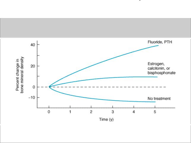
respond to it throughout the period of treatment. In contrast, estrogen, calcitonin, and bisphosphonates block bone resorption. This leads to a transient increase in bone mineral density because bone formation is not initially decreased. However, with time, both bone formation and bone resorption are decreased and bone mineral density reaches a new plateau.
Despite early promise that fluoride might be useful in the prevention or treatment of postmenopausal osteoporosis, this form of therapy remains controversial. A new formulation of fluoride (slow release, lower dose) appears to avoid much of the toxicity of earlier formulations and may reduce fracture rates. This formulation is under consideration for approval by the FDA. Teriparatide, the recombinant form of PTH 1-34, has recently been approved for treatment of osteoporosis. Teriparatide is given in a dosage 20  g subcutaneously daily. Like fluoride, teriparatide stimulates new bone formation, but unlike fluoride this new bone appears structurally normal and is associated with a substantial reduction in the incidence of fractures.
Calcitonin is approved for use in the treatment of postmenopausal osteoporosis. It has been shown to increase bone mass and reduce fractures, but only in the spine. It does not appear to be as effective as bisphosphonates or teriparatide.
Bisphosphonates are potent inhibitors of bone resorption. They increase bone density and reduce the risk of fractures in the hip, spine, and other locations. Alendronate and risedronate are approved for the treatment of osteoporosis, using either daily dosing schedules: alendronate 10 mg/d, risedronate 5 mg/d; or weekly schedules: alendronate 70 mg/wk, risedronate 35 mg/wk. These drugs are effective in men as well as women and for various causes of osteoporosis.
X-Linked & Autosomal Dominant Hypophosphatemia
These disorders are manifested by the appearance of rickets and hypophosphatemia in children, though they may first present in adults. X-linked hypophosphatemia is caused by mutations in a gene encoding a protein called PHEX, which appears to be an endopeptidase. Mutations in the gene responsible for the autosomal dominant form target a newly discovered member of the fibroblast growth factor (FGF) family, FGF23. The current concept is that FGF23 blocks the renal uptake of phosphate and blocks 1,25(OH)2D3 production. Normally PHEX cleaves and inactivates FGF23. Mutations in PHEX inactivate this enzyme, allowing FGF23 to accumulate. Similarly, mutations in FGF23 can prevent cleavage by PHEX. In either case, intact and biologically active FGF23 accumulates, leading to phosphate wasting in the urine and hypophosphatemia.
Phosphate is critical to normal bone mineralization; when phosphate stores are deficient, a clinical and pathologic picture resembling vitamin D-deficient rickets develops. However, such children fail to respond to the usual doses of vitamin D employed in the treatment of nutritional rickets. A defect in 1,25(OH)2D production by the kidney has also been noted, because the serum 1,25(OH)2D levels tend to be low relative to the degree of hypophosphatemia observed. This combination of low serum phosphate and low or low-normal serum 1,25(OH)2D provides the rationale for treating such patients with oral phosphate (1–3 g daily) and calcitriol (0.25–2 
 g daily). Reports of such combination therapy are encouraging in this otherwise debilitating disease.
Vitamin D-Dependent Rickets Types I & II
These distinctly different autosomal recessive diseases present as childhood rickets that does not respond to conventional doses of vitamin D. Type I vitamin D-dependent rickets is due to an isolated deficiency of 1,25(OH)2D production caused by mutations in 25(OH)D-1
 -hydroxylase. This condition can be treated with vitamin D (4000 units daily) or calcitriol (0.25–0.5 g daily).