

Hart C. Anthony, Shears Paul. Color Atlas of Medical Microbiology.pdf
.pdfNosocomial Infections 345
hospital. The prevalence of nosocomial infections increases with the size of the hospital. Within a particular hospital, the infection rate is always highest in the intensive care units.
Sources of Infection, Transmission Pathways
Nosocomial infections originate either from the patient’s own flora (endogenous infections) or from external sources (exogenous infections). Endogenous infections are the more frequent type. In such cases, the patient may have brought the pathogens into the hospital. It is, however, frequently the case that a patient’s skin and mucosa are colonized within one to two days
by bacteria of the hospital flora, which often shows multiple resistance to 4 antibiotics and replaces the patient’s individual flora, and that most endogen-
ous infections are then actually caused by the specific hospital flora. The source of infection for exogenous infections is most likely to lie with the medical staff. In most cases, the pathogens are transmitted from patient to patient during medical and nursing activities. Less frequently, the staff is either also infected or colonized by the hospital flora. Another important cause of nosocomial infections is technical medical measures that facilitate passage of the pathogens into the body. All invasive diagnostic measures present infection risks. The patient’s surroundings, i.e., the air, floor, or walls of the hospital room, are relatively unimportant as sources of infection.
Control
The measures taken to control and prevent nosocomial infections correspond in the wider sense to the general methods of infection control. The many different individual measures will not be listed here. The infection control program varies depending on the situation in each particular hospital and can be summarized in three general groups:
Operational measures. This category includes all measures pertaining to treatment and care of patients and cleaning measures. This includes asepsis, disinfection, sterilization, and cleaning. Further precautionary operational measures include isolation of patients that would be sources of infection and the economical and specific administration of antibiotic therapies.
Organizational measures. The organization of hospital infection control must be adapted to the structure of each particular hospital. Realization of the necessary measures, which of course always involve working time and expense, is best realized by establishing an infection control committee charged with the following tasks: determination and analysis of the situation,

346 4 Bacteria as Human Pathogens
definition of measures required to improve infection control by issuing binding guidelines, cooperation in the planning and acquisition of operational and structural facilities, cooperation on functional procedures in the various sections of the hospital, contributions to staff training in matters of hospital infection control. In order to carry out these tasks efficiently, the committee should have access to a working group of specialists. In larger hospitals, a hospital epidemiologist, and staff as required, are retained for these functions.
Structural measures.
These measures refer above all to new structures, which must be built in accordance with hygienic criteria. It is therefore the obligation of the planning
4architect to consult experts when planning the hygienically relevant parts of a construction measure. Hygienic aspects must of course also be considered in reconstruction and restoration of older building substance.

III
Mycology
Absidia corymbifera

348
5General Mycology
F. H. Kayser
|
|
General Characteristics of Fungi |
|
|
|
|
|
|
& Fungi are eukaryotic microorganisms (domain eucarya) that occur ubiqui- |
||
|
tously in nature. Only about 200 of the thousands of species have been |
||
|
identified as human pathogens, and among these known pathogenic species |
||
|
fewer than a dozen are responsible for more than 90% of all human fungal |
||
|
|||
5 |
infections. |
|
|
|
|
The basic morphological element of filamentous fungi is the hypha and a |
|
|
web of intertwined hyphae is called a mycelium. The basic form of a unicel- |
||
|
|||
|
lular fungus is the yeast cell. Dimorphic fungi usually assume the form of |
||
|
yeasts in the parasitic stage and the form of mycelia in the saprophytic stage. |
||
|
The cell walls of fungi consist of nearly 90% carbohydrate (chitin, glucans, |
||
|
mannans) and fungal membranes are rich in sterol types not found in other |
||
|
biological membranes (e.g., ergosterol). Filamentous fungi reproduce either |
||
|
asexually (mitosis), by hyphal growth and tip extension, or with the help of |
||
|
asexual spores. Yeasts reproduce by a process of budding. Sexual reproduc- |
||
|
|
tion (meiosis) on the other hand, produces sexual spores. Fungi imperfecti |
|
|
|
or deuteromycetes are the designation for a type of fungi in which the fruc- |
|
|
|
tification forms are either unknown or missing entirely. |
& |
|
|
|
|
Definition and Taxonomy
Fungi are microorganisms in the domain eucarya (see. p. 5). They show less differentiation than plants, but a higher degree of organization than the prokaryotes bacteria (Table 5.1). The kingdom of the fungi (Mycota) comprises over 50 000 different species, only about 200 of which have been identified as human pathogens. Only about a dozen of these “pathogenic” species cause 90% of all human mycoses. Many mycotic infections are relatively harmless, for instance the dermatomycoses. In recent years, however, the increasing numbers of patients with various kinds of immune defects have resulted in more life-threatening mycoses.

General Characteristics of Fungi 349
Table 5.1 Some Differences between Fungi and Bacteria
Properties |
Fungi |
Bacteria |
|
|
|
|
|
|
|
Nucleus |
Eukaryotic; nuclear |
Prokaryotic; no membrane; |
|
|
|
membrane; more than one |
nucleoid; only one “chromo- |
|
|
|
chromosome; mitosis |
some” |
|
|
Cytoplasm |
Mitochondria; endoplasmic |
No mitochondria; |
|
|
|
reticulum; 80S ribosomes |
no endoplasmic reticulum; |
|
|
|
|
70S ribosomes |
|
|
Cytoplasmic |
Sterols (ergosterol) |
No sterols |
|
|
membrane |
|
|
|
|
Cell wall |
Glucans, mannans, chitin, |
Murein, teichoic acids |
|
|
|
chitosan |
(Gram-positive), proteins |
|
|
Metabolism |
Heterotrophic; |
Heterotrophic; obligate |
|
|
|
5 |
|||
|
mostly aerobes; |
aerobes and anaerobes, |
|
|
|
|
|
||
|
no photosynthesis |
facultative anaerobes |
|
|
Size, mean diameter |
Yeast cells: 3–5–10 lm. |
1–5 lm |
|
|
|
|
|||
|
Molds: indefinable |
|
|
|
Dimorphism |
In some species |
None |
|
|
|
|
|
|
|
The taxonomy of the fungi is essentially based on their morphology. In medical mycology, fungi are classified according to practical aspects as dermatophytes, yeasts, molds, and dimorphic fungi. Molds grow in filamentous structures, yeasts as single cells and dermatophytes cause infections of the keratinized tissues (skin, hair, nails, etc.). Dimorphic fungi can appear in both of the two forms, as yeast cells or as mycelia (see the following pages).
Fungi are carbon heterotrophs. The saprobic or saprophytic fungi take carbon compounds from dead organic material whereas biotrophic fungi (parasites or symbionts) require living host organisms. Some fungi can exist in both saprophytic and biotrophic forms.
Morphology
Two morphological forms of fungi are observed (Fig. 5.1):
& Hypha: this is the basic element of filamentous fungi with a branched, tubular structure, 2–10 lm in width.
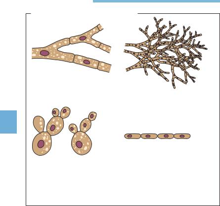
350 5 General Mycology
Basic Morphological Elements of Fungi
a |
b |
5
c |
d |
Fig. 5.1 |
There are two basic morphological forms: hypha and yeast. |
a Hypha, septate, or nonseptate.
b Mycelium: web of branched hyphae.
c Yeast form, budding (diameter of individual cell 3–5 lm).
dPseudomycelium.
&Mycelium: this is the web or matlike structure of hyphae. Substrate mycelia (specialized for nutrition) penetrate into the nutrient substrate, whereas aerial mycelia (for asexual propagation) develop above the nutrient medium.
&Fungal thallus: this is the entirety of the mycelia and is also called the fungal body or colony.
&Yeast: the basic element of the unicellular fungi. It is round to oval and 3– 10 lm in diameter. Several elongated yeast cells chained together and resembling true hyphae are called pseudohyphae.
&Dimorphism: some fungal species can develop either the yeast or the mycelium form depending on the environmental conditions, a property called dimorphism. Dimorphic pathogenic fungi take the form of yeast cells in the parasitic stage and appear as mycelia in the saprophytic stage.

General Characteristics of Fungi 351
Metabolism
All fungi are carbon heterotrophs, which means they are dependent on exogenous nutrient substrates as sources of organic carbon, and with a few exceptions, fungi are obligate aerobes. Many species are capable of maintaining metabolic activity in the most basic of nutrient mediums. The known metabolic types of fungi include thermophilic, psychrophilic, acidophilic, and halophilic species. The metabolic capabilities of fungi are exploited in the food industry (e.g., in the production of bread, wine, beer, cheese, or single-cell proteins) and in the pharmaceutical industry (e.g., in the production of antibiotic substances, enzymes, citric acid, etc.). The metabolic activity of fungi can also be a damaging factor. Fungal infestation can destroy foods, wooden structures, textiles, etc. Fungi also cause numerous plant diseases, in particular diseases of crops.
5
Reproduction in Fungi
Asexual reproduction. This category includes the vegetative propagation of hyphae and yeasts as well as vegetative fructification, i.e., formation of asexual spores.
&Hyphae elongate in a zone just short of the tip in which the cell wall is particularly elastic. This apical growth process can also include formation of swellings that develop into lateral hyphae, which can in turn also branch out.
&Yeasts reproduce by budding. This process begins with an outgrowth on the mother cell wall that develops into a daughter cell or blastoconidium. The isthmus between the two is finally cut off by formation of a septum. Some yeasts propagate in both the yeast and hypha forms (Fig. 6.2, p. 362).
&Vegetative fructification. A type of propagative form, the asexual spores, is formed in this process. These structures show considerable resistance to exogenous noxae and help fungi spread in the natural environment. Asexual spores come in a number of morphological types: conidia, sporangiospores, arthrospores, and blastospores. These forms rarely develop during the para-
sitic stages in hosts, but they are observed in cultures. The morphology of the asexual spores of fungi is an important identification characteristic.
Sexual fructification. Sexual reproduction in fungi perfecti (eumycetes) follows essentially the same patterns as in the higher eukaryotes. The nuclei of two haploid partners fuse to form a diploid zygote. The diploid nucleus then undergoes meiosis to form the haploid nuclei, finally resulting in the haploid

352 5 General Mycology
sexual spores: zygospores, ascospores, and basidiospores. Sexual spores are only rarely produced in the types of fungi that parasitize human tissues.
Sexual reproduction structures are either unknown or not present in many species of pathogenic fungi, known as fungi imperfecti (deuteromycetes).
General Aspects of Fungal Disease
& Besides fungal allergies (e.g., extrinsic allergic alveolitis) and mycotoxicoses (aflatoxicosis), fungal infections are by far the most frequent fungal diseases. Mycoses are classified clinically as follows:
—Primary mycoses (coccidioidomycosis, histoplasmosis, blastomycoses).
—Opportunistic mycoses (surface and deep yeast mycoses, aspergillosis,
5mucormycoses, phaeohyphomycoses, hyalohyphomycoses, cryptococcoses; penicilliosis, pneumocystosis).
—Subcutaneous mycoses (sporotrichosis, chromoblastomycosis, Madura foot (mycetoma).
—Cutaneous mycoses (pityriasis versicolor, dermatomycoses).
Little is known about fungal pathogenicity factors. The natural resistance of the macroorganism to fungal infection is based mainly on effective phagocytosis whereas specific resistance is generally through cellular immunity. Opportunistic mycoses develop mainly in patients with immune deficiencies (e.g., in neutropenia). Laboratory diagnostic methods for fungal infections mostly include microscopy and culturing, in order to detect the pathogens directly, and identification of specific antibodies. Therapeutics for treatment of mycoses include polyenes (above all amphotericin B), azoles (e.g., itraconazole, fluconazole, voriconazole), allylamines, antimetabolites (e.g., 5-flu- orocytosine), and echinocandins (e.g., caspofungin). Antimycotics are often administered in combination. &
Fungal Allergies and Fungal Toxicoses
Mycogenic Allergies
The spores of ubiquitous fungi continuously enter the respiratory tract with inspirated air. These spores contain potent allergens to which susceptible individuals may manifest strong hypersensitivity reactions. Depending on the localization of the reaction, it may assume the form of allergic rhinitis, bron-

General Aspects of Fungal Disease 353
chial asthma, or allergic alveolitis. Many of these allergic reactions are certified occupational diseases, i.e., “farmer’s lung,” “woodworker’s lung,” and other types of extrinsic allergic alveolitis.
Mycotoxicoses
Some fungi produce mycotoxins, the best known of which are the aflatoxins produced by the Aspergillus species. These toxins are ingested with the food stuffs on which the fungi have been growing. Aflatoxin B1 may contribute to primary hepatic carcinoma, a disease observed frequently in Africa and Southeast Asia.
Mycoses |
5 |
|
Data on the general incidence of mycotic infections can only be approximate, since there is no requirement that they be reported to the health authorities. It can be assumed that cutaneous mycoses are among the most frequent infections worldwide. Primary and opportunistic mycoses are, on the other hand, relatively rare. Opportunistic mycoses have been on the increase in recent years and decades, reflecting the fact that clinical manifestations are only observed in hosts whose immune disposition allows them to develop. Increasing numbers of patients with immune defects and a high frequency of invasive and aggressive medical therapies are the factors contributing to the increasing significance of mycoses. Table 5.2 provides a summary view of the most important human mycoses. The categorization of the infections used here disregards taxonomic considerations to concentrate on practical clinical aspects.
Host-pathogen interactions
The factors that determine the onset, clinical picture, severity, and outcome of a mycosis include interactions between fungal pathogenicity factors and host immune defense mechanisms. Compared with the situation in the field of bacteriology, it must be said that we still know little about the underlying causes and mechanisms of fungal pathogenicity.
Humans show high levels of nonspecific resistance to most fungi based on mechanical, humoral, and cellular factors (see Table 1.6, p. 22). Among these factors, phagocytosis by neutrophilic granulocytes and macrophages is the most important. Intensive contact with fungi results in the acquisition of spe-

354 5 General Mycology
Table 5.2 Overview of the Most Important Mycoses in Humans
|
|
Disease |
Etiology |
Remarks |
|
|
|
|
|
|
|
Primary mycoses |
|
|
|
|
(do not occur endemic in Europe) |
|
|
|
|
Coccidioidomycosis |
Coccidioides immitis |
Pulmonary mycosis. Inhalation |
|
|
|
|
of spores. Southwestern US and |
|
|
|
|
South America |
|
|
Histoplasmosis |
Histoplasma capsulatum |
Pulmonary mycosis. Inhalation |
|
|
|
|
of spores. Dissemination into |
|
|
|
|
RES. America, Asia, Africa |
|
|
North American |
Blastomyces dermatitidis |
Primary pulmonary mycosis. |
|
|
Blastomycoses |
|
Secondary dissemination |
|
|
|
|
(dermal). |
5 |
|
|
|
|
|
|
|
North America, Africa |
|
|
|
South American |
Paracoccidioides brasiliensis |
Primary pulmonary mycosis. |
|
|
Blastomycoses |
|
Secondary dissemination |
|
|
|
||
|
|
|
|
|
|
|
Opportunistic mycoses |
|
|
|
|
Candidiasis (soor) |
Candida albicans, other |
Endogenous infection. Primary |
|
|
|
Candida sp. |
infection of mucosa and skin |
|
|
|
|
with secondary dissemination |
|
|
Aspergillosis |
Aspergillus fumigatus |
Aspergilloses of the respiratory |
|
|
|
(90%); other |
tract, endophthalmitis; aspergil- |
|
|
|
Aspergillus sp. |
losis of CNS; septic aspergillosis |
|
|
Cryptococcosis |
Cryptococcus neoformans |
Aerogenic infection. Pulmonary |
|
|
|
(yeast; thick capsule) |
cryptococcosis. Secondary disse- |
|
|
|
|
mination into CNS |
|
|
Mucormycoses |
Mucor spp.; |
Rhinocerebral, pulmonary, gas- |
|
|
(zygomycoses) |
Rhizopus spp.; |
trointestinal, cutaneous mucor- |
|
|
|
Absidia spp.; |
mycosis |
|
|
|
Cuninghamella spp., |
|
|
|
|
and others |
|
|
|
Phaeohyphomycoses |
Over 100 species |
Subcutaneous infections, parana- |
|
|
(caused by |
discovered to date, e.g., |
sal sinus infections, infections of |
|
|
“dematious” or |
Curvularia spp.; Bipolaris |
the CNS, sepsis also possible |
|
|
“black” fungi) |
spp.; Alternaria spp. |
|
|
|
|
Melanin integrated in |
|
|
|
|
cell wall |
|
|
|
Pneumocystosis |
Pneumocystis carinii |
Defective cellular immunity |
|
|
|
|
|