


174 Cytology of Organ Biopsies and Exudates
In this guide to morphology, only a basic indication can be given of the materials that may be drawn upon for a cytological diagnosis and what basic kinds of information cytology is able to give.
For specialized cytological organ diagnostics, the reader should refer to a suitable cytology atlas. Often appropriately prepared samples are often sent away to a hematological–cytological or a pathoanatomic laboratory for analysis. Thus, the images in this chapter are intended particularly to help the clinician understand the interpretation of samples that he or she has not investigated in person.
In principle, all parenchymatous organs can be accessed for material for cytological analysis. Of particular importance are thyroid biopsy (especially in the region of scintigraphically “cold” nodules), liver and spleen biopsy (under laparascopic guidance) in the region of lumps lying close to the surface, and breast and prostate biopsy. Again, the cytological analysis is usually made by a specialist cytologist or pathologist.
Lymph node cytology, effusion cytology (pleura, ascites), cerebrospinal fluid cytology, and bronchial lavage are usually the responsibility of the internist with a special interest in morphology and are closely related to hemato-oncology.
Lymph Node Cytology
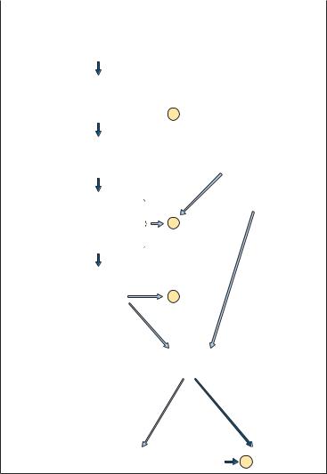
The diagnosis of enlarged lymph nodes receives special attention here because lymph nodes are as important as bone marrow for hematopoiesis. While in most instances abnormalities in the bone marrow cell series can be detected from the peripheral blood, this is very rarely the case for lymphomas. For this reason, lymph node cytology, a relatively simple and well-tolerated technique (p. 24), is critically important for the guidance it can give about the cause of enlarged lymph nodes. Figure 62 offers a diagnostic flow chart.
|
Lymph Node Cytology |
175 |
|
|
|
|
|
|
Anamnesis – sudden fast swelling |
slow onset, unclear |
|
– possible onset in youth |
symptoms: subfebrile, |
|
– possible contact |
night sweat, weight |
|
with animals |
loss |
|
– possible fever |
|
|
Findings |
– pressure pain |
|
– focal, or distri- |
|
buted over several |
|
locations |
Blood |
– relative lympho- |
analysis |
cytosis with |
|
stimulated forms |
Serology/ |
– mononucleosis test |
immunology – toxoplasmosis |
|
|
– rubella |
|
– syphilis |
|
– tuberculin skin test |
or Search |
– e.g. teeth |
for disease |
– sinuses |
focus |
– infections of |
|
the genitalia |
|
– if findings are |
|
negative or un- |
|
successful therapy |
|
after 1 week |
considered reactive if the lymph node swelling does not recede after
2 weeks or the cause is found
indolent often in one location, possibly localization of
D  primary tumor
specific cell presentation (e.g. CLL, immunocytoma, ALL and others)
or unspecific signs
D
D
Lymph node biopsy
suspicion of tumor or malignant lymphoma
attempt to clarify |
diagnosis based |
the diagnosis after histology possibly |
D on histology |
Fig. 62 Diagnostic flow chart for cases of lymph node enlargement. (D) Diagnosis.

176 Cytology of Organ Biopsies and Exudates
Reactive Lymph Node Hyperplasia and
Lymphogranulomatosis (Hodgkin Disease)
Reactive lymph node hyperplasia of whatever etiology is characterized by a confused mixture of small, middle-sized, and large lymphocytes. When the latter have a nuclear diameter at least three times the size of the predominating small lymphocytes and have a fair width of basophilic cytoplasm, they are called immunoblasts (lymphoblasts). Cells with deeply basophilic, eccentric cytoplasm and dense nuclei are called plasmablasts, and cells with a narrow cytoplasmic seam are centroblasts. Lymphocytes can also to varying degrees show a tendency to appear as plasma cells, e.g., as plasmacytoid lymphocytes with a relatively wide seam of cytoplasm. Monocytes and phagocytic macrophages are also seen (Fig. 63).
Table 30 (p. 178) summarizes the different forms of reactive lymphadenitis. The basic cytological findings in all of them is always a complete mixture of small to very large lymphocytes. Occasionally more specific findings may indicate the possibility of mononucleosis (increased immature monocytes) or toxoplasmosis (plasmablasts, phagocytic macrophages, and possibly epithelioid cells).
Any enlargement of the lymph nodes that persists for more than two weeks should be subjected to histological analysis unless the history, clinical findings, serology, or CBC offer an explanation.
At first sight, the confusion visible in the cytological findings of lymphogranulomatosis (Hodgkin disease) is reminiscent of the picture in reactive hyperplasia (something which may be important for an understanding of the pathology of this disease compared with other malignant neoplasms). However, some cells elements show signs of a strong immunological “over-reaction” in which large, immunoblast-like cells form with welldeveloped nucleoli (Hodgkin cells). Sporadically, some of these cells are found to be multinucleated (Reed–Sternberg giant cells); infiltrations of eosinophils and plasma cells may also be found. Findings of this type always require histological analysis, which can distinguish between four prognostically relevant histological subtypes. In addition to this, the very lack of a clear demarcation between Hodgkin disease and reactive conditions is reason enough to conduct a histological study of every lymph node that appears reactive if does not regress completely within two weeks.
In cases of histologically verified Hodgkin disease, cytological analysis is especially useful in the assessment of new lymphomas after therapy.

Reactive lymph node hyperplasia and lymphogranulomatosis (Hodgkin disease): a polymorphous mixture of cells
a 
b 
 c
c
Fig. 63 Reactive lymph node hyperplasia and lymphogranulomatosis. a Lymph node cytology in severe reactive hyperplasia. Large blastic cells alongside small lymphocytes (if it fails to regress, histological analysis is required). b Hodgkin disease: a giant mononuclear cell with a large nucleolus (arrow) and wide cytoplasmic layer (Hodgkin cell), surrounded by small and medium-sized lymphocytes. c Hodgkin disease: giant binuclear cell (Reed–Sternberg giant cell).
177
Table 30 Sequence of steps in the diagnosis of reactive lymphoma |
|
|
|
|
|
|||
|
|
|
|
|
|
|
178 |
|
Anamnesis |
Symptoms |
Tentative diagnosis |
Diagnostic studies |
Cytology |
Histology |
|
||
|
|
|
|
|
|
|
|
|
Rapid progression |
Localized, painful |
Perifocal lym- |
|
Search for disease |
(!) Non-specific |
|
|
|
(fever) |
|
phadenitis |
|
focus, non-specific |
lymphadenitis |
|
|
Cytology |
|
|
|
|
changes in CBC and |
|
|
|
|
|
|
|
|
ESR |
|
|
|
|
|
|
|
|
|
|
|
|
|
Rapid prog- |
Diffuse, painfult; |
Mononucleosis |
|
Pfeiffer cells in the |
(!) Adenitis with |
|
|
of |
ression, fever, |
angina, possibly |
|
|
blood analysis, EBV |
histiocytosis |
|
|
|
|
|
|
|
Organ |
||||
sore throat |
spleen |
|
|
serology (1) |
|
|
|
|
|
|
|
|
|
|
|
|
|
Most children |
Nuchal lymph |
Rubella |
|
Plasma cells in the |
|
|
|
Biopsies |
|
nodes, later |
|
|
blood, |
|
|
|
|
|
exanthema |
|
|
rubella-AHT "(2) |
|
|
|
|
|
|
|
|
|
|
|
|
|
Ingestion of raw |
Diffuse |
Toxoplasmosis |
|
Serology (3) |
(!) With |
Epithelioid cell |
|
and |
meat |
|
|
|
|
epithelioid cells and |
lymphadenitis |
|
Exudates |
Contact with cats |
|
|
|
|
macrophages |
|
|
|
|
|
|
|
|
|
|
|
|
Pharyngitis |
Local |
Common viruses, |
|
Complement fixation |
|
|
|
|
(! conjunctivitis) |
Neck area |
specific adeno- |
|
reaction (adeno- |
|
|
|
|
|
|
viruses |
|
viruses, less com- |
|
|
|
|
|
|
|
|
monly coxsackie- |
|
|
|
|
|
|
|
|
viruses) (4) |
|
|
|
|
|
|
|
|
|
|
|
|
|
Slow growth, |
Inflamed lymph |
Tuberculosis |
|
Thorax, local primary |
(!) Epithelioid |
|
|
|
general malaise |
nodes, possibly |
|
|
infections, skin test, |
cells, Langhans |
|
|
|
|
fistulae |
|
|
pathogen in biopsy |
giant cells |
|
|
|
|
|
|
|
aspirate, possibly PCR |
|
|
|
|
|
|
|
|
|
|
|
|
|
Slow growth |
Hard; possibly skin |
Sarcoidosis (Boeck |
|
Thorax, tuberculin |
!Epithelioid cells |
Epithelioid cell |
|
|
|
infiltrations |
disease) |
|
test usually negative, |
|
fibrosis |
|
|
|
|
|
||||||
|
|
|
|
ACE |
|
|
|
|
|
|
|
|
|
|
|
|
|

Contact with |
Tonsillitis, neck |
Listeriosis |
Agglutination test, |
(!) It may be possible |
|
animals |
lymph nodes |
|
complement fixation |
to determine the |
|
|
|
|
reaction |
pathogen from the |
|
|
|
|
|
biopsy |
|
|
|
|
|
|
|
Contact with |
Fever, spleen |
Brucellosis |
In case of fever: |
|
|
animals |
|
|
pathogen in blood, |
|
|
Milk intake |
|
|
serology |
|
|
|
|
|
|
|
|
Open wound, |
Local primary lesion |
Cat-scratch dis- |
Leukocytosis, lym- |
!Epithelioid cells, |
Perforating |
possibly from a |
|
ease |
phocytosis, comple- |
giant cells |
lymphadenitis |
cat |
|
|
ment fixation reaction |
|
|
|
|
|
|
|
|
Contact with |
Local primary lesion |
Tularemia (rabbit |
Agglutination test |
|
|
wildlife |
|
fever) |
|
|
|
|
|
|
|
|
|
Little sense of |
Inflamed hard infil- |
Actinomycosis |
Leukocytosis, left shift |
!“Gland” tissue in |
Therapeutic |
illness |
trates, possibly |
|
|
the biopsy material |
excision |
|
fistulae |
|
|
|
|
|
|
|
|
|
|
Symptomatic |
Joints, spleen, |
Collagen disease |
Antinuclear factor |
|
|
joints |
possibly kidney |
(PCP, LE) Felty syn- |
|
|
|
|
|
drome, Still dis- |
|
|
|
|
|
ease |
|
|
|
|
|
|
|
|
|
No symptoms |
Submandibular |
Branchial cyst |
|
!Epithelial cells, |
Therapeutic |
|
swelling, no irrita- |
|
|
macrophages, and |
excision |
|
tion |
|
|
granulocytes |
|
|
|
|
|
|
|
(!) = Optional step; != usually diagnostic step, if there is no arrow, the diagnosis can be made on the basis of preceding steps. (1) = Positive from day 5;
(2) = 1–4 days after exanthema, may be as much as 1 month; (3) = from week 4; (4) = from day 10.
179 Cytology Node Lymph