

Predominance of Mononuclear Round to Oval Cells |
63 |
|
|
Predominance of Mononuclear Round
to Oval Cells (Table 5)
The cell counts (per microliter) show a wide range of values and depend on a variety of conditions even in normal blood. Any disease may therefore result in higher (leukocytosis) or lower cell counts (leukopenia). The assessment of cell counts requires the knowledge of normal values and their ranges (spreads) (Table 2, p. 12). The most important diagnostic indicator therefore is not the cell count but the cell type. In a first assessment, mononuclear (round to oval) and polynuclear (segmented) cells are compared. This is the first step in the differential diagnosis of the multifaceted spectrum of blood cells.
Absolute or relative predominance of mononuclear cells points to a defined set of diagnostic probabilities. The differential diagnostic notes opposite can help with a first orientation.
Consequently, the most important step is to distinguish between lymphatic cells and myeloid blasts. The nuclear morphology makes it possible to distinguish between the two cell types. In lymphatic cells the nucleus usually displays dense coarse chromatin with slate-like architecture and lighter zones between very dense ones. In contrast, the immature cells in myeloid leukemias contain nuclei with a more delicate reticular, sometimes sand-like chromatin structure with a finer, more irregular pattern.

64 Abnormalities of the White Cell Series
Table 5 Diagnostic work-up for abnormalities in the white cell series with mononuclear cells predominating
Clinical findings |
Hb |
MCH |
Leukocytes |
Segmented |
Lympho- |
Other cells |
|
|
|
|
cells |
cytes (%) |
|
Fever, lymph nodes, |
n |
n |
↓/n |
↓ |
↑ |
– |
possibly exanthema |
|
|
|
|
Stimulated |
|
|
|
|
|
|
forms |
|
Fever, severe lym- |
n |
n |
↑ |
↓ |
↑ |
Large blastic |
phoma, possibly |
|
|
|
|
|
stimulated cells |
spleen ↑ |
|
|
|
|
|
(DD: lympho- |
|
|
|
|
|
|
blasts) |
Slowly progressing |
↓/n |
↓/n |
↑ |
↓ |
↑↑ |
– |
lymph node enlarge- |
|
|
|
|
|
|
ment in several loca- |
|
|
|
|
|
|
tions |
|
|
|
|
|
|
|
|
|
|
|
|
|
Night sweat, slowly |
↓ |
↓ |
n/↑ |
↓ |
↑ |
Plasmacytoid |
progressing lym- |
|
|
|
|
|
cells, possibly |
phoma (!spleen), |
|
|
|
|
|
rouleaux forma- |
possibly fever |
|
|
|
|
|
tion of the |
|
|
|
|
|
|
erythrocytes |
|
|
|
|
|
|
|
Slowly progressing |
↓ |
n/↓ |
n/↑/↓ |
n/↓ |
Possibly ↑ |
Possibly cells |
lymph node enlarge- |
|
|
|
|
|
with grooves |
ment in one or a few |
|
|
|
|
|
|
locations |
|
|
|
|
|
|
|
|
|
|
|
|
|
Isolated severe |
↓ |
↓ |
↓/↑ |
↓ |
Hairy cells |
|
splenomegaly |
|
|
|
|
|
|
|
|
|
|
|
|
|
Fever, angina |
n |
n |
↓ |
↓ |
↑ |
Monocytes ↑ |
|
|
|
|
|
|
|
Diffuse general |
↓ |
↓ |
↑ |
↓ |
n |
Monocytes ↑ |
symptoms |
|
|
|
|
|
|
|
|
|
|
|
|
|
Pale skin, fever |
↓ |
↓ |
↑/n/↓ |
↓ |
↓ |
Atypical round |
(signs of hemor- |
|
|
|
|
|
cells predomi- |
rhage) |
|
|
|
|
|
nate |
|
|
|
|
|
|
|
Fatigue, night |
↓ |
↓ |
n/↓ |
n |
n |
Possibly rouleaux |
sweat, possibly |
|
|
|
|
|
formations of |
bone pain |
|
|
|
|
|
erythrocytes |
|
|
|
|
|
|
|
Diagnostic steps proceed from left to right. | The next step is usually unnecessary; !optional step, may not progress the diagnosis; → the next step is obligatory.

|
Predominance of Mononuclear Round to Oval Cells |
65 |
|||||||
|
|
|
|
|
|
|
|
|
|
|
|
|
|
|
|
|
|
|
|
Thrombo- |
Electro- |
|
Tentative |
|
Evidence/advanced |
Bone mar- |
Ref. page |
|
|
cytes |
phoresis |
|
diagnosis |
|
diagnostics |
row |
|
|
|
n |
n/Y↑ |
|
Virus infec- |
|
Clinical developments, |
|
p. 66 |
|
|
|
|
|
tion, |
|
possibly serological |
|
|
|
|
|
|
|
e.g., rubeola |
|
tests |
|
|
|
|
n |
Y↑ |
|
Mono- |
|
Mononucleosis quick |
|
p. 68 |
|
|
|
|
|
nucleosis |
|
test, EBV serology |
|
|
|
|
|
|
|
|
|
(cytomegaly titer?) |
|
|
|
|
|
|
|
|
|
|
|
|
|
|
n/↓ |
n/Y↓ |
|
Leukemic |
|
Marker analysis of |
Always |
p. 74 |
|
|
|
|
|
non-Hodgkin |
|
peripheral lymphocytes |
involved in |
|
|
|
|
|
|
lymphoma, |
|
is sufficient in typical |
CLL, not |
|
|
|
|
|
|
esp. CLL |
|
disease presentations; |
always in |
|
|
|
|
|
|
|
|
lymph node histology |
other lym- |
|
|
|
|
|
|
|
|
for treatment decisions, |
phomas |
|
|
|
|
|
|
|
|
if necessary |
. |
|
|
|
|
|
|
|
|
|
|
|
|
|
n/↓ |
Possibly |
|
Immunocy- |
|
Immunoelectrophore- |
Usually |
p. 78 |
|
|
|
Y↑ |
|
toma (incl. |
|
sis; marker analysis for |
involved |
|
|
|
|
|
|
Walden- |
|
lymphocytes in blood |
|
|
|
|
|
|
|
ström dis- |
|
and bone marrow; |
|
|
|
|
|
|
|
ease) |
|
lymph node histology in |
|
|
|
|
|
|
|
|
|
case of doubt |
. |
|
|
|
|
|
|
|
|
|
|
|
|
|
n/↓ |
n |
|
Non-Hodgkin |
|
Lymph node histology |
For the pur- |
p. 78 |
|
|
|
|
|
lymphoma, |
|
|
pose of stag- |
|
|
|
|
|
|
e.g. follicular |
|
|
ing, possibly |
|
|
|
|
|
|
lymphoma |
|
|
. positive |
|
|
|
↓ |
n |
|
Hairy cell |
|
Cell surface markers |
Always |
p. 80 |
|
|
|
|
|
leukemia |
|
|
involved, dis- |
|
|
|
|
|
|
|
|
|
crete in the |
|
|
|
|
|
|
|
|
|
. beginning |
|
|
|
n |
n |
|
Agranulo- |
|
→ |
Maturation |
p. 86 |
|
|
|
|
|
cytosis |
|
|
arrest |
|
|
|
n |
Possibly |
|
Reactive |
|
Determine disease |
|
p. 88 |
|
|
|
|
|
|
|
|||||
|
a2↑ |
|
monocytosis |
|
origin |
|
|
|
|
|
|
|
in chronic |
|
|
|
|
|
|
|
|
|
infection or |
|
|
|
|
|
|
|
|
|
tumor |
|
|
|
|
|
|
↓ |
n |
|
Acute |
|
Cytochemistry, marker |
Marker for |
p. 90ff |
|
|
|
|
|
leukemia |
|
analysis, possibly cyto- |
blasts |
|
|
|
|
|
|
|
|
genetics |
|
|
|
|
|
|
|
|
|
→ |
|
|
|
|
|
|
|
|
|
|
|
|
|
|
n/↓ |
Sharp |
|
Multiple |
|
Immunoelectrophore- |
Bone marrow |
p. 82 |
|
|
|
peaks ↑ |
|
myeloma |
|
sis, skeletal X-ray |
contains |
|
|
|
|
|
|
|
|
→ |
plasma cells |
|
|
|
|
|
|
|
|
|
|
|
|
|

66 Abnormalities of the White Cell Series
Reactive Lymphocytosis
Lymphatic cells show wide variability and transform easily. This is usually seen as enlarged nuclei, a moderately loose, coarse chromatin structure, and a marked widening of the basophilic cytoplasmic layer.
Clinical findings, which include acute fever symptoms, enlarged lymph nodes, and sometimes exanthema, help to identify a lymphatic reactive state. Unlike the case in acute leukemias, erythrocyte and thrombocyte counts are not significantly reduced. Although the granulocyte count is relatively reduced, its absolute value (per microliter) rarely falls below the lower limit of normal values.
Morphologically normal lymphocytes predominate in the blood analyses in the following diseases:
Whooping cough (pertussis) with clear leukocytosis and total lymphocyte counts up to 20 000 and even 50 000/µl; occasionally, slightly plasmacytoid differentiation.
Infectious lymphocytosis, a pediatric infectious disease with fever of short duration. Lymphocyte counts may increase to "50 000/µl.
Chickenpox, measles, and brucellosis, in which a less well-developed relative lymphocytosis is found, and the counts remain within the normal range.
Hyperthyroidism and Addison disease, which show relative lymphocytosis.
Constitutional relative lymphocytosis, which can reach up to 60% and occurs without apparent reason (mostly in asthenic teenagers).
Absolute granulocytopenias with relative lymphocytosis (p. 86).
Chronic lymphocytic leukemia (CLL), which is always accompanied by absolute and relative lymphocytosis, usually with high cell counts.
Transformed, “stimulated” lymphocytes (“virocytes”) predominate in the CBC in the following diseases with reactive symptoms:
Lymphomatous toxoplasmosis does not usually involve significant leukocytosis. Slightly plasmacytoid cell forms are found.
In rubella infections the total leukocyte count is normal or low, and the lymphocytosis is only relatively developed. The cell morphology ranges from basophilic plasmacytoid cells to typical plasma cells.
In hepatitis the total leukocyte and lymphocyte counts are normal. However, the lymphocytes often clearly show plasmacytoid transformation.
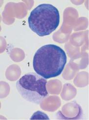
The most extreme lymphocyte transformation is observed in mononucleosis (Epstein–Barr virus [EBV] or cytomegalovirus [CMV] infection) (p. 68).
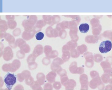
During lymphatic reactive states, variable cells with dense, round nuclei (e.g., virocytes) dominate the CBC
a
b 
 c
c
d 
 e
e
Fig. 21 Lymphatic reactive states. a–e Wide variability of the lymphatic cells in a lymphotropic infection (in this case cytomegalovirus infection). Some of the cells may resemble myelocytes, but their chromatin is always denser than myelocyte chromatin.
67

68 Abnormalities of the White Cell Series
Examples of Extreme Lymphocytic Stimulation:
Infectious Mononucleosis
Epstein–Barr virus infection should be considered when, after a prodromal fever of unknown origin, there are signs of enlarged lymph nodes and developing angina, and the blood analysis shows predominantly mononuclear cells and a slightly, or moderately, elevated leukocyte count. Varying proportions of the mononuclear cells (at least 20%) may be rather extensively transformed round cells (Pfeiffer cells, virocytes). Immunological markers are necessary to ascertain that these are stimulated lymphocytes (mostly T-lymphocytes) defending the B-lymphocyte stem population against the virus attack. The nuclei of these stimulated lymphocytes are twoto three-fold larger than those of normal lymphocytes and their chromatin has changed from a dense and coarse structure to a looser, more irregular organization. The cytoplasm is always relatively wide and more or less basophilic with vacuoles. Granules are absent. A small proportion of cells appear plasmacytoid. In the course of the disease, the degree of transformation and the proportions of the different cell morphologies change almost daily. A slight left shift and elevated monocyte count are often found in the granulocyte series.
Acute leukemia is often considered in the differential diagnosis in addition to other viral conditions, because the transformed lymphocytes can resemble the blasts found in leukemia. Absence of a quantitative reduction of hematopoiesis in all the blood cell series, however, makes leukemia unlikely, as do the variety and speed of change in the cell morphology. Finally, serological tests (EBV antigen test, test for antibodies, and, if indicated, quick tests) can add clarification.
Where serological tests are negative, the cause of the symptoms is usually cytomegalovirus rather than EBV.
Characteristics of Infectious Mononucleosis
Age of onset: School age
Clinical findings: Enlarged lymph nodes (rapid onset), inflammations of throat and possibly spleen !
CBC: Leukocytes !, stimulated lymph nodes, partially lymphoblasts (hematocrit and thrombocytes are normal)
Further diagnostics: EBV serological test (IgM +), transaminases usually !
Differential diagnosis: Lymphomas (usually without fever), leukemia (usually Hb ", thrombocytes ") Persistent disease (more than 3 weeks): possibly test for blood cell markers, lymph node cytology (#histology)
Course, treatment: Spontaneous recovery within 2–4 weeks; general palliation

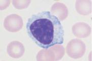
Extreme transformation of lymphocytes leads to blast-like cells: mononucleosis
a 
 b
b
c 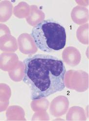
 d
d
Fig. 22 Lymphocytes during viral infection. a “Blastic,” lymphatic reactive form (Pfeiffer cell), in addition to less reactive virocytes in Epstein–Barr virus (EBV) infection. This phase with blastic cells lasts only a few days. b Virocyte (1) with homogeneous deep blue stained cytoplasm in EBV infection, in addition to normal lymphocyte (2) and monocyte (3). c Virus infection can also lead to elevated counts of large granulated lymphocytes (LGL) (1). Monocyte (2). d Severe lymphatic stress reaction with granulated lymphocytes. A lymphoma must be considered if this finding persists.
69

70 Abnormalities of the White Cell Series
Diseases of the Lymphatic System
(Non-Hodgkin Lymphomas)
Malignant diseases of the lymphatic system are further classified as Hodgkin and non-Hodgkin lymphomas (NHL). NHL will be discussed here because most NHLs can be diagnosed on the blood analysis.
Non-Hodgkin lymphomas arise mostly from small or blastic B-cells.
Small-cell NHL cells are usually leukemic and relatively indolent, of the type of chronic lymphocytic leukemia and its variants.
Blastic NHL (precursor lymphoma) is not usually leukemic. An exception is the lymphoblastic lymphoma, which takes its course as an acute lymphocytic leukemia.
Plasmacytoma is an osteotropic B-cell lymphoma that releases not its cells but their products (immunoglobulins) into the bloodstream.
Malignant lymphogranulomatosis (Hodgkin disease) cannot be diagnosed on the basis of blood analysis. It is therefore discussed under lymph node cytology (p. 176).
The modern pathological classification of lymphomas is based on morphology and cell immunology. The updated classification system according to Lennert (Kiel) has been adopted in Germany and was recently modified to reflect the WHO classification. The definitions of stages I–IV in NHL agree with the Ann Arbor classification for Hodgkin’s disease.
Table 6a Classification of non-Hodgkin: comparison of the relevant classes in the Kiel and WHO classifications
WHO |
Kiel |
Clinical |
Marker* |
|
|
|
|
Characteristics |
|
Precursor lymphomas/leukemias |
|
|
|
|
Precursor-B-lymphoblastic |
B-lymphoblastic |
Aggressive |
Tdt, |
|
|
lymphoma |
lymphoma |
|
CD19, 22, |
Precursor B-cell active lympho- |
B-ALL |
|
79a |
|
|
blastic leukemia |
|
|
|
Mature B-cell lymphoma |
|
|
|
|
|
Chronic lymphocytic leukemia |
B-CLL |
Usually indolent |
CD5, 19, |
|
– contains the lymphoplasma- |
Lymphoplasmacytoid |
Usually indolent |
20, 23 |
|
cytoid immunocytoma |
immunocytoma |
|
tris 12 or |
|
|
|
|
13q-, |
|
|
|
|
11q-, 6q-, |
|
B-cell prolymphocytic leukemia |
|
Aggressive |
p53 |
|
|
|
|
|
|
|
Predominance of Mononuclear Round to Oval Cells |
71 |
||||
|
|
|
|
|
|
|
|
Table 6a |
Continued |
|
|
|
|
|
|
|
|
|
|
|
|
|
|
WHO |
|
Kiel |
Clinical |
Marker* |
|
|
|
|
|
|
|
Characteristics |
|
|
|
|
Lymphoplasmacytic leukemia |
Lymphoplasmacytic |
Sometimes IgM |
–/+ CD5 |
|
|
|
|
|
|
immunocytoma |
paraprotein |
|
|
|
|
|
|
|
(Waldenström |
|
|
|
|
|
|
|
disease) |
|
|
|
|
Mantle cell lymphoma |
Centrocytic lymphoma |
Usually aggres- |
– CD23, |
|
|
|
|
|
|
|
sive |
CD5 |
|
|
|
|
|
|
|
t(11;14) |
|
|
|
|
|
|
|
bcl-1 |
|
|
|
|
|
|
|
rearr. |
|
|
|
Marginal zone lymphoma |
|
Usually aggres- |
–CD5, |
|
|
|
|
– |
Nodal |
Monocytoid lymphoma |
sive |
CD23 |
|
|
|
– Extranodal in mucosa (MALT) |
MALT (mucosa-assoc. |
Often indolent |
|
|
|
|
|
|
|
lymphoid tissue) lym- |
|
|
|
|
|
|
|
phoma |
|
|
|
|
|
– |
Lienal (splenic) |
Lymphoma with |
|
|
|
|
|
|
|
splenomegaly |
|
|
|
|
|
Follicular lymphoma |
Centroblastic/centro- |
Usually indolent |
–CD5, |
|
|
|
|
– |
Grade 1, 2 |
cytic lymphoma; |
|
! CD10 |
|
|
|
– Grade 3 (a, b) |
centroblastic lymphoma |
Highly malignant |
t(14;18) |
|
|
|
|
|
|
(a), secondary (b), follic- |
|
bcl2 |
|
|
|
|
|
ular |
|
|
|
|
|
Hairy cell leukemia |
Hairy cell leukemia |
Usually indolent |
–CD103, |
|
|
|
|
|
|
|
|
CD11c, |
|
|
|
|
|
|
|
CD25 |
|
|
|
|
|
|
|
t(2;8) |
|
|
|
|
|
|
|
t(8;14) |
|
|
Plasma cell myeloma (plasma- |
[Plasmacytoma is not |
|
CD 138 |
|
|
||
|
cytoma) |
included in the Kiel |
|
|
|
|
|
|
– |
Monoclonal hypergamma- |
classification] |
|
|
|
|
|
|
globulinemia (GUS) |
|
|
|
|
|
–Solitary bone plasmacytoma
–Extraosseous bone plasmacytoma
–Primary amyloidosis
–Heavy-chain disease
Primary large-cell lymphoma
|
Diffuse large-cell B-cell |
Centroblastic |
Extremely malig- |
CD20, |
|
|
lymphoma |
Immunoblastic |
nant |
79a, 19, |
|
|
– |
Centroblastic |
Large-cell anaplastic |
Extremely malig- |
22 |
|
– |
Immunoblastic |
|
nant |
|
|
– |
Large-cell anaplastic |
|
Extremely malig- |
|
|
|
|
|
nant |
|
|
Burkitt lymphoma |
Burkitt lymphoma |
Extremely malig- |
t(2;8) |
|
|
|
|
|
nant |
t(8;14) |
|
|
|
|
|
myc |
|
|
|
|
|
|
* Cited markers are positive, absent markers are indicated with a minus sign.

72 Abnormalities of the White Cell Series
Table 6b T-cell lymphomas (since T-cell lymphomas make up only 10% of all NHLs, this table gives just a brief characterization; for markers see Table 7)
WHO |
|
Clinical characteris- |
|
(! Kiel) |
|
tics |
|
Classification |
Manifestation |
|
|
|
T-prolymphocytic leukemia |
Leukemic |
Aggressive |
Large granular lymphocyte leukemia (LGL) |
Leukemic |
Sometimes indolent |
|
|
T-cell lymphoblastic leukemia |
Leukemic |
Aggressive |
Sézary syndrome, mycosis fungoides |
Cutaneous |
Chronic, progressive |
|
Angioimmunoblastic T-cell lymphoma (AILD) |
Nodal and ENT |
Usually aggressive |
|
Lymphoblastic T-cell lymphoma |
Nodal |
Aggressive |
|
T-cell zone lymphoma (nonspecific peripheral |
Nodal |
Sometimes slowly |
|
|
lymphoma) |
|
progressive |
Lennert lymphoma with multifocal epithelioid |
Nodal |
Sometimes slowly |
|
|
cells |
|
progressive |
Large-cell anaplastic lymphoma (ki1) |
Nodal |
Aggressive |
|
Differentiation of the Lymphatic Cells and Cell Surface Marker Expression in Non-Hodgkin Lymphoma Cells
Non-Hodgkin lymphoma cells derive monoclonally from specific stages in the B- or T-cell differentiation, and their surface markers reflect this. The surface markers are identified in immunocytological tests (Table 7) carried out on heparinized blood or bone marrow spicules.
The blastic lymphomas will not be discussed further in the context of diagnostics based on blood cell morphology. The findings in the primarily leukemic forms of the disease, such as lymphoblastic lymphoma, resemble those for ALL (p. 104). Other blastic lymphomas can usually only be diagnosed on the basis of lymph node tissue (Fig. 65). Of course, despite all the progress in the analysis of blood cell differentiation, often analysis of histological slides in conjunction with the blood analysis is required for a confident diagnosis.

Table 7 Cell surface markers of lymphatic cells in leukemic, low-grade malignant non-Hodgkin lymphoma
Marker |
B-CLL |
B-PLL |
HCL |
FL |
MCL |
SLVL |
T-CLL |
GL |
SS |
T-PLL |
ATLL |
|
|
|
|
|
|
|
|
|
|
|
|
S Ig |
(+) |
++ |
++ |
++ |
++ |
++ |
– |
– |
– |
– |
– |
CD 2 |
– |
– |
– |
– |
– |
– |
+ |
+ |
+ |
+ |
+ |
CD 3 |
– |
– |
– |
– |
– |
– |
+ |
– |
+ |
+ |
+ |
CD 4 |
– |
– |
– |
– |
– |
– |
– |
– |
+ |
+/– |
+ |
CD 5 |
++ |
– |
– |
– |
+ |
– |
– |
– |
+ |
+ |
+ |
CD 7 |
– |
– |
– |
– |
– |
– |
– |
– |
+/– |
+ |
–/+ |
CD 8 |
– |
– |
– |
– |
– |
– |
+ |
+/– |
+/– |
+/– |
+/– |
CD 19/20/24 |
++ |
++ |
++ |
+ |
+ |
++ |
– |
– |
– |
– |
– |
CD 22 |
+/– |
++ |
++ |
+ |
+ |
++ |
– |
– |
– |
– |
– |
CD 10 |
– |
– |
– |
+/– |
– |
– |
– |
– |
– |
– |
– |
CD 25 |
– |
– |
++ |
– |
– |
+/– |
– |
– |
– |
– |
+ |
CD 56 |
– |
– |
– |
– |
– |
– |
– |
+ |
– |
– |
– |
CD 103 |
– |
– |
++ |
– |
– |
– |
– |
– |
– |
– |
– |
|
|
|
|
|
|
|
|
|
|
|
|
CLL chronic lymphocytic leukemia; PLL prolymphocytic leukemia; HCL hairy cell leukemia; FL follicular lymphoma; MCL mantle cell lymphoma; SLVL splenic lymphoma with villous lymphocytes; LGL large granular lymphocyte leukemia; SS Sézary syndrome; ATLL adult T-cell lymphoma.
73 Cells Oval to Round Mononuclear of Predominance

74 Abnormalities of the White Cell Series
Chronic Lymphocytic Leukemia (CLL) and Related Diseases
A chronic lymphadenoma, or chronic lymphocytic leukemia, can sometimes be clinically diagnosed with some certainty. An example is the case of a patient (usually older) with clearly enlarged lymph nodes and significant lymphocytosis (in 60% of the cases this is greater than 20 000/µl and in 20% of the cases it is greater than 100 000/µl) in the absence of symptoms that point to a reactive disorder. The lymphoma cells are relatively small, and the nuclear chromatin is coarse and dense. The narrow layer of slightly basophilic cytoplasm does not contain granules. Shadows around the nucleus are an artifact produced by chromatin fragmentation during preparation (Gumprecht’s nuclear shadow). In order to confirm the diagnosis, the B-cell markers on circulating lymphocytes should be characterized to show that the cells are indeed monoclonal. The transformed lymphocytes are dispersed at varying cell densities throughout the bone marrow and the lymph nodes. A slowly progressing hypogammaglobulinemia is another important indicator of a B-cell maturation disorder.
Transition to a diffuse large-cell B-lymphoma (Richter syndrome) is rare: B-prolymphocytic leukemia (B-PLL) displays unique symptoms. At least 55% of the lymphocytes in circulating blood have large central vacuoles. When 15–55% of the cells are prolymphocytes, the diagnosis of atypical CLL, or transitional CLL/PLL is confirmed. In some CLL-like diseases, the layer of cytoplasm is slightly wider. B-CLL was defined as lymphoplasmacytoid immunocytoma in the Kiel classification. According to the WHO classification, it is a B-CLL variation (compare this with lymphoplasmacytic leukemia, p. 78). CLL of the T-lymphocytes is rare. The cells show nuclei with either invaginations or well-defined nucleoli (T-prolym- phocytic leukemia). The leukemic phase of cutaneous T-cell lymphoma (CTCL) is known as Sézary syndrome. The cell elements in this syndrome and T-PLL are similar.
Fig. 23 CLL. a Extensive proliferation of lymphocytes with densely structured ! nuclei and little variation in CLL. Nuclear shadows are frequently seen, a sign of the fragility of the cells (magnification #400). b Lymphocytes in CLL with typical coarse chromatin structure and small cytoplasmic layer (enlargement of a section from 23 a, magnification #1000); only discreet nucleoli may occur. c Slightly eccentric enlargement of the cytoplasm in the lymphoplasmacytoid variant of CLL.
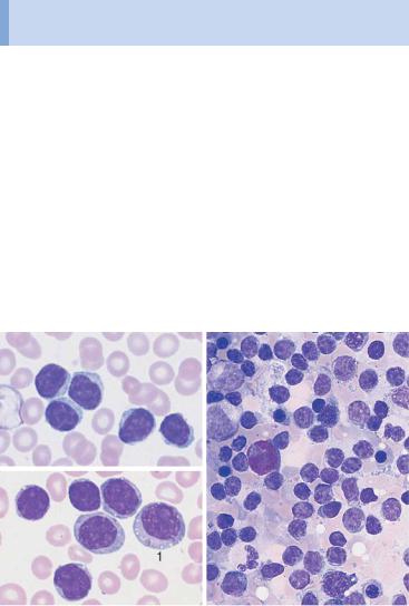
Monotonous proliferation of small lymphocytes suggests chronic lymphocytic leukemia (CLL)
a 
 b
b
c |
|
d |
e |
Fig. 23 d Proliferation of atypical large lymphocytes (1) with irregularly structured nucleus, well-defined nucleolus, and wide cytoplasm (atypical CLL or transitional form CLL/PLL). e Bone marrow cytology in CLL: There is always strong proliferation of the typical small lymphocytes, which are usually spread out diffusely.
75

76 |
Abnormalities of the White Cell Series |
||
|
|||
Table 8 Staging of CLL according to Rai (1975) |
|||
|
|
|
|
Stage |
|
|
Identifying criteria/definition |
(Low risk) |
0 |
Lymphocytosis " 15000/µl |
|
|
|
|
Bone marrow infiltration " 40% |
(Intermediate |
I |
Lymphocytosis and lymphadenopathy |
|
risk) |
|
II |
Lymphocytosis and hepatomegaly and/or spleno- |
|
|
|
megaly (with or without lymphadenopathy) |
(High risk) |
III |
Lymphocytosis and anemia (Hb $ 11.0 g/dl) (with or |
|
|
|
|
without lymphadenopathy and/or organomegaly) |
|
|
IV |
Lymphocytosis and thrombopenia ($ 100000/µl) |
|
|
|
(with or without anemia, lymphadenopathy, or |
|
|
|
organomegaly) |
|
|
|
|
Table 9 Staging of CLL according to Binet (1981)
Stage |
Identifying criteria/definition |
|
|
AHb " 10.0 g/dl, normal thrombocyte count $ 3 regions with enlarged lymph nodes
B |
Hb " |
10.0 g/dl, normal thrombocyte count |
|
" 3 regions with enlarged lymph nodes |
|
|
|
|
C |
Hb $ |
10.0 g/dl and/or thrombocyte count $ 100000/µl |
|
independent of the number of affected locations |
|
|
|
|
Characteristics of CLL
Age of onset: Mature adulthood
Clinical presentation: Gradual enlargement of all lymph nodes, usually moderately enlarged spleen, slow onset of anemia and increasing susceptibility to infections, later thrombocytopenia
CBC: In all cases absolute lymphocytosis; in the course of the disease Hb ", thrombocytes ", immunoglobulin "
Further diagnostics: Lymphocyte surface markers (see pp. 68ff.); bone marrow (always infiltrated); lymph node histology further clarifies the diagnosis
Differential diagnosis: (a) Related lymphomas: marker analysis, lymph node histology; (b) acute leukemia: cell surface marker analysis, cytochemistry, cytogenetics (pp. 88ff.)
Course, therapy: Individually varying, usually fairly indolent course; in advanced stages or fast progressing disease: moderate chemotherapy (cell surface marker, see Table 7)
|
|
Atypical lymphocytes are not part of B-CLL |
|
a |
|
|
b |
|
|
||
c |
|
|
d |
|
|
|
e |
Fig. 24 Lymphoma of the B-cell and T-cell lineages. a Prevalence of large lymphocyteswithclearlydefinednucleoliandwidecytoplasm:prolymphocyticleukemiaof theB-cellseries(B-PLL). b Thepresenceoflargeblasticcells(arrow)inCLLsuggesta rare transformation (Richter syndrome). c The rare Sézary syndrome (T-cell lymphoma of the skin) is characterized by irregular, indented lymphocytes. d Prolymphocytic leukemia of the T-cell series (T-PLL) with indented nuclei and nucleoli (rare). e Bone marrow in lymphoplasmacytic immunocytoma: focal or diffuse lymphocyte infiltration (e.g., 1), plasmacytoid lymphocytes (e.g., 2) and plasma cells (e.g., 3). Red cell precursors predominate (e.g., basophilic erythroblasts, arrow).
77

78 Abnormalities of the White Cell Series
The pathological staging for CLL is always Ann Arbor stage IV because the bone marrow is affected. In the classifications of disease activity by Rai and Binet (analogous to that for leukemic immunocytoma), the transition between stages is smooth (Tables 8 and 9).
Lymphoplasmacytic Lymphoma
The CBC shows lymphocytes with relatively wide layers of cytoplasm. The bone marrow contains a mixture of lymphocytes, plasmacytic lymphocytes, and plasma cells. In up to 30% of cases paraprotein is secreted, predominantly monoclonal IgM. This constitutes the classic Waldenström syndrome (Waldenström macroglobulinemia). The differential diagnosis may call for exclusion of the rare plasma cell leukemia (see p. 82) and of lymphoplasmacytoid immunocytoma, which is closely related to CLL (see p. 74).
Characteristics
Lymphoplasmacytoid immunocytoma: This is a special form of B- CLL in which usually only a few precursors migrate into the bloodstream (a lesser degree of malignancy). A diagnosis may only be possible on the basis of bone marrow or lymph node analysis.
Lymphoplasmacytic lymphoma: Few precursors migrate into the bloodstream (i.e., bone marrow or lymph node analysis is sometimes necessary). There is often secretion of IgM paraprotein, which can lead to hyperviscosity.
Further diagnostics: Marker analyses in circulating cells, lymph node cytology, bone marrow cytology and histology, and immunoelectrophoresis. Plasmacytoma cells migrate into the circulating blood in appreciable numbers in only 1–2% of all cases of plasma cell leukemia. Therefore, paraproteins must be analyzed in bone marrow aspirates (p. 82).
Facultative Leukemic Lymphomas
(e.g., Mantle Cell Lymphoma and Follicular Lymphoma)
In all cases of non-Hodgkin lymphoma, the transformed cells may migrate into the blood stream. This is usually observed in mantle cell lymphoma: The cells are typically of medium size. On close examination, their nuclei show loosely structured chromatin and they are lobed with small indentations (cleaved cells). Either initially, or, more commonly, during the course of the disease, a portion of cells becomes larger with relatively enlarged nuclei (diameter 8–12 µm). These larger cells are variably “blastoid.” Lymphoid cells also migrate into the blood in stage IV follicular lymphoma.

Deep nuclear indentation suggests follicular lymphoma or mantle cell lymphoma
a 
 b
b
 c
c
Fig. 25 Mantle cell lymphoma. a Fine, dense chromatin and small indentations of the nuclei suggest migration of leukemic mantle cell lymphoma cells into the blood stream. b Denser chromatin and sharp indentations suggest migration of follicular lymphoma cells into the blood stream. c Diffuse infiltration of the bone marrow with polygonal, in some cases indented lymphatic cells in mantle cell lymphoma. Bone marrow involvement in follicular lymphoma can often only be demonstrated by histological and cytogenetic studies.
79

80 Abnormalities of the White Cell Series
“Monocytoid” cells with a wide layer of only faintly staining cytoplasm occur in blood in marginal zone lymphadenoma (differential diagnosis: lymphoplasmacytic immunocytoma).
Lymphoma, Usually with Splenomegaly
(e.g., Hairy Cell Leukemia and Splenic Lymphoma with Villous Lymphocytes)
Hairy cell leukemia (HCL). In cases of slowly progressive general malaise with isolated splenomegaly and pancytopenia revealed by CBC (leukocytopenia, anemia, and thrombocytopenia), the predominating mononuclear cells deserve particular attention. The nucleus is oval, often kidney beanshaped, and shows a delicate, elaborate chromatin structure. The cytoplasm is basophilic and stains slightly gray. Long, very thin cytoplasmic processes give the cells the hairy appearance that gave rise to the term “hairy cell leukemia” used in the international literature. The disease affects the spleen, liver, and bone marrow. Severe lymphoma is usually absent. Aside from the typical hairy cells with their long, thin processes, there are also cells with a smooth plasma membrane, similar to cells in immunocytoma. A variant shows well-defined nucleoli (HCL-V, hairy cell leukemia variant). A bone marrow aspirate often does not yield material for an analysis (“punctio sicca” or “empty tap”) because the marrow is very fibrous. Apart from the bone marrow histology, advanced cell diagnostics are therefore very important, in particular in the determination of blood cell surface markers (immunophenotyping). This analysis reveals CD 103 and 11c as specific markers and has largely replaced the test for tartrateresistant acid phosphatase.
Splenic lymphoma with villous lymphocytes (SLVL). This lymphatic system disease mostly affects the spleen. There is little involvement of the bone marrow and no involvement of the lymph nodes. The blood contains lymphatic cells, which resemble hairy cells. However, the “hairs,” i.e., cytoplasmic processes, are thicker and mostly restricted to one area at the cell pole, and the CD 103 marker is absent.
Splenomegaly may develop in all non-Hodgkin lymphomas. In hairy cell leukemia, the rare splenic lymphadenoma with villous lymphocytes (SLVL) and marginal zone lymphadenoma may be seen. These mostly affect the spleen.
 Cytoplasmic processes the main feature of hairy cell leukemia
a 
 b
b
 d
d
c |
e |
Fig. 26 Hairy cell leukemia and splenic lymphoma. a and b Ovaloid nuclei and finely “fraying” cytoplasm are characteristics of cells in hairy cell leukemia (HCL). c Occasionally, the hairy cell processes appear merely fuzzy. d and e When the cytoplasmic processes look thicker and much less like hair, diagnosis of the rare splenic lymphoma with villous lymphocytes (SLVL) must be considered. Here, too, the next diagnostic step is analysis of cell surface markers.
81

82 Abnormalities of the White Cell Series
Monoclonal Gammopathy (Hypergammaglobulinemia),
Multiple Myeloma*, Plasma Cell Myeloma, Plasmacytoma
Plasmacytoma is the result of malignant transformation of the most mature B-lymphocytes (Fig. 1, p. 2). For this reason the diagnostics of this disease will be discussed here, even though migration of its specific cells into the blood stream (plasma cell leukemia) is extremely rare (1–2%).
Immunoelectrophoresis of serum and urine is performed when electrophoresis shows very discrete gammaglobulin, or globulin, fractions, or when hypogammaglobulinemia is found (in light-chain plasmacytoma). A wide range of possibilities arises for the differential diagnosis of monoclonal transformed cells (Table 10).
The presence of more than 10% of plasma cells, or atypical plasma cells in the bone marrow, is an important diagnostic factor in the diagnosis of plasmacytoma. For more criteria, see p. 84.
Table 10 Differential diagnosis of monoclonal hypergammaglobulinemia
Type |
|
Characteristics |
Benign disorders |
|
|
Essential hypergammaglobuline- |
Usually in advanced age |
|
mia = MGUS (monoclonal gam- |
$ 10%, plasma cells found in the |
|
mopathy of unknown significance) |
bone marrow, no progression, |
|
|
|
normal polyclonal Ig |
Symptomatic hypergammaglobu- |
All ages (otherwise as above) |
|
linemia, secondary to |
|
|
– |
Infections |
|
– |
Tumors |
|
– |
Autoimmune disease |
|
Malignant diseases
Plasmocytoma
(usually IgG, A or light-chain [Bence Jones protein], rarely IgM, D, E) 90% disseminated (multiple myeloma), 5% solitary, 5% extramedullary (like a lymphoma or ENT tumor)
–Osteolysis or X-ray with severe osteoporosis
–Plasmocytosis of the bone marrow " 10%
–Monoclonal gammaglobulin in serum/urine with progression
Lymphoma |
– Enlarged lymph nodes |
e. g., immunocytoma, CLL |
– Usually blood lymphocytosis |
(potentially all lymphomas of the |
– Monoclonal immunoglobulin, |
B-cell series) |
usually IgM |
|
|
*The current WHO classification suggests “multiple myeloma” (MM) for generalized plasmacytoma and “plasmacytoma” only for the rare solitary or nonosseous form of plasmacytoma.

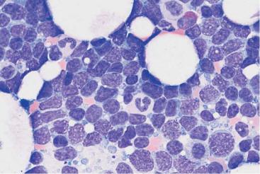
Plasmacytoma cannot be diagnosed without bone marrow analysis
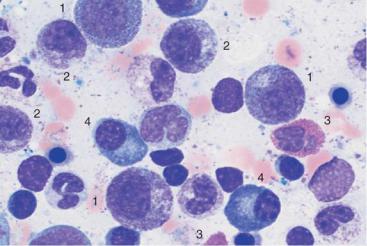 a
a
 b
b
Fig. 27 Reactive plasmacytosis and plasmacytoma. a Bone marrow cytology with clear reactive features in the granulocyte series: strong granulation of promyelocytes (1) and myelocytes (2), eosinophilia (3), and plasma cell proliferation (4): reactive plasmacytosis (magnification #630). b Extensive (about 50%) infiltration of the bone marrow of mostly well-differentiated plasma cells: multiple myeloma (magnification #400).
83

84 Abnormalities of the White Cell Series
Variability of Plasmacytoma Morphology
It is not easy to visually distinguish malignant cells from normal plasma cells. Like lymphocytes, normal plasma cells have a densely structured nucleus. Plasma cell nuclei with radial chromatin organization, known as “wheel-spoke nuclei,” are mostly seen during histological analysis.
The following attributes suggest a malignant character of plasma cells: the cells are unusually large (Fig. 28c), they contain crystalline inclusions or protein inclusions (“Russell bodies”) (Fig. 28b), or they have more than one nucleus (Fig. 28c).
In the differential diagnosis, they must be distinguished from hematopoietic precursor cells (Fig. 28d), osteoblasts (Fig. 20c), and blasts in acute leukemias (see Fig. 31, p. 97).
Bone marrow involvement may be focal or in rare cases even solitary. Aside from cytological tests, bone marrow histology assays are therefore indicated. Sometimes, the biopsy must be obtained from a clearly identified osteolytic region.
Although plasmacytomas progress slowly, staging criteria are available (staging according to Salmon and Durie) (Table 11).
Therapy may be put on hold in stage I. Smoldering indolent myeloma can be left for a considerable time without the introduction of therapy stress. However, chemotherapy is indicated once the myeloma has exceeded the stage 1 criteria.
Table 11 Staging of plasmacytomas according to Salmon and Durie
Stage I
All the following are present:
–Hb " 10 g/dl
–Serum calcium is normal
–X-ray shows normal bone structure or solitary skeletal plasmacytoma
–IgG $ 5 g/dl* IgA $ 3 g/dl*
Light chains in the urine* $ 4 g/24 h
Stage II
Findings fulfill neither stage I nor stage III criteria
Stage III
One or more of the following are present:
–Hb $ 8.5 g/dl
–Serum calcium is elevated
–X-ray shows advanced bone lesions
–IgG " 7 g/dl* IgA " 5 g/dl*
Light chains in the urine: " 12 g/24 h
* Monoclonal in each case.
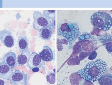
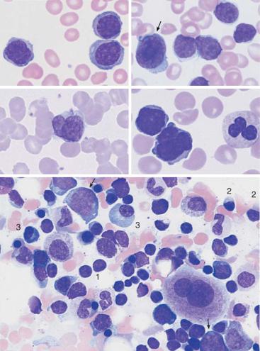
Atypias and differential diagnoses of multiple myeloma
a |
b |
c 
 d
d
Fig. 28 Atypical cells in multiple myeloma. a Extensive infiltration of the bone marrow by loosely structured, slightly dedifferentiated plasma cells with wide cytoplasm in multiple myeloma. b In multiple myeloma, vacuolated cytoplasmic protein precipitates (Russell bodies) may be seen in plasma cells but are without diagnostic significance. c Binuclear plasma cells are frequently observed in multiple myeloma (1). Mitotic red cell precursor (2). d Differential diagnosis: red cell precursor cells can sometimes look like plasma cells. Proerythroblast (1) and basophilic erythroblast (2).
85