

Practical Plastic Surgery
.pdfChapter 82
Replantation
Zol B. Kryger and John Y.S. Kim
Indications
General indications for replantation include amputation of multiple fingers, the thumb, the hand, or any part in a child. Loss of a single finger (excluding the thumb) is a relative indication for replantation; many surgeons will attempt replantation of distal digital injuries if at least 4 mm of intact skin proximal to the nailfold is present. Replantation of ring finger avulsions may be attempted, especially if one of the flexor tendons remains intact.
Contraindications for replantation:
•Significant systemic illness or comorbid conditions
•Concomitant life-threatening injuries
•Self-mutilation injuries
•Severe crush or avulsion injuries
•Extreme contamination
•Multiple level injuries
•Forearm or arm amputations with greater than 6 hours of warm ischemia time
Classification
Some authors classify the amputation by the flexor tendon zone (see Chapter 90). An alternative classification of amputation level has been introduced by Tamai:
Level I Amputation at the proximal nail fold Level II Amputation at the DIP joint
Level III Amputation at the middle phalanx Level IV Amputation at the proximal phalanx
Level V Amputation at the superficial palmar arch
Preoperative Considerations
Ischemia Time
Amputated parts should be cooled as soon as possible since they can tolerate significantly longer “cold ischemia” than “warm ischemia” time. Ideally, the part should be wrapped in saline-soaked, cold gauze sponge and placed in a bag on ice. Digits can survive for 24-36 hours cold, compared to 8 hours warm. A hand has been successfully replanted after 54 hours of cold ischemia time. The forearm can tolerate up to 10 hours of cold ischemia and 4-6 hours of warm ischemia.
Radiographs of the amputated part and the residual extremity should be obtained to determine if there are any missing segments of bone. The patient should be consented for possible tissue grafting or free flap coverage in addition to replantation. Prior to surgery, the patient should be hydrated and warmed.
Practical Plastic Surgery, edited by Zol B. Kryger and Mark Sisco. ©2007 Landes Bioscience.

Replantation |
493 |
Intraoperative Considerations |
|
Preparation of the Amputated Part |
|
Devitalized tissue is carefully debrided, and the vessels are dissected out under |
|
the microscope. Once a vessel is identified, it is marked with a tag. Arteries are |
|
closely inspected for signs of stretching or avulsion, which is suggested by a |
|
corkscrew-like appearance termed the “ribbon” sign. Bruising along the course of |
|
the digital vessel can also be a sign of avulsion injury. Nerves and tendons are iden- |
|
tified and tagged. The exposed bone is then minimally debrided. |
|
Preparation of the Stump |
|
After identification of the important vessels, nerves and tendons, the devitalized |
|
soft tissue of the stump is carefully debrided and irrigated with antibiotic solution. |
|
Large bone fragments are saved for possible grafting. The proximal bone stump is |
|
debrided. In the palm or finger, adequate exposure of the structures to be repaired |
|
may require Bruner zigzag incisions. |
|
The Order of Repair |
|
The order in which structures are repaired varies. The following outlines a com- |
82 |
monly used progression: |
|
1.Bone shortening followed by rigid fixation—if ischemia time is an issue, circulation should be restored prior to bony fixation
2.Flexor tendon repair—some authors will repair only the profundus tendon
3.Extensor tendon repair
4.Arterial anastomosis—at least one artery is usually required
5.Venous anastomosis—at least two veins for more proximal digital amputations. Level I amputations can be replanted without venous anastomosis
6.Nerve repair—this is optional, and may be delayed for a secondary procedure
7.Soft tissue coverage—skin grafts are sometimes required, especially in avulsion injuries. Vein grafts can be harvested with overlying skin as a composite graft
Bone Fixation
Fixation of an amputated digit can usually be achieved with crossed Kirschner wires passed retrograde through the amputated digit into the finger stump. Amputations through the proximal phalanx (Level IV) may require plate fixation for early postoperative mobilization. Transmetacarpal, transcarpal, and forearm amputations also require screw and plate fixation. This should be followed by periosteal repair, when indicated, to minimize tendon adhesion to the plates.
Vascular Repair
The arteries and veins must be trimmed back until normal intima is apparent. In order to minimize spasm, topical papavarine or concentrated lidocaine can be administered. Alternatively, a Fogarty catheter can be used to break a spasm. In crush or avulsion injuries, a vein graft may be required. Vein grafts can be taken from the dorsum of the foot, from the volar wrist, or from the dorsum of the hand. Alternatively, the lesser or greater saphenous systems can be used. The length of donor veins should be measured in situ since they shrink after harvest. Vein grafts should not be excessively long, since this can lead to kinking.

494 |
Practical Plastic Surgery |
Arterial repair is usually done first. Prior to venous repair, the appearance of the replanted part should be assessed for several minutes after completion of the arterial anastomosis to confirm adequate perfusion. After the replant has warmed up, bleeding from the veins to be anastomosed should be brisk.
Level I digital amputations can be replanted without a venous anastomosis. At least two veins are required for proximal digital amputations. If necessary, digital veins can be transferred from an adjacent digit. Venous drainage can also be achieved with a proximally based cross-finger flap. If the contralateral digital artery has retrograde blood flow, it can be anastomosed to a vein to provide outflow. Finally, if no vein can be identified, the nail is removed and a heparin-soaked sponge is placed on the nail bed, or leeches are applied to the tip of the digit.
Tendon Repair
Debridement of the tendon ends should be minimized to avoid the need for tendon grafting. Most surgeons repair the extensor tendons first, and many postpone repair of the flexor tendons. In Zone II flexor tendon amputations, some surgeons will elect to repair only the FDP tendon. A core suture technique is used; this is described in detail in Chapter 90.
82 Nerve Repair
Nerves should be repaired primarily whenever possible with 8-0 or 9-0 suture. A description of primary nerve repair is discussed in the nerve repair chapter. When necessary, nerve grafts can be taken from a number of sites: a nonsalvageable digit, the posterior interosseus nerve, the lateral femoral cutaneous nerve, the superficial peroneal nerve, and the sural nerve. Vascularized nerve grafts have also been described. Difficult nerve repairs may be delayed after the nerve ends are identified and tagged. Sensory outcomes of digital replantation are improving. Two-point discrimination of 10 mm or less has been reported.
Soft Tissue Coverage
In order to avoid compression of the vascular anastomoses, the skin must be closed in a tension-free fashion. If this cannot be accomplished, a split-thickness skin graft should be used. In forearm or more proximal amputations, soft tissue loss may be extensive, requiring local or free flap coverage.
Special Considerations
Ring Avulsions
Outcomes for ring-avulsion replants are not as good as for amputations. Success rates range from 30-70%, depending on the extent of injury. Ring avulsions range in severity, from tearing of the soft tissue with intact circulation to complete degloving of the digit. In degloving injuries, vein and nerve grafts are required. Repair of thumb avulsions should always be attempted, while fingers avulsed through the PIP joint should be amputated.
Hand and Forearm Amputations
Amputations through the carpal region have generally good functional outcomes. Transmetacarpal amputations, however, have a much poorer long-term prognosis. This is in part due to the many small vessels that are severed in these injuries. In any

Replantation |
495 |
case, chronic swelling of the hand is common so many surgeons will release the carpal tunnel and dorsal interosseous compartments at the time of surgery.
Of all the tissue in the upper extremity, muscle is least tolerant of ischemia. In proximal amputations, restoration of blood flow is the first priority since more muscle is involved. Prophylactic forearm fasciotomies are often performed if warm ischemia time is prolonged. Proximal amputations will usually require secondary procedures such as nerve grafts and tendon transfers.
Postoperative Care and Monitoring
At the completion of the procedure, the extremity should be placed in a well-padded splint without any circumferential dressings since swelling is inevitable. The fingertips should be exposed so that they can be monitored postoperatively.
The patient must be kept in a warm room with the extremity elevated above the heart. A continuous axillary anesthetic block can help with pain control and act as a chemical sympathectomy. Aspirin should be given since it has a potent anti-platelet effect. Although many centers use some form of anticoagulation, no randomized control trials have demonstrated a benefit to any regimen. The patient should be hydrated with intravenous fluids to avoid hypotension.
Monitoring the circulation after replantation is similar to a free-flap as discussed 82 in the chapter on free-flap monitoring. Early detection of microvascular failure is critical for salvage of the replant. The most reliable means of monitoring a replanted
digit is a clinical exam by an experienced individual combined with pulse oximetry or Doppler monitoring. SpO2 values above 95% are normal; a saturation below 85% indicates a venous problem, and a complete lack of signal indicates and arterial problem. Arterial and venous Doppler monitoring is used in many centers; both pencil and laser Doppler can be used. Finally, removal of the nail with monitoring of nail bed bleeding provides a rudimentary means of monitoring arterial circulation.
Outcomes
The most common early complications after replantation are bleeding, infection and loss of the replant. Late complications include cold intolerance, bony nonunion, nerve or tendon adhesions requiring neurolysis or tenolysis, and late necrosis of the replanted part.
Centers with a highly experienced replantation team report survival rates of 90-100% for digital replants. Level II amputations generally do better than Level I amputations. In terms of sensory recovery, two-point discrimination of 10 mm or less has been reported in a number of series. In both adults and children, persistent cold intolerance is common.
Pediatric Replantation
Many surgeons agree that unless a contraindication exists, almost all amputations in children should undergo replantation. Long-term follow-up at 15 years has demonstrated return of normal sensation, strength, and bone growth in up to 90% of digital replants in children. Distal finger tip amputations have been shown to survive with composite grafting when microvascular replantation is not possible. Even in these cases, sensory recovery can be excellent, presumably due to spontaneous neurotization. In children, bone shortening should be undertaken with caution to protect the epiphyseal growth plate so that the replanted bone will continue to grow normally.

496 |
Practical Plastic Surgery |
Pearls and Pitfalls
Three of the leading replant teams, the Kleinert-Kutz group, the Buncke group, and the Tamai group, have emphasized several important points in replantation technique:
•Bone shortening to reduce tension on the vessels, tendons, and nerves
•Repair of both the artery and vein before tourniquet release
•Heparin bolus after completion of the anastomosis
•Washing out the intravascular clots from crushed digits or hands
•The use of “spare parts” from unreplantable digits
•Transposing an amputated digit onto a different stump in cases of multiple digit amputations
Suggested Reading
1.Buncke Jr HJ. Microvascular hand surgery—Transplants and replants—Over the past 25 years. J Hand Surg 2000; 25A:415.
2.Kim JYS, Brown RJ, Jones NF. Pediatric upper extremity replantation. Clin Plast Surg 2005; 32:1.
3.Kleinert HE, Jablon M, Tsai TM. An overview of replantation and results of 347 replants in 245 patients. J Trauma 1980; 29:390.
824. Lee BI, Chung HY, Kim WK et al. The effects of the number and ratio of repaired arteries and veins on the survival in digital replantation. Ann Plast Surg 2000; 44:288.
5.Pederson WC. Replantation. Plast Reconstr Surg 2001; 107:823.
6.Soucacos PN. Indications and selection for digital amputation and replantation. J Hand Surg (Br) 2001; 26B:572.
7.Tamai S, Michon J, Tupper J et al. Report of the subcommittee on replantation. J Hand Surg 1983; 8:730.
8.Weinzweig N, Sharzer LA, Startker I. Replantation and revascularization at the transmetacarpal level: Long-term functional results. J Hand Surg 1996; 21:877.

Chapter 83
Fractures of the Distal Radius and Ulna
Craig Birgfeld and Benjamin Chang
Introduction
The radius and ulna form the bony structure of the forearm. They articulate with the humerus at the elbow and the proximal carpal row at the wrist. These bones form the framework upon which the long flexor and extensor muscles of the forearm take origin. The radius lies on the thumb side of the forearm (the “radial” side) and the ulna lies on the little finger side (the “ulnar” side) of the forearm. The anatomic shape of these bones and their relationship at the joints provides for a high degree of mobility. However, the price of this mobility is propensity for injury and degenerative disorders.
Anatomy
The elbow is formed by articulations between the radius and humerus and the ulna and humerus. The ulnar-humeral joint is a stable hinge joint, allowing only flexion and extension from 0-120 degrees. The radial-humeral joint is a pivot joint, which allows the radius to rotate on the humerus. This joint provides the mobility which allows the pronation and supination of the forearm through 180 degrees.
The radius and ulna are joined by an interosseous membrane, which is flexible enough to allow rotation of the radius around the ulna in pronation and supination, but strong enough to tether the two bones in a stable relationship. Pronator and supinator muscles course between the two bones and the long flexor and extensor muscles of the wrist and fingers originate from these bones in the forearm. At the wrist, the radius and ulna support the proximal row of carpal bones. The radius articulates with the lunate and scaphoid at the lunate fossa and the scaphoid fossa. Ligaments join the scaphoid and lunate to the radius and these articulations can also be the source of arthritic pain. The distal ulna articulates with the triquetrum through the triangular fibrocartilage complex (TFCC). The radius and ulna meet at the sigmoid notch and are joined by the TFCC forming the distal-radial-ulnar-joint (DRUJ), a frequent site of injury and arthritis. The ulna styloid lies most distally and laterally and is frequently involved in fractures of the distal radius.
Radiographs
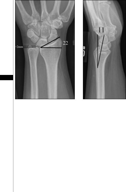
Relationships seen on normal radiographs are important to remember as these will be disrupted in fractures to the distal radius and ulna. All radiographs of the wrist should be evaluated for ulnar variance, inclination and volar tilt.
•Ulnar variance: on the normal AP view, the distal ulna is within ± 1-2 mm of the distal radius (Fig. 83.1).
•Inclination: the radius tilts toward the ulna at an angle of 22˚ when measured from a line perpendicular to its long axis on AP view (Fig. 83.1).
•Volar tilt: on lateral view, the radius tilts in a volar direction 11˚ when measured from a line perpendicular to its long axis (Fig. 83.2).
Practical Plastic Surgery, edited by Zol B. Kryger and Mark Sisco. ©2007 Landes Bioscience.
498 |
Practical Plastic Surgery |
83
Figure 83.1. An AP radiograph showing |
Figure 83.2. A lateral radio- |
normal ulnar variance of +2 mm and |
graph demonstrating the normal |
normal radius inclination of 22˚. These |
11˚ volar tilt of the radius |
two terms are defined in the text. |
when measured from a line per- |
|
pendicular to its long axis. |
Traumatic disruptions of these relationships can destroy the normal kinematics of the wrist and lead to reduced range of motion and degenerative arthritis.
Fractures
A variety of fractures can occur in the radius and ulna after the common traumatic history of, “fall on an outstretched hand”. These are amongst the most common skeletal injuries and occur more frequently in children and the elderly. Fractures of the distal radius are best described in terms of their comminution, articular involvement, displacement and angulation with particular attention paid to the measurements of ulnar variance, inclination, and volar tilt. Eponyms are frequently used for expediency, though these can lead to confusion for inexperienced evaluators. Fractures of the ulna are often multiple and can have varying degrees of clinical significance. Recall that the radius and ulna essentially form a ring between the elbow and wrist and, as is seen with the mandible, a fracture of one side of the ring will usually result in a fracture at the other.
The most common fracture, is a dorsally angulated, extra-articular fracture of the distal radius, otherwise known as a Colles’ fracture. A Smith’s fracture is a volarly

Fractures of the Distal Radius and Ulna |
499 |
angulated, extra-articular fracture and generally occurs from a fall onto the dorsum of the hand. A Barton’s fracture is an intra-articular fracture and can be either volarly or dorsally angulated. A Chauffeur’s fracture is an intra-articular fracture of the radial styloid, usually caused by forced extension and radial deviation of the wrist. The name originates from the chauffeurs who were sent to start the old Model-T Fords with the hand cranks, which would occasionally backlash onto the wrist, fracturing the radial styloid. Occasionally, a fall on an outstretched hand can occur with such force as to cause a Monteggia fracture, which is an unstable fracture of the proximal ulna with dislocation of the radial head. These fractures must be repaired operatively with reduction of the radial head and plate fixation of the ulnar fracture.
Evaluation
The evaluation of injuries to the radius and ulna always begins with a thorough history and physical exam of the hand. Though the injury may be immediately obvious, it is imperative that the entire hand and arm be evaluated in a careful, systematic manner so that all associated injuries are identified and treated. A thorough history of the injury is taken, including hand dominance and occupation. Ask about previous injuries to the extremity as well as relevant medical problems such as arthritis or carpal tunnel syndrome. A detailed description of the mechanism of injury can be helpful as well as associated symptoms such as numbness and tingling.
Next, inspect the arm from shoulder to fingertip and note any bruising, lacera- 83 tions, and deformities. Take note of the manner in which the patient holds the
arm at rest and note any pallor, cool skin or lack of sweating. Then, gently palpate the arm along its entire length to determine areas of tenderness and deformity. Evaluate the range of motion of all joints, taking into account local edema or arthritis which could limit mobility. Remember to evaluate pronation and supination of the forearm. Next, systematically check each long flexor and extensor of the wrist and fingers, as well as the intrinsic muscles of the hand. Finally, confirm that the radial, ulnar, and median nerves are intact and functioning. Entrapment or compression of these nerves can change a simple fracture of the wrist into a medical emergency requiring prompt reduction and decompression before permanent nerve damage occurs.
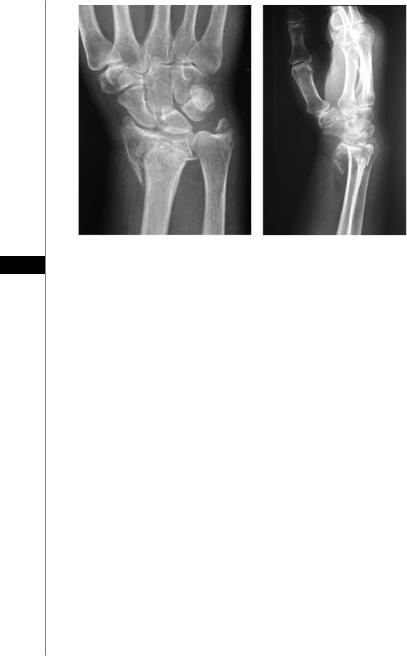
Example of a Correct Fracture Description
Figure 83.3 illustrates a common fracture and the correct means of describing it. An unacceptable call to one’s chief resident to describe the above fracture would go something like this: “The patient has broken her right wrist”. A professional call to the chief resident would be more along the lines of: “The patient is an 86 year old, right hand dominant female with a history of osteoporosis who fell on an outstretched right hand today while walking her dog. She has an obvious, painful deformity of her right wrist with no open wounds and no vascular compromise or loss of sensation. Her radiographs display an unstable, severely comminuted, intra-articular fracture of her distal radius and ulna styloid. The fracture is volarly displaced with an ulnar positive variance of 5 mm. She has an inclination of 14˚ and a volar tilt of -4˚. There is significant articular incongruity.
Treatment
The aim of treatment is to return the fracture as nearly as possible to a normal anatomic alignment with minimal morbidity to the patient restoring adequate length (ulnar variance) and volar tilt are key. The choice of treatment depends entirely on an
500 |
Practical Plastic Surgery |
|
|
|
|
|
|
|
83Figure 83.3. AP and lateral radiographs illustrating an example of a comminuted, intra-articular fracture of the distal radius and ulna styloid.
accurate assessment of the injury. A simple, minimally displaced fracture can be treated with cast immobilization for a period of 3-4 weeks. A displaced Colles’ fracture can be treated with closed reduction, followed by application of a sugar tong splint, then cast immobilization for 4 weeks. Unstable fractures, intra-articular fractures and comminuted fractures frequently require more rigid fixation. Closed reduction with K-wire fixation is minimally invasive and adequately treats most unstable fractures that are not comminuted. However, most hand surgeons have largely shifted towards the use of a volar plate for rigid fixation. Comminuted and intra-articular fractures often need to be reduced openly in the OR, then rigidly fixed with a volar plate to hold optimal alignment. The advantage of this is an early return to activity and avoidance of joint stiffness. The downside is a trip to the OR and its inherent risks. Sometimes, the fracture is so comminuted that it won’t hold a plate or the bone is too osteoporotic to hold screws and an external fixator is required. These are bulky and cumbersome but neutralize the compression force and help to maintain length.
Volar Plate Technique for Distal Radius Fractures
After exsanguination of the arm, a longitudinal incision is made over the distal portion of flexor carpi radialis (FCR). The incision is carried through the tendon sheath down to the tendon. Care must be taken to avoid injury to nearby structures such as the radial artery and palmar cutaneous branch of the median nerve. The FCR tendon is retracted radialy, and the pronator quadratus is visualized. The muscle is incised sharply, leaving a cuff of tissue on the radial side so that the muscle can be sewn back into place to cover the plate. It is then elevated in the subperiosteal plane off the distal radius with care to preserve its integrity. The periosteum of the distal radius including the fracture site is carefully elevated. The fracture is exposed, curetted, and reduced under fluoroscopy. Radial length and volar tilt must be restored. A multitude of companies produce volar plates. All are essentially locking plates with

Fractures of the Distal Radius and Ulna |
501 |
a prefabricated bend in the plate that matches the normal volar tilt. Once all the screws are placed, fluoroscopy is used to confirm the final result. The site is irrigated and the pronator reapproximated to cover as much of the plate as possible. The skin is closed with 4-0 nylon horizontal mattress sutures. A bulky dressing and volar splint are applied. The rigid fixation provided by the volar plate allows for early postoperative motion.
Pearls and Pitfalls
•X-ray all injured extremities including one joint proximal and distal to the level of injury.
•Reduction before fixation.
•When one forearm bone is fractured, look for another fracture in the other bone or a disruption of the joint at the wrist or elbow.
•Look for associated carpal injuries such as a scaphoid fracture or scpaholunate dissociation (tear of the scapholunate ligament).
Suggested Reading
1. Chang B. Principles of upper limb surgery. Chapter 65. Grabb and Smith’s Plastic Surgery. 5th ed. Philadelphia: Lippincott-Raven, 1997.
2. Fernandez DL, Palmer AK. Fractures of the distal radius. Chapter 29. Green’s Opera- 83 tive Hand Surgery. 4th ed. Philadelphia: Churchill Livingstone, 1999.
3.Leibovic SJ. Fixation for distal radius fractures. Hand Clin 1997; 13:665.
4.Trumble TE, Schmitt SR, Vedder NB. Factors affecting functional outcome of displaced intra-articular distal radius fractured. J Hand Surg 1994; 19:325.