
Подготовка у универсиаде 2012 / Lentiviral Vectors biosafety
.pdfLentiviral Vectors
Information and Biosafety Considerations
Introduction
Viral vectors are becoming increasingly popular in both clinical and non-clinical research. The evolution of new specialized molecular mechanisms and the ability of viruses to deliver recombinant DNA molecules (transgenes) into host cells has led them to be desirable for many applications. Common applications for viral vectors include restoration of functional genes in genetic therapy studies, standard cellular biology research involving a wide range of both in vivo and in vitro experimental designs, oncological research, and number of other studies. The viral vectors used in laboratory and clinical use are based on murine and human RNA and DNA viruses possessing very different genomic structures and host ranges. The great majority of current clinical gene therapy trials worldwide involve viral vectors derived from retroviruses (including lentiviruses), adenoviruses, adeno-associated viruses, herpes viruses and poxviruses (Walther and Stein 2000). While these viral vector constructs each feature differing mechanisms for targeting infection, all possess high rates of infectivity and the ability to infect both dividing and non-dividing differentiated cell populations. The aim of several ongoing studies involving these vectors is to refine their safety, improve targeting of gene transfer, and to provide efficient expression of desired transgenes.
Lentiviral vector constructs have proven to be very productive in terms of transduction due to their ability to infect both replicating and non-replicating cells, including stem cells. Lentiviral vectors are becoming the vectors of choice for short-interfering RNA (siRNA) delivery (Sachdeva et al., 2007). The increased use of lentiviral vector constructs in established and novel research applications makes it essential for laboratory workers to understand and protect themselves from related exposure hazards. The purpose of this document is to provide principal investigators (PIs) and laboratory technicians with information regarding risk assessment, proper work methods, containment levels, suitable engineering controls option, and personal protective equipment for development of research protocols that effectively reduce the risk of occupational exposure to lentivirus vectors as prescribed by National Institutes of Health (NIH) documents::
"Guidelines for Research Involving Recombinant DNA Molecules" and Recombinant DNA Advisory Committee: "Guidelines for Research Involving Lentiviral Vectors".
Background: Basic Biology of Lentiviruses
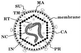
Lentiviruses are composed of 2 copies of RNA, a nuclear capsid (NC), a Capsid (CA) a membrane associated matrix (MA), envelope proteins such as surface glycoproteins (SU) and transmembrane proteins (TM) and enzymes such as integrase (IN), protease (PR), reverse transcriptase (RT) and accessory proteins (Figure 1). The role of these proteins is summarized in Table 1.
|
|
Figure 1. |
TABLE 1.: Roles and Functions of retroviral proteins |
||
|
|
|
Denotation |
Protein Name |
Function |
CA |
Capsid |
gag gene; protects the core |
IN |
Integrase |
pol gene; needed for integration of the provirus |
MA |
Matrix |
gag gene; lines envelope |
NC |
Nuclear Capsid |
gag gene; protects the genome and forms the core |
PR |
Protease |
Essential for gag protein cleavage during maturation |
RT |
Reverse |
Reverse transcribes the RNA genome |
|
Transcriptase |
|
SU |
Surface Glycoprotein |
The outer envelope glycoprotein; major virus |
|
|
antigen |
TM |
Trans Membrane |
The inner component of the mature envelope |
|
Protein |
glycoprotein |
Accessory |
Ex: Nef, Vif, Vpu, |
Play a role for infectivity and pathogenicity of HIV |
Proteins |
Vpr |
|
(HIV) |
|
|
The replication cycle of retroviruses commences by first binding to a receptor on the host cell via the surface glycoprotein ligand of the virus to a surface receptor of the host cell. This is followed by fusion of the membranes of the envelope of the virus and the membrane of the host cell which allows for entry. Immediately following entry, uncoating and reverse transcription takes place which leads to the formation of a pre-integration complex (PIC). The exact composition of PIC remains unknown, but it has been shown to contain double stranded DNA (a product of reverse transcription), RT, IN, Vpr (or Vpx in HIV-2) NC, and some copies of the MA (Suzuki and Craigie 2007, Depienne et al., 2000, Bukrinsky et al., 1993 and Miller et al., 1997). Lentiviruses are unique in the fact that they can infect undividing cells by actively entering the nucleus of a cell through the nuclear envelope via the PIC. Other retroviruses such as the Moloney virus require cell division for infection due to the fact that it cannot enter the nuclear envelope of a nondividing cell. This is one of the reasons why lentiviruses are becoming increasingly popular as vectors. Once the provirus enters the nuclear envelope, it integrates itself with the host genome. The normal cellular functions of transcription and translation are followed by assembly of structural viral proteins with viral RNA which precedes budding. The newly formed virus matures outside of the cell (Figure 2).
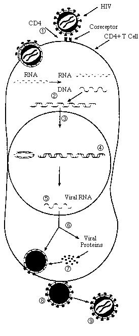
Steps in Viral Replication
1.Attachment/Entry
2.Reverse Transcription and DNA
Synthesis
3.Transport to Nucleus
4.Integration
5.Viral Transcription
6.Viral Protein Synthesis
7.Assembly of Virus
8.Release of Virus
9.Maturation
Figure 2.
Background: History of Retroviral Vectors
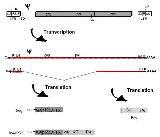
Retroviral vectors require a packaging cell line to incorporate the transgene of interest, and to delete any genes that would render the virus replicative competent. The Moloney Murine Leukemia Virus (MoMuLv) was the original retrovirus vector and the engineering involved in creating a packaging system was developed from MoMuLv. Figure 3 shows how genes of the MoMuLv are transcribed, translated and eventually packaged into a new virus.
Figure 3.
The Psi symbol represents the packaging sequence which allows the transcribed viral RNA to be incorporated into the assembly of the new virus. This is the RNA sequence which separates it from normal host RNA. The structural proteins, enzymes and envelope proteins are translated and along with the viral RNA are then assembled into a new virus.
Figure 4 outlines the 2 plasmid system of the original retroviral system; the helper plasmid and the engineered vector plasmid with the gene of interest.
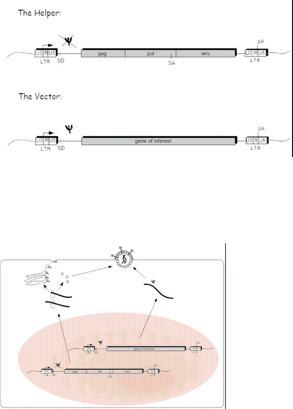
Figure 4.
The Helper plasmid includes all the structural proteins needed in order to package a new virus within the packaging cell. Due to the Psi deletion within the helper plasmid, this transcribed RNA does not get incorporated into the new recombinant virus, only the plasmid with the gene of interest gets incorporated because of the included packaging sequence (Psi). The LTR sequence contains the promoter and integration sequences to insert the new gene of interest in the host cell DNA. Figure 5 shows a schematic of the packaging cell line with this two plasmid system.
Figure 5
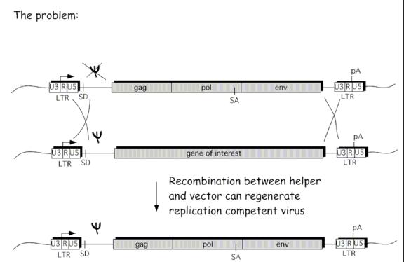
The helper plasmid transcribes and translates the structural genes required for the assembly of a new virus, where as the vector plasmid gets transcribed due to the packaging sequence, but since the vector plasmid lacks any of the other structural genes, it cannot form a replicative competent virus in the host cell. The end result is a replicative incompetent recombinant retrovirus that includes the transgene, but that lacks any of the structural genes to form a new virus in the host target cell.
Even though this construct was efficient at delivering transgenes, problems have occurred. The MoMuLv vector has been shown to become replicative competent via a recombination events in the packaging cell. A double crossover event between the vector plasmid and the helper plasmid had caused the packaging sequence (Psi) to be placed on the helper package, completing the sequence for a replicative competent virus. Instead of the transgene being packaged, a whole replicative competent virus was instead produced (see Figure 6).
Figure 6
A solution to this problem was developed by splitting the genomes of the helper plasmid into multiple plasmids so that multiple recombination events would be required in order to form a replicative competent virus. One method was to separate the gag and the pol genes from the env gene and create two separate plasmids for each, so that not only would there have to be recombination between the vector plasmid (which would give the packaging sequence), but also between the two split plasmids. Figure 7 outlines this solution.
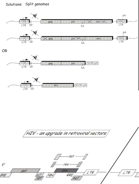
Figure 7.
The problems associated with the MoMuLv vector and the consequent engineering of a better vector have led to go a step further and to utilize the HIV lentivirus as a potential viral vector. HIV has many accessory proteins that are required to create a replicative competent lentivirus (RCL) (Figure 8). By deleting these accessory proteins and by splitting the genome into multiple plasmids, you can create a safer lentiviral vector that has extremely reduced capabilities of forming an RCL (Figure 9).
Figure 8
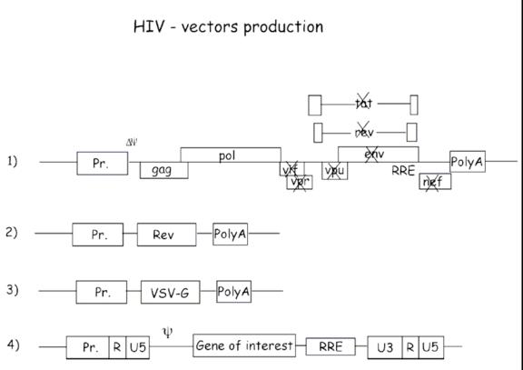
Figure 9
A 4 plasmid vector system is used in a third generation lentiviral vector. The accessory proteins such as tat, vrf, vpr and nef are deleted from the original plasmid. Also, the env gene is replaced with a VSV-G (Vesicular Stomatitis Virus Envelope Protein) and is placed on a third helper plasmid. Pseudotyping increases tropism which is beneficial but may also pose some challenges and safety concerns regarding the types of cells the virus can infect. By splitting the vector system into 4 plasmids (3 helper and 1 vector), the number of recombination events required to form a complete replicative competent virus increases. To date there are no known cases where this type of construct has produced RCLs.
The lentiviruses have an intrinsic problem that is exploited for viral vector production. When the viral DNA is transcribed to RNA the virus looses sequences at the 5’ end and 3’ end (the promoter sequence and the Poly A signal). During reverse transcription, the virus then copies sequences on the 3’ end and places them on the 5’end through “jumps”. The U3 segment (see figure 9 and 10) contains a primer and if that primer is deleted, that deletion will also get copied onto the 5’ end of the final RNA. Therefore, in order for an RCL event to occur, the virus would have to pick up a promoter as well. The only promoter that dictates transcription of cDNA is the internal promoter in the vector plasmid.
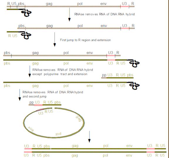
Figure 10
These safety features enable lentiviruses to be used safely while increasing tropism and transduction effectively. There have been no known recombination events associated with 3rd generation lentiviral vectors that have created RCLs. More work is being conducted to ensure safety and transduction efficiency with lentiviral vectors. For example placing the gag and pol genes on separate plasmids would increase the recombination events required to produce an RCL therefore increasing safety.
Risk Assessment:
A.Two major risks need to be considered when working with lentiviruses: 1. The potential for generation of RCL
2.The potential for oncogenesis.
3. The mitigation of these risks can be achieved through proper engineering of viral vectors (such as multiple plasmids), however; they may be exacerbated through the nature of the transgene (ex: oncogene).
B.When conducting a risk assessment the Recombinant DNA Advisory Committee (RAC) recommends the general criteria for risk assessment of lentivirus vectors:
1.The nature of the vector system and the potential for regeneration of replication competent virus from the vector components
2.The nature of the transgene insert (e.g., known oncogenes or genes with high oncogenic potential may merit special care)
3.The vector titer and the total amount of vector
4.The potential for HIV positive individuals who’s native virus may recombine with the vector (this is more of a problem with lower generation viruses)
5.The inherent biological containment of the animal host, if relevant, negative RCL testing.
C.The potential for the generation of RCL from HIV-1 based lentivirus vectors depends upon several parameters:
1.The number of recombination events necessary to reassemble a replication competent virus genome
2.The number of essential genes that have been deleted from the vector/packaging system.
C. It is always recommended to use later generation vector systems (i.e., 3rd generation) which are likely to provide a greater margin of safety than earlier vectors. This is due to the fact that they use a heterologus coat protein (VSV-G) in place of the native HIV-1 envelope protein (this however, broadens the host cell and tissue tropism which should be considered in the overall safety assessment). The use of 4 or more plasmids to separate vector and packaging functions also increases safety as well as the deletion of certain structural proteins (i.g., deletion of Tat gene which is essential for replication of wild-type HIV-1).
General Containment ConsiderationsBiosafety Levels: It is important to
conduct a proper risk assessment when working with lentiviruses. Once that is completed, proper containment levels can be decided. Generally, BSL-2 or BSL-2+ (enhanced BSL-2 containment including BSL-3 work practices and PPE) is often appropriate in the laboratory setting for higher generation lentivirus vectors with multiple safety features (four or more plasmids). Enhanced BSL-2 may include in addition to sharps safety, the use of PPE intended to reduce the potential for mucosal exposure to the vector (such as an N-95 respirator), especially if working with high volumes (>10L).
Animal Studies
A. Some animals cannot support replication of infectious HIV-1. As a result, the potential for shedding of RCL is very low. It may be prudent to consider the biosafety issues associated with
