

Подготовка у универсиаде 2012 / Lentiviral vectors basic to translational
.pdfwww.biochemj.org
Biochem. J. (2012) 443, 603–618 (Printed in Great Britain) doi:10.1042/BJ20120146 |
603 |
REVIEW ARTICLE
Lentiviral vectors: basic to translational
Toshie SAKUMA, Michael A. BARRY and Yasuhiro IKEDA1
Department of Molecular Medicine, Mayo Clinic College of Medicine, 200 First Street SW, Rochester, MN 55905, U.S.A.
More than two decades have passed since genetically modified HIV was used for gene delivery. Through continuous improvements these early marker gene-carrying HIVs have evolved into safer and more effective lentiviral vectors. Lentiviral vectors offer several attractive properties as gene-delivery vehicles, including: (i) sustained gene delivery through stable vector integration into host genome; (ii) the capability of infecting both dividing and non-dividing cells; (iii) broad tissue tropisms, including important geneand cell-therapy- target cell types; (iv) no expression of viral proteins after vector transduction; (v) the ability to deliver complex genetic elements, such as polycistronic or intron-containing sequences; (vi) potentially safer integration site profile; and (vii) a relatively easy system for vector manipulation and production. Accordingly, lentivector technologies now have widespread use in basic biology and translational studies for stable transgene overexpression,
persistent gene silencing, immunization, in vivo imaging, generating transgenic animals, induction of pluripotent cells, stem cell modification and lineage tracking, or site-directed gene editing. Moreover, in the present high-throughput ‘-omics’ era, the commercial availability of premade lentiviral vectors, which are engineered to express or silence genome-wide genes, accelerates the rapid expansion of this vector technology. In the present review, we assess the advances in lentiviral vector technology, including basic lentivirology, vector designs for improved efficiency and biosafety, protocols for vector production and infection, targeted gene delivery, advanced lentiviral applications and issues associated with the vector system.
Key words: cell targeting, central polypurine tract (cPPT), gene therapy, HIV-1, Lentivirus, self-inactivating (SIN).
BASIC LENTIVIROGY
Lentiviruses are members of the viral family Retroviridae (retroviruses) that are characterized by their use of viral RT (reverse transcriptase) and IN (integrase) for stable insertion of viral genomic information into the host genome. Unlike other retroviruses, lentiviruses can replicate in non-dividing cells [1,2] and cause slowly progressive diseases, including immunodeficiency, anaemia, pneumonitis and encephalitis, in their specific hosts (human, monkey, cat, horse, cow, goat and sheep) [3–9]. Lentiviruses from different species have distinct properties in their genome structure, receptor usage and pathogenicity. Since the majority of lentiviral vectors are based on HIV-1, we start by providing basic information on this virus, including its genomic structure, viral proteins and life cycle.
HIV-1 genome
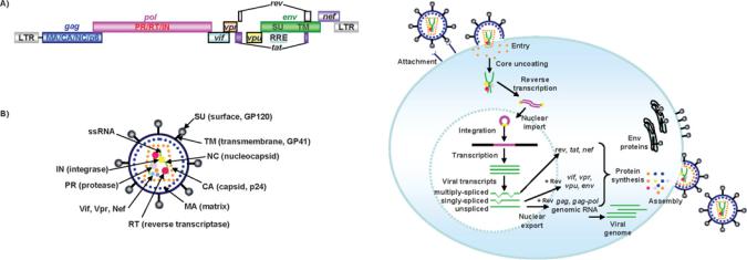
HIV has a single-stranded positive-sense RNA genome of approximately 9 kb in length that encodes nine viral proteins (Figure 1A). The three largest open-reading frames encode its three major structural proteins: Gag, Pol and Env. The gag gene encodes viral core proteins. The pol gene encodes a set of enzymes required for viral replication. The env gene encodes the viral surface glycoprotein gp160. In addition to these major
proteins, the viral genome also encodes the regulatory proteins Tat and Rev, which activate viral transcription and control the splicing and nuclear exports of viral transcripts respectively. Four other genes encode accessory proteins Vif, Vpr, Vpu and Nef. The viral genome is flanked by LTRs (long terminal repeats) that are required for viral transcription, reverse transcription and integration (Figure 1A). The genome dimerization and packaging signal ‘ ’ is located between the 5 -LTR and the gag gene.
HIV-1 viral particles (virions)
HIV core proteins are encoded by gag and pol genes that are synthesized from the same transcripts by a ribosomal frameshift [10–12]. These make up the core structure of the infectious viral particle, called the virion (Figure 1B). The Gag-Pol precursor protein is also packaged and proteolytically cleaved into three viral enzymes, PR (protease), RT and IN, in virions. Env gp160 protein is cleaved into gp120 and gp41, the outer membrane proteins of HIV virions. Gp120 is referred to as the surface subunit or SU and gp41 is referred to as the TM (transmembrane) subunit. Both gp120 and gp41 are essential for normal infection of CD4 cells by wild-type virus. In many cases in vectors, the Env function is replaced with another protein to expand vector tropism. Once encapsidated, each virion contains Gag, Pol and Env proteins, accessory proteins Vif, Vpr and Nef, and two copies of viral genomic RNA (Figure 1B).
Abbreviations used: BL, Biosafety Level; CMV, cytomegalovirus; cPPT, central polypurine tract; cHS4, chicken hypersensitive site-4; EIAV, equine infectious anaemia virus; FIV, feline immunodeficiency virus; HEK, human embryonic kidney; IN, integrase; IRES, internal ribosome entry site; LTR, long terminal repeat; MA, matrix protein; miRNA, microRNA; MLV, murine leukaemia virus; NF-κB, nuclear factor κB; OCT4, octamer 4; PPT, polypurine tract; PR, protease; RCL, replication-competent lentivirus; RCR, replication-competent retrovirus; RNAi, RNA interference; RRE, Rev-responsive element; RSV, rous sarcoma virus; RT, reverse transcriptase; rtTA, reverse tetracylin-controlled transactivator; shRNA, short hairpin RNA; SIN, self-inactivating; siRNA, short interfering RNA; SIV, simian immunodeficiency virus; SLC20, solute carrier family 20; SOX2, sex-determining region Y-box 2; Sp1, specificity protein 1; SU, surface; TAR, transactivation-response element; TetR, Tet repressor; TM, transmembrane; UCOE, ubiquitously acting chromatin-opening element; VP16, viral protein 16; VSV-G, vesicular stomatitis virus envelope glycoprotein G; WHV, woodchuck hepatitis virus; WPRE, WHV post-transcriptional regulatory element; X-SCID, X-linked severe combined immunodeficiency; ZFN, zinc finger nuclease.
1 To whom correspondence should be addressed (email ikeda.yasuhiro@mayo.edu).
c The Authors Journal compilation c 2012 Biochemical Society
Biochemical Journal
604 |
T. Sakuma, M. A. Barry and Y. Ikeda |
|
|
Figure 1 Schematic representation of the HIV-1 viral genome and structure of HIV-1 virion
(A) The viral genome encodes three structural (i.e. gag, pol and env), regulatory (i.e. rev and tat ) and accessory (i.e. vif , vpr, vpu and nef ) genes flanked by LTRs. The Gag precursor consists of MA, capsid protein (CA), nucleocapsid protein (NC) and p6. The Gag-Pol precursor protein encodes three essential replication enzymes: RT, IN and PR. The envelope glycoprotein of HIV-1 encodes the polyprotein envelope precursor (gp160), which is eventually cleaved by PR to generate viral SU and TM domains. The RRE, which is located in the env region of the HIV genome, is also indicated. (B) Two single-stranded viral RNAs, RT, IN, PR and CA as well as accessory proteins are surrounded by CA. Inner viral membrane and outer viral membranes are coated with MA and Env respectively. A three-dimensional model of the virion is available online at http://www.BiochemJ.org/bj/443/bj4430603add.htm.
HIV-1 infection
The natural HIV infection cycle is initiated by attachment of SU to its primary receptor CD4 and to its co-receptor CXCR4 (CXC chemokine receptor 4), expressed on T-lymphocytes or CCR5 on monocytes/macrophages, dendritic cells and activated T-lymphocytes. Upon receptor recognition, TM changes conformation to facilitate membrane fusion of HIV with the host cell, leading to viral entry (Figure 2). After cell entry, capsid proteins are uncoated, releasing the viral genome and MA (matrix protein), RT, IN and Vpr proteins into the cytoplasm. The positive sense RNA strand is converted into double-stranded DNA by viral RT. This proviral DNA is then imported into the nucleus and integrated into the host genome by viral IN [13–15].
HIV-1 production
Once proviral DNA is integrated, the LTRs capping the ends of the viral genome regulate transcription and polyadenylation of viral mRNAs. The LTR at the 5 -end of the genome acts as a combined enhancer and promoter for transcription by host cell RNA polymerase II. The LTR at the 3 -end of the genome stabilizes these transcripts by mediating their polyadenylation. Basal promoter activity by the 5 -LTR is minimal in the absence of viral transactivator Tat. Initial transcription in the absence of Tat is inefficient and produces viral mRNAs that are multiply spliced into short transcripts. These short transcripts encode the non-structural proteins Tat, Rev and Nef that facilitate subsequent events in the viral life cycle. Newly synthesized Tat binds to TAR (transactivation-response element) on the 5 -end of HIV-1 mRNAs and transactivates and amplifies transcription of other structural viral proteins [16]. Meanwhile, Rev binds to the RRE (Rev-responsive element) on the viral transcripts to facilitate nuclear export of singly spliced or non-spliced viral transcripts and genomes [17]. Singly spliced transcripts encode Env, Vif, Vpr and Vpu, whereas non-spliced viral RNAs are used for
Figure 2 Schematic diagram of the HIV-1 life cycle
Upon cell entry, capsid proteins are uncoated, resulting in the release of RNA genome and viral enzymes (RT, IN and PR). The positive sense RNA (green) is converted by RT into double-stranded DNA (dark pink) in the cytoplasm and imported into the nucleus followed by integration into the host genome. After transcription, viral mRNAs are processed by cellular machinery. During the early viral life cycle, only fully spliced viral mRNAs (i.e. tat , nef and rev) can be exported from the nucleus to the cytoplasm. After Rev is synthesized, the Rev protein is imported into the nucleus, and singly spliced (i.e. vif , vpr, vpu and env) and unspliced (i.e. gag and gag-pol) mRNA, which contains RRE as a cis-element, are exported from the nucleus through interaction with the Rev protein. Once the viral mRNAs are synthesized (most viral proteins are synthesized in the cytosol and Env proteins are synthesized through the endoplasmic reticulum), viral genome and proteins are assembled at the plasma membrane. New HIV particles are then released from the host cell. Immediately after virus budding, the multimerization of Gag and Gag-Pol activate the viral PR, which leads to the structural rearrangements and gives rise to the mature infectious virions. An animation of this Figure is available online at http://www.BiochemJ.org/bj/443/0603/bj4430603add.htm.
translation of Gag and Pol and as the genomic RNAs for progeny viruses. Exported viral genomes and proteins are assembled at the plasma membrane. After release from the host cell, multimerization of Gag and Gag-Pol activates the viral PR that converts these immature virions into mature infectious viruses [13,18].
TRANSFORMING THE AIDS VIRUS INTO LENTIVIRAL VECTORS
Most currently available lentiviral vectors/packaging constructs are based on the secondor third-generation lentiviral vectors. In this section, we will review the transition of HIV-1 from a wild pathogenic virus to a transgene-carrying virus. We will then describe these highly sophisticated secondor third-generation lentiviral vectors that have been optimized for improved safety and infectivity.
Early HIV-1 vectors
The earliest lentiviral vectors were replication-competent viruses carrying transgenes. These were of use to lentivirologists to track viral replication in vitro and in vivo and as platforms to screen for anti-HIV-1 drugs. The first replication-competent HIV-1 vector was constructed by insertion of the CAT (chloramphenicol acetyltransferase) gene in the place of nef [20].
To make these vectors safer, HIV vectors have evolved through a series of modifications to separate viral sequences needed for packaging and production from those encoding viral proteins. The first prototypes separated virus elements into two plasmids:
(i) a plasmid encoding HIV-1 proviral DNA with a deletion in the env gene; and (ii) a plasmid expressing Env [21,22]. Trans-complementation of Env protein from the separate plasmid
c The Authors Journal compilation c 2012 Biochemical Society
Lentiviral technology |
605 |
|
|
allowed production of viruses that could undergo a single round of infection, but not a second round, since they do not carry the env gene. These early HIV vectors had transgenes inserted in nef or env with their expression driven by the 5 -LTR [21–23]. Later, more sophisticated HIV-1-based vectors were generated carrying essential cis-acting elements for genome packaging, reverse transcription, and integration (LTRs, and RRE), but no viral proteins [24]. In most cases, expression of the foreign genes in these vectors is driven by a heterologous internal promoter such as CMV (cytomegalovirus) or others.
Expanded tropism through pseudotyping with VSV-G (vesicular stomatitis virus envelope glycoprotein G)
Since HIV-1 Env recognizes human CD4 as a primary receptor, early HIV vectors could only infect human cells expressing CD4. In parallel to HIV vector development, Burns et al. [25] ‘pseudotyped’ MLV (murine leukaemia virus)-based retroviral vectors by replacing the retroviral Env glycoprotein with the viral attachment protein of VSV-G. These VSV-G-pseudotyped retroviral vectors had two primary advantages over unmodified vectors. First, VSV-G is substantially more stable than retroviral or lentiviral envelopes, allowing pseudotyped viruses to be concentrated by ultracentrifugation to higher titres than ever before. Secondly, although the receptor for VSV-G is controversial [26], it has been known that VSV-G binds the ubiquitous membrane component phosphatidylserine, allowing these vectors to transduce a markedly wider set of cells, even including nonmammalian cells (fish). Given these benefits, Akkina et al. [27] used VSV-G to pseudotype HIV-1 vectors and demonstrated production of highly concentrated vectors that mediated highefficiency gene transfer into CD34+ haematopoietic stem cells. Most lentiviral vectors are now pseudotyped with VSV-G, giving them robust transduction into many cell types. This also increases that possibility of unintended transduction of users, so care needs to be taken in their use.
First-generation HIV-1-based lentiviral vectors with increased safety achieved through splitting vector components into three plasmids
Lentiviral vectors are derived from the pathogen HIV-1. Therefore there are safety considerations inherent to developing and using these as gene delivery vectors. Particular consideration must be given to the possibility of generating RCLs (replicationcompetent lentiviruses) with pathogenic potential when one intends to deliver a replication-defective gene delivery vector. HIV-1 is a RG3 (risk group 3) virus. Therefore vectors derived from it are typically handled in the U.S.A. under BL3 (Biosafety Level 3) or modified BL2 + level containment in consideration of the finite risk of pathogen production. Although there are increased biosafety considerations with lentiviral vectors, unlike other retroviruses they can mediate stable gene transfer into both dividing and non-dividing cells [28], making them potent vectors for basic and translational research.
To reduce the likelihood of the production of RCLs in vector preparations, many laboratories have developed a number of ‘generations’ of lentiviral vector to reduce this risk. Lentiviral vector ‘generation’ is a loose terminology that does not consider the early replication-competent prototypes of HIV vectors described above as ‘first generation’. Rather, first-generation vectors are referred to as those vectors that first split the system into three separate plasmids to increase safety.
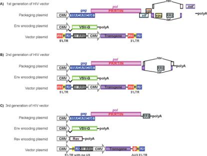
First-generation replication-deficient recombinant HIV-1 vectors are produced from three separate elements: (i) a
packaging construct; (ii) an Env plasmid encoding a viral glycoprotein; and (iii) a transfer vector genome construct (Figure 3A). The packaging construct expresses HIV Gag, Pol and regulatory/accessory proteins from a strong mammalian promoter to generate viral particles. The Env plasmid expresses a viral glycoprotein, such as VSV-G, to provide the vector particles with a receptor-binding protein. These two plasmids have been specifically engineered without either a packaging signal or LTRs to avoid their transmission into vector particles and to reduce the production of RCL in vector preparations.
The transfer vector plasmid contains the transgene(s) and all of the essential cis-acting elements (LTRs, and RRE) for packaging/reverse transcription/integration, but expresses no HIV proteins. Since the transactivator Tat is not encoded by the transfer genome, the promoter activity by the 5 -LTR is minimal. Instead, transfer genomes use an internal promoter to express transgenes in transduced cells.
This three-plasmid system allows the delivery of a gene of interest without expressing viral proteins in target cells. Splitting the vector components into three plasmids means at least two recombination events are required to yield a replication-competent HIV-1-like virus during vector production. The use of VSV- G, rather than HIV-1 Env, also reduces recombination, since it eliminates homologous sequences between the Env and transfer vector plasmids.
Second-generation lentiviral vectors with no viral accessory proteins (Vif, Vpu, Vpr or Nef)
First-generation lentiviral vectors provided a new level of safety for these potent gene-delivery vehicles. To increase safety further, second-generation vectors have been developed by modifying accessory genes in the system (Figure 3B).
HIV-1 Vif, Vpu, Vpr and Nef are called accessory proteins because they can be deleted without affecting viral replication in certain human lymphoid cell lines. However, these proteins are actually essential for efficient HIV-1 propagation/virulence in primary cells or in vivo. For example, lymphocytes are resistant to vif -deficient HIV-1 replication [29]. Vif is necessary to inactivate a host antiviral factor, APOBEC3G (apolipoprotein B mRNAediting enzyme-catalytic polypeptide-like 3G), to ensure efficient virus production [30]. Similarly, Vpu neutralizes another cellular antiviral factor, called Tetherin [31,32]. On the other hand, Nef promotes the degradation of host proteins, such as MHC class I and CD4, to augment virus production and facilitate immune evasion [33].
Therefore, although these accessory genes are important for HIV as a pathogen, they can be deleted in second-generation lentivectors [34] (Figure 3B). By replacing HIV-1 Env with VSV- G, these second-generation vectors include only four of the nine HIV genes: gag, pol, tat and rev [34].
SIN (self-inactivating) vectors with a deletion in the U3 region of the 3 -LTR
Conventional lentiviral vectors integrate transgene cassettes flanked by two LTRs into the host genome (Figure 4). Under normal circumstances this should be a dead-end integration event. However, if replication-competent recombinant lentiviruses are produced, they may be able to replicate in a similar manner to that of wild-type viruses. An alternative problem could arise if vector-transduced cells are subsequently infected by a wild-type lentivirus. In this case, the wild-type virus can act as a helper virus to rescue the integrated vector into new viral particles to spread transduction beyond the original target cell.
c The Authors Journal compilation c 2012 Biochemical Society
606 |
T. Sakuma, M. A. Barry and Y. Ikeda |
|
|
Figure 3 Schematic representation of HIV vectors
(A) The first generation of HIV vectors includes all of the viral proteins, except Env protein, in a packaging plasmid. VSV-G is provided by a different plasmid. HIV vector plasmid contains LTRs and the transgene is expressed under a strong viral promoter such as the CMV promoter. (B) For the second generation of HIV vectors, all of the accessory proteins are excluded from the packaging plasmid. Similar to the first generation of HIV vectors, expression of glycoprotein and transgene are provided by different plasmids. (C) The third generation of HIV vectors requires four different plasmids. In addition to the three plasmids (i.e. a packaging plasmid, an Env-encoding plasmid and a vector plasmid), Rev protein is provided by a different plasmid. The vector plasmid is also modified by deleting the U3 region from 5 -LTR and partially deleting 3 -LTR to reduce the possible production of replication-competent viruses, and a strong viral promoter such as RSV or CMV is inserted for expression of the vector.
Another serious issue is the undesired activation of cellular genes by integrated vectors. Since LTRs have an enhancer [binding sites for host transcription factors, including Sp1 (specificity protein 1) or NF-κB (nuclear factor κB)] and promoter regions, integration of LTRs into the genome can activate adjacent cellular genes. If semi-random integration of the transgene occurs near a proto-oncogene, these enhancers/promoters have the potential to activate transcription of these genes, moving that cell towards oncogenesis. Given these issues, SIN lentivectors have been developed.
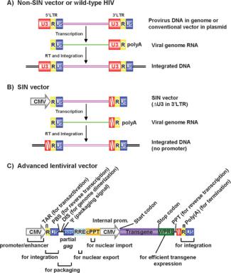
A standard vector genome is flanked by two LTRs that each contain three regions: U3, R and U5 (Figure 4). U3 acts as a viral enhancer/promoter and R in the 3 -LTR acts as the polyadenylation signal. The 5 U3 and the 3 U5 element are therefore not present in mRNA from the provirus. Instead, the R region caps both ends (Figure 4). Duplication of LTR elements occurs during reverse transcription prior to integration when U3 in the 3 -LTR is copied and transferred to the 5 -LTR. As first shown in MLV vectors, if part of U3 in the 3 -LTR is deleted, its duplication will transfer the same deletion into the 5 -LTR’s promoter/enhancer region. This deletion therefore results in transcriptional inactivation of potentially packageable viral genomes in the transduced cell [35].
On the basis of success in MLV, this SIN approach was applied to HIV vectors by deletion of 3 -LTR elements, including its TATA-box-, Sp1-, NF-κB- and NFAT (nuclear factor of activated T-cells)-binding sites [36–38]. This SIN modification reduces the likelihood of: (i) propagation of spontaneously produced replication-competent recombinant HIV-like viruses; (ii) insertional activation of cellular oncogenes by residual promoter activities of integrated LTRs; (iii) mobilization of integrated vectors by a wild-type virus; and (iv) transcriptional interference and suppression by LTRs.
Third-generation Tat-independent vectors from four plasmids
HIV-1 uses regulatory proteins Tat and Rev for viral transcription and nuclear export of intron-containing transcripts. Unlike its accessory proteins, Tat and Rev are absolutely required for HIV- 1 replication [39,40]. To increase safety, third-generation vectors have been designed to be Tat-independent with Rev provided from a separate plasmid (Figure 3C). Tat-independence is achieved by replacing the U3 promoter region of the 5 -LTR in the transfer vector with strong viral promoters from CMV or RSV (rous sarcoma virus) [41,42]. The four plasmids used to generate third-generation vectors are: (i) a packaging construct containing only gag and pol genes; (ii) a plasmid expressing Rev; (iii) an Env (VSV-G) plasmid; and (iv) a transgene plasmid driven by a heterologous strong promoter. The enhancer/promoter region (U3) of 3 -LTR is also removed to add the SIN property.
This vector system has only three of the nine genes of HIV, increasing its predicted biosafety. Since the vector elements are split into four plasmids, at least three recombination events are required to generate a replication-competent HIV-1-like virus. Even if these occurred, the resulting viruses would have only HIV- 1 Gag, Pol, Rev and VSV-G proteins, with no active LTRs, Tat or accessory proteins. Increased safety of third-generation vectors is supported by the detection of no RCL within 1.4×10¹⁰ transducing units of vector produced from ten independent 14 litre production lots [43]. When compared with other three-plasmid systems, the vector yields of third-generation vectors are typically lower.
Introduction of a cis-acting cPPT [central PPT (polypurine tract)] for increased vector transduction efficiency
During reverse transcription of HIV-1, plus strand DNA synthesis starts from the PPT and the cPPT. This leads to a plus strand
c The Authors Journal compilation c 2012 Biochemical Society
Lentiviral technology |
607 |
|
|
Figure 4 Schematic representation of HIV vector constructs and integrated proviruses
(A) Non-SIN vectors or wild-type HIV contains LTRs at both the 3 - and 5 -ends. Viral transcription starts at the U3/R region in the 5 -LTR and terminates at the R/U5 region in the 3 -LTR. Integrated viral DNA contains a cis-element (such as the TATA box and binding site for transcriptional factors Sp1 and NF-κB) of the LTR, thus leading to the potential activation of proto-oncogenes by random integration of the vector/virus. (B) With the SIN vector, the 5 -LTR U3 region has been replaced with a CMV promoter, and the 3 -LTR U3 region, which contains the cis-element, has also been partially deleted. As a result, the viral transcript contains no complete U3 sequence, and reduces the possible generation of replication-competent virus or activation of proto-oncogenes after the vector integration. (C) The cis-acting elements of advanced lentivectors. The location and role of each cis-element are displayed. DIS, dimerization-initiation site; PBS, primer-binding site; Psi, (packaging signal).
overlap called the central DNA flap [44]. It has been proposed that the central DNA flap enhances nuclear import of HIV-1 proviral DNA [45], although this is debated [46]. In practice, introduction of cPPT into HIV-based vectors significantly increases vector transduction efficiency in vitro and in vivo [47–50].
Using WPRE [WHV (woodchuck hepatitis virus) post-transcriptional regulatory element] for increased transgene expression
Another cis-acting element that has been used to improve lentiviral vector expression is the WPRE sequence. WPRE increases the amount of unspliced RNA in both nuclear and cytoplasmic compartments [51,52]. Introduction of WPRE into lentiviral vectors significantly increases transgene expression in target cells [52–54]. Although increased expression is useful, the use of WPRE may raise safety concerns, since it contains a truncated form of the WHV X gene, which has been implicated in animal liver cancer [55]. WPRE safety has subsequently been improved by a mutation of the open reading frame of the X gene [56].
FURTHER MODIFICATIONS FOR IMPROVED BIOSAFETY AND VECTOR PERFORMANCE
The second and third packaging systems, VSV-G envelope, selfinactivating features, cPPT and WPRE elements, have been widely used in lentiviral vectors. In this section we describe additional modifications that further reduce the risk of generation of RCLs during vector production.
The mechanisms of genetic recombination have been extensively studied in γ -retroviruses. Molecular analysis of the RCR (replication-competent retrovirus) has revealed that as little as 10 bp of nucleotide identity between a packaging construct and a vector constructs can mediate homologous recombination and RCR production [57]. Homologous recombination can occur when two different RNAs are packaged into one virion followed by RT-mediated strand transfer (template switching). Viral RT-mediated recombination is also observed in HIV-1 reverse transcription [58].
Given that HIV-1-based vectors have multiple overlapping cis- acting sequences that are shared between vector and packaging constructs, recombination between these sequences is formally possible. Some of these sequences are difficult to impossible to remove from all constructs. For example, the RRE sequence must be on both constructs. The gag gene must of course be present in the Gag expression plasmid, but a part of gag gene must also be included in the transfer vector for it to be efficiently packaged. In addition, the cPPT element in the pol gene can be on both constructs. Accordingly, packaging signal and gag recombinants have been frequently detected in HIV-1-based vector preparations [58–60], although no RCL has been observed in secondor thirdgeneration HIV-1 vectors. However, it should be noted that an artificial replication-competent recombinant lentivirus with VSV- G and accessory genes vif and vpr has been produced [61].
To minimize the possible risk of generating RCLs, we can:
(i) further reduce the homologous sequences between vector and helper sequences; and (ii) further divide helper functions into multiple plasmids. On the basis of those concepts, additional modifications have been introduced into lentiviral vectors, which we summarize below, along with additional modifications leading to enhanced lentiviral vector performance in certain applications.
Codon optimization for Rev-independent Gag-Pol expression
The binding of HIV-1 Rev to RRE in HIV-1 transcripts results in the nuclear export of those intron-containing transcripts. Some studies have found AU-rich destabilizing sequences in the gag gene, which can be stabilized by the Rev–RRE interaction [62]. Indeed, the HIV-1 genome, especially gag, pol and env genes, is highly AU-rich, and this imparts a codon bias that is highly different from the one used by human genes. Intriguingly, codon optimization of the HIV-1 gag-pol gene not only increases Gag-Pol protein expression, but also makes these genes Revindependent [63,64]. Since many of the third bases are changed in codons to increase expression, this also fortuitously disrupts homology of the synthetic gag sequence with the native gag sequence needed for packaging of the transfer construct. This results in a significantly lower rate of recombination between the packaging and vector genome constructs [60].
Rev-independent transfer vectors
In contrast with the successful Rev-independent Gag-Pol expression, generating a Rev-independent transfer vector construct has been challenging. The CTE (constitutive transport element) from Mason–Pfizer monkey virus has been used to
c The Authors Journal compilation c 2012 Biochemical Society
608 |
T. Sakuma, M. A. Barry and Y. Ikeda |
|
|
replace the RRE/Rev mRNA transport mechanism. Although CTE elements can rescue the Rev/RRE-independent Gag-Pol and/or vector genome expression, the resulting vector titres are generally low [42,65–67].
Trans-lentiviral vector system
The trans-lentiviral vector system has been developed through splitting the Gag/Gag-Pol packaging element into two separate plasmids, one that expresses Gag and PR, and another that expresses RT and IN. For efficient encapsidation of the RT and IN polyprotein, the RT–IN polyprotein is expressed as a fusion protein with the virion-associated accessory protein Vpr [58,68]. These trans-lentiviral vectors are currently produced from four separate vector components: (i) Gag-, Pro-, Vif-, Tatand Rev-expressing packaging construct; (ii) Vpr–RT–IN-expressing packaging construct; (iii) VSV-G/Env-expressing construct; and (iv) Tat-dependent transfer vector [58].
Lentiviral vectors from other primate lentiviruses
Chromatin-insulator elements and chromatin-opening elements
If a lentiviral vector integrates into a transcriptionally inactive region of the chromosome, their transgene expression may also be repressed by surrounding chromatin. In addition, epigenetic position effects can also affect the levels of transgene expression. Insulators are DNA sequences that can shield enhancers and promoters from the activation or silencing by adjacent chromatin (reviewed in [86]). Given this, chromatininsulator sequences have been incorporated into lentiviral vectors to increase the efficiency of transgene expression. The cHS4 (chicken hypersensitive site-4) insulator has been used to increase vector transgene expression [87,88], although some studies found minimal effects with this modification [89]. Similar elements used to prevent vector silencing are the enhancerless UCOEs (ubiquitously acting chromatin-opening elements). Introduction of human HNRPA2B1-CBX3 UCOE (A2UCOE) has demonstrated highly reproducible and stable transgene expression in haematopoietic stem cells [90] through blocking DNA methylation-mediated silencing of lentiviral vectors [91].
To reduce safety concerns related to the generation of RCLs, White et al. [69] developed a hybrid lentiviral vector system, where HIV-1 vector genome constructs are packaged by the viral proteins of a non-virulent SIV (simian immunodeficiency virus) strain, SIVmac 1A11. This design increases the predictable biosafety owing to the reduced homology between HIV-1 and SIVmac. Lentiviral vectors derived from other primate lentiviruses (i.e. HIV-2 and SIV) have also been developed and demonstrated efficient gene transduction of dividing and nondividing cells [70,71]. Although capsid-dependent post-entry restriction of SIV vectors has been observed in rodent cells [72], SIV vectors have demonstrated efficient gene transfer into haematopoietic stem cells in non-human primate models [73,74].
Lentiviral vectors derived from non-primate lentiviruses
Non-primate lentiviruses, including FIV (feline immunodeficiency virus) and EIAV (equine infectious anaemia virus) have highly restricted species tropisms and cannot replicate in human cells. Their narrow host ranges are primarily due to the lack of their functional receptors on human cells [75], whereas an additional intracellular block has been observed in human cells [76]. A series of non-primate lentiviral vectors have been developed with expanded tropisms through pseudotyping with the VSV-G glycoprotein and by the introduction of a strong internal promoter into the constructs [77–80]. A variety of dividing or non-dividing cells can be transduced by nonprimate lentiviral vectors, including primary aortic smooth muscle cells, hepatocyes, dendritic cells and neuronal cells [77–79,81], although some studies have observed post-entry restrictions to FIVor EIAV-based vectors in some human cells [82,83].
It has been proposed that non-primate lentiviral vectors may be safer alternatives to HIV-based vectors, because they should not be able to replicate in humans even if replication-competent viruses are produced. Conversely, it has also been proposed that these lentiviral vectors may be able to replicate in humans if the VSV-G gene is incorporated into the recombinant virus. If so, it may well be safer to develop vectors from HIV-1, whose molecular biology and pathogenesis are better understood, and for which multiple drug therapies are available [84]. In this light, it should be noted that in utero and neonatal gene transfer in mice with EIAV, but not HIV-based vectors, has led to a very high incidence of liver and lung tumours [85]. The causal mechanisms leading to this oncogenesis by the EIAV vectors remain elusive.
Packaging elements from different HIV isolates for altered vector tropism
Gag is essential for HIV genome packaging, virion assembly and budding, whereas Pol is necessary for virion maturation, reverse transcription and viral integration. Although there are many genetically divergent HIV-1 strains with diverse biological properties, most HIV-1 vectors have been derived from only a few well-characterized clones of laboratory-adapted T-cell-tropic HIV-1. Therefore it is likely that more robust lentivectors or vector elements can be found in the wealth of HIV-1 genetic diversity beyond the common strains. Indeed, we and others have shown that naturally occurring substitutions in gag can actually significantly alter vector performance, vector titres and the ability to infect traditionally refractory cells [92,93]. On the basis of this, new lentiviral vector systems based on new strains are being developed. For example, new vectors based on a macrophagetropic HIV-1 YU-2 clone have demonstrated comparable gene transfer efficiency with widely available vectors [67].
Lentiviral vectors with a SIV accessory protein Vpx for improved transduction of monocytes and dendritic cells
Several primate lentiviruses have an accessory protein called Vpx. VSV-G-pseudotyped SIV vectors generated with only Gag, Pol, Tat and Rev helper functions cannot transduce human monocytes and dendritic cells [94]. However, this defect can be rescued when Vpx protein is expressed in vector-producing cells [95]. Similarly, Vpx-containing lentivectors, based on a pathogenic sooty mongabey monkey isolate (SIVsmm-PBj), can efficiently transduce freshly isolated human monocytes [96]. Recent studies identified SAMHD1 (sterile α-motif domain and HD domain 1) as the dendriticand myeloid cell-specific HIV-1 restriction factor and that this factor is counteracted by Vpx [97]. Codelivery of Vpx-containing viral-like particles not only enhances SIV vector transduction, but also potently enhances HIV-1- based vector infection of monocyte-derived dendritic cells [98].
TARGETING LENTIVIRAL VECTORS
Targeted gene delivery and expression to organs and tissues of interest is the ultimate goal for many gene-delivery applications. There are two steps in lentivector infection, which can be modified for targeted vector transduction, i.e. vector entry and
c The Authors Journal compilation c 2012 Biochemical Society
Lentiviral technology |
609 |
|
|
gene expression. In this section, we will review strategies to generate tissue-specific lentivectors.
Limited vector entry through pseudotyping with heterologous viral glycoproteins
Combining viral particles with a foreign envelope glycoprotein can alter the host tropism. Because of the successful incorporation into HIV-1-like particles and its high transduction efficiency in various cell types, lentivirus vectors are commonly pseudotyped with VSV-G. However, VSV-G-pseudotyped vectors can show cytotoxicity at high concentrations, whereas VSV-G-mediated non-specific gene delivery into undesired cell types poses a safety concern for their use in the clinic.
Many viruses use specific receptors for viral entry, which can be exploited for selective lentiviral vector delivery. γ - retroviruses, such as amphotropic (broad host range) MLVs, gibbon ape leukaemia virus, feline endogenous retrovirus RD114 and a xenotropic MLV-related virus XMRV (xenotropic MLVrelated virus), have been used to target their original receptors PIT2 [SLC20A2 (solute carrier family 20 member 2)], GLVR1 (SLC20A1), neutral amino acid transporter (SLC1A5 gene) and XPR1 (xenotropic and polytropic retrovirus receptor 1), which are broadly, but less ubiquitously, expressed in gene therapy target cells [99–102]. Pseudotyping with the viral surface glycoproteins from other RNA viruses, including Ross River virus, Semliki Forest virus, lymphocytic choriomeningitis virus, rabies virus, Mokola virus and Ebola virus, also yields hightitre vectors [78,103–107]. Intriguingly, amphotropic MLV Env or RD114 Env-pseudotyped lentiviral vectors show efficient transduction of human CD34+ haematopoietic stem cells [108,109], whereas Ebola virus glycoprotein-pseudotyped vectors efficiently transduce airway epithelia from the apical surface [110]. One caveat for the use of non-VSV-G glycoproteins for pseudotyping is that resulting vectors can be more fragile/unstable than VSV-G-pseudotypes. For instance, γ - retroviral Env-pseudotyped HIV-1 vectors are generally more sensitive to freeze and thaw [100]. We have also observed large amounts of particle-free amphotropic MLV Env in vector preparations competing with MLV-pseudotyped vectors during vector transduction [100].
Targeting lentiviral vectors with bioengineered ligand/antibody-displaying Envs
Significant efforts have been devoted to generate targeting lentiviral vectors using a ligand protein or antibody fused to viral glycoproteins to retarget the lentiviral particles to specific cell-surface molecules [111]. For instance, pseudotyping with an MLV Env glycoprotein engineered to display anti-CD3 single-chain antibody significantly improves transduction of primary lymphocytes [112]. Similarly, lentiviral vectors coated with a modified chimaeric Sindbis virus envelope conjugated with anti-P-glycoprotein antibody can selectively transduce P- glycoprotein on metastatic melanoma [113]. One of the most promising re-targeting envelope platforms is based on the F and H glycoproteins of measles virus that allows targeted lentiviral entry into CD20-positive B-lymphocytes or quiescent T-cells [114,115].
Selective vector transgene expression through introduction of a tissue-specific promoter
Use of a |
cell-type-specific regulatory element/promoter as |
an internal |
promoter can target vector transgene expression |
to the cells of interest. Unlike the envelope-mediated entrytargeting strategies, vectors with a tissue-specific promoter can basically enter and integrate in any cell types. However, their transgene expression is limited to a certain cell type by the internal promoter. Various tissue-specific promoters have been incorporated into lentiviral vectors, including neuron-specific [116], dendritic cell-specific [117], tumour angiogenesis-specific [118], vascular differentiation-targeted [119] or hepatocytespecific [120] promoters.
Selective vector transduction through miRNA (microRNA)-mediated silencing in undesired cells
A total of 21–22 nucleotides of non-coding miRNAs mediate post-transcriptional gene regulation [121]. Lentivector-carrying miRNA vector has been developed as a new approach to limit undesired vector transgene expression. For instance, to minimize off-target transgene expression in antigenpresenting cells, lentivectors have been engineered to carry a haematopoietic-specific miRNA, miR-142-3p, which leads to reduced transduction of antigen-presenting cells and antitransgene immune responses [122].
LENTIVIRAL VECTOR PRODUCTION, CONCENTRATION, TITRATION AND EX VIVO INFECTION
Transient vector production
Lentiviral vectors are typically produced in HEK (human embryonic kidney)-293T or HEK-293T-derived cell clones, such as HEK-293T/17 (A.T.C.C. CRL-11268), through transient transfection of multiple vector plasmids. Various transfection methods have been successfully used. Resulting vector titres can vary, depending on the condition of the HEK-293T cells, vector packaging system (second, third or trans-lentiviral systems) and the design of the transfer vector. As more plasmids are introduced into a transfection, each is proportionately less efficiently delivered into the cells. Therefore four-plasmid systems produce relatively lower titre vectors than three-plasmid systems. Vectors with complex structures, such as multiple expression cassettes, long polycistronic transgene or siRNA (short interfering RNA) expression constructs, tend to produce lower titres of vectors than systems with simpler designs (single expression cassette, short transgene or no siRNA expression). Inclusion of the cPPT in vector constructs frequently increases vector titres/transduction efficiency [49]. Another factor which can strongly affect vector performance is the internal promoters used to drive trangenes. Tissue-specific promoters can be silent in different types of cells, whereas commonly used constitutively active promoters, such as the CMV promoter, can be less efficient in certain cell types [82,123,124].
Stable packaging cell lines for lentiviral vectors
One can deliver all the elements needed for lentiviral production by transient transfection or engineer stable cell lines to produce one or more viral proteins in trans, reducing the number of plasmids needed for transfection. Stable lentivector packaging cell lines should allow continuous reproducible generation of large vector batches, which would be preferable for the production of clinical-grade lentiviral vectors. Towards this goal, inducible packaging cell lines for VSV-G-pseudotyped HIV-based lentiviral vectors have been constructed without accessory genes [125– 128]. As an alternative approach, retroviral vector-mediated
c The Authors Journal compilation c 2012 Biochemical Society
610 |
T. Sakuma, M. A. Barry and Y. Ikeda |
|
|
introduction of vector-packaging elements has been used to establish continous packaging cell lines stably expressing codonoptimized Gag-Pol, Rev, Tat and a γ -retrovirus Env from four independent constructs [60]. However, those packaging cell lines generate non-SIN vectors [125–129] or conditional SIN vectors [130]. A clinically applicable SIN lentiviral producer cell line has been generated through retroviral delivery of packaging elements and concatemeric array transfection of a SIN vector genome construct [131].
Vector concentration
Various concentration methods have been successfully used to increase vector titres, including ultracentrifugation [(1–3)×105 g for 1–4 h at 4 ◦C], high-speed long-term centrifugation ( 104 g for 8–12 h at 4 ◦C using a high-speed centrifuge), precipitation with calcium phosphate, or centrifugal filtration using a 100 kDa molecular-mass cut-off filter [100,132–134]. The efficiency can be affected by the serum concentrations in vector preparations, especially for high-speed centrifugation or centrifugal filtration methods.
Vector titration
Before vector infection of target cells, titration of vectors produced is required to adjust vector doses. Vector titres are also critical to evaluate transduction efficieincy. Various methods have been used, including: (i) measuring the quantity of vector particle components (physical particle numbers/p24 capsid concentration/RT activity/genome copy numbers) in vector preparations; (ii) measuring proviral vector DNA copy numbers in infected cells; or (iii) measuring transgenes expressed in cells (by flow cytometry or immunostaining).
The quantification of viral components in culture supernatants has been broadly used in HIV-1 research. One major problem of these methods is the equal detection of infectious and noninfectious vectors. Indeed, titration with viral genomic RNA copy numbers and p24 concentrations has been shown to be rather poor in predicting vector transduction efficiency [135]. In contrast, detecting proviral DNA copy numbers in infected cells by real-time PCR can evaluate infectious vector copy numbers. We and others have successfully used real-time PCR with primer and probe sequences specific to the HIV-1 packaging signal (ψ ) [135,136] that is relatively well-conserved among HIV-1 vectors. However, it has to be noted that VSV-G-pseudotyped lentiviral vectors can harbour high levels of plasmid DNAs from vectorproducing HEK-293T cells [137] and only a small proportion of the reverse-transcribed vector DNA can successfully integrate into the host genome. Use of total cellular DNA isolated from cells after one or two passages can minimize the detction of contaminated plasmid DNA and defective non-infectious proviral DNAs in target cells. This method is suitable if the vector has no marker gene or the transgene is driven by a certain tissue-specific promoter, although high infectious vector copy numbers do not necessarily indicate expression of vector transgenes in target cells. Finally, measuring transduced cells by transgene expression is the most reliable method to evaluate infectious lentiviral titres. This method can be used for vectors with reliable marker genes [GFP (green fluorescent protein), puromycin resistance or neo gene etc.] or when specific antibodies against transgenes are available. Since this method measures transgene expression, vector titres can vary depending on the vector permissivity of the cells used for vector titration. In addition, this method may not be reliable when measuring the expression of transgenes driven by certain tissue-specific promoters.
Strategies to enhance ex vivo vector transduction
Various strategies have been used to improve vector infection. Although it shows some cytotoxicity, polybrene, which reduces the charge repulsion between virions and the target cell surface, has been widely used to enhance lentiviral vector infection [138]. Retronectin is also used to increase lentiviral transduction of haematopoietic stem cells, particularly those with γ -retroviral Envs [139]. In addition to those reagents, spinoculation, where vectors are centrifuged down on to monolayers of cells, has been used to increase vector transduction efficiency of various cell types [54,100].
ISSUES ASSOCIATED WITH LENTIVIRAL VECTOR INTEGRATION
As mentioned above, integrating vectors can insert into repressed regions of the chromosome. They can also integrate randomly into regions of the genome that can be problematic. This reality has long been recognized by the field, but did not come to the forefront until the clinical trials that used retroviral gene therapy for X- SCID (X-linked severe combined immunodeficiency). In these trials, patients with X-SCID received CD34+ haematopoietic stem cells that had been genetically modified with γ -retroviral vectors expressing the common γ -chain from IL2RG (interleukin 2 receptor γ ) [140–142]. Although this work demonstrated successful gene therapy in humans [141–144], a subset of these patients subsequently developed a leukaemia-like disease that appeared to be due to insertional mutagenesis by the vector [140,145]. Given that both γ -retroviruses and lentiviruses integrate semi-randomly into the genome, this side effect is relevant to the lentiviral vectors discussed here. In this section, we summarize ongoing efforts to minimize the risks associated with lentiviral vector integration into the host genome.
Distinct target site preferences between HIV-1 and γ -retrovirus MLV
Characterization of the integration site preferences of HIV-1 and MLV may help to understand the possible risks associated with lentiviral vector integration and to improve vector designs for safer gene therapy to avoid the risk of insertional mutagenesis. Mapping of HIV and MLV integration sites on the human genome sequence found distinct target site preferences between the two viruses. MLV vectors appear to be biased towards integration near transcription start sites or CpG islands [146,147], whereas HIV appears to favour integration within active transcription regions [147,148]. Cellular lens epithelium-derived growth factor (LEDGF/p75), which binds both chromosomal DNA and HIV-1 IN [149] plays a role in controlling the location of virus integration in human cells [150].
Low oncogenic potential of lentiviral vector integration
Haematopoietic stem cell gene transfer in a tumour-prone mouse model has revealed that prototypical lentiviral vectors have lower oncogenic potential than conventional retroviral vectors [151]. Similarly, in a sensitive in vitro immortalization assay to quantify the risk of haematopoietic cell transformation, the insertion pattern of lentiviral vectors was found to be approximately 3- fold lower than that mediated by retroviral vectors to trigger transformation of primary haematopoietc cells [152].
SIN vector design reduces the insertional mutagenesis potential
Insertional mutagenesis has become a major hurdle for clinical applications of integrating vectors. As described above, the
c The Authors Journal compilation c 2012 Biochemical Society
Lentiviral technology |
611 |
|
|
SIN vector design (deletion of the U3 region in 3 -LTR) can eliminate the enhancers/promoter elements from the both 3 - and 5 -LTRs in integrated proviral DNA. In a mouse model of haematopoietic stem cell gene therapy, introduction of the SIN mutation increased the safety of retroviral and lentiviral vectors [153]. In vitro immortalization assays indicate that the risk of insertional transformation by SIN lentiviral vectors depends on the type of the internal promoter. Constructing SIN lentivectors with a tissue-specific promoter appears unable to trigger cell transformation in these models [152]. Nevertheless, SIN lentiviral vectors have been shown to induce leukaemia in a mouse model of X-SCID using the IL2RG gene [154,155]. Woods et al. [154] concluded that the therapeutic IL2RG gene itself can act as a ‘hit’ in leukaemogenesis that may amplify this effect in X-SCID, but perhaps not in other types of genetic diseases. Ginn et al. [155] have also proposed the existence of other risk factors, unrelated to insertional mutagenesis or IL2RG gene overexpression, that may have exacerbated this side effect.
Integration-deficient lentiviral vectors for non-dividing cells
If cell division is prevented, unintegrated lentiviral DNAs can express transgenes [156]. Integration-deficient lentiviral vector therefore represents an interesting alternative approach to reduce insertional mutagenesis. Integration-deficient HIV-1 vectors typically have mutations in the IN gene [157] that amplify the formation of episomal genomes in the nucleus, with minimal vector integration. Integration can be reduced up to 104-fold with an integration-deficient HIV-1 vector [158]. An integrationdeficient HIV-1 vector can transduce non-dividing cells, such as muscle cells [159], brain cells [160] and ocular cells [161] in vivo. Clearly, integration-deficient lentivectors increase predictable vector safety, although their long-term gene expression is limited to non-dividing cells and their levels of transgene expression are often less than those of conventional integrating lentiviral vectors.
Self-deleting vectors
Self-excising lentiviral vectors have been developed on the basis of the Cre/loxP system. Self-deletion is achieved by expression of the Cre recombinase from vectors with a loxP site into the U3 region of the 3 -LTR. Duplication of the U3 region of the 3 - LTR during reverse transcription generates a proviral DNA with one loxP site in both LTRs. After integration and subsequent Cre expression, Cre-mediated recombination of these two loxP sites deletes most of the integrated vector genome with the exception of the flanking U sequences and one loxP site [162]. Similarly, lentiviral vectors with loxP sites have been used for conditional deletion of integrated vector sequences (LTR to LTR or transgene-expressing cassette only) in combination with other Cre-expressing vectors [163].
Site-directed integration using ZFNs (zinc finger nucleases) and non-integrating lentiviral vectors
ZFNs that recognize and cleave unique genomic sequences in living cells can be used for targeted gene editing and mutagenesis [164]. Naldini and co-workers have incorporated the ZFN technology into non-integrating lentiviral vectors to express ZFNs and provide the template DNA for gene correction [164a]. This system achieves high levels of site-directed gene addition in a panel of human cell lines, including embryonic stem cells, allowing rapid selection-free isolation of clonogenic cells with the desired genetic modification.
LENTIVIRAL VECTOR APPLICATIONS
Lentiviral vectors have been used successfully to transduce various cell types, such as neurons, hepatocytes, haematopoietic stem cells, retinal cells, dendritic cells, myocytes and islet cells [28,37,49,77]. Notable pre-clinical animal studies include successful correction of β-thalassaemia and sickle cell anaemia [165–167], haemophilia B [122] and ZAP-70 (ζ -chain-associated protein kinase of 70 kDa) immunodeficiency [168], whereas amelioration of other major genetic disorders has been seen with lentiviral vector-mediated interventions, including Parkinson’s disease [169], cystic fibrosis [170] and spinal muscular atrophy [171]. In this section, we describe various lentiviral applications for basic and translational researches.
Delivery of complex genetic structures
Lentiviral vectors typically encode an internal promoter and a polyA-less cDNA in the sense orientation. However, owing to the Rev/RRE-mediated nuclear export of unspliced vector genome RNAs, they can efficiently transfer complex genetic structures by putting introns into the vector by placing the transgene expression cassette in backwards [166]. For generation of multigene lentiviral vectors, two strategies are typically employed to express multiple genes; through incorporation of multiple expression cassettes or construction of a polycistronic transcript driven by a single internal promoter. The use of multiple expression cassettes allows independent gene expression driven by different internal promoters [172]. Some studies successfully used bi-directional promoters to express multiple genes [173]. Owing to the limited packaging capacity of lentiviral vectors (< 9–10 kb in total), those strategies are only suitable for the expression of relatively short expression cassettes. Another strategy is to deliver multiple genes from a single expression cassette encoding a bicistronic, or polycistronic, transcript. Typically, cDNA sequences are linked with an IRES (internal ribosome entry site), which attract ribosomes for translational initiation [174]. One common problem of multigene lentiviral vectors with IRES elements has been their biased expression of two transgenes: the IRES-dependent second gene expression is often weaker than the first gene expression. We have also experienced impaired first gene expression in a cell-type-specific manner, especially in lymphoid cell lines and primary dendritic cells (Y. Ikeda, unpublished work). Indeed, various viral and mammalian IRES sequences have demonstrated tissue-specific biased expression profiles in vitro and in vivo [175]. As an alternative approach to achieve comparable protein expressions from a single expression cassette, retroviral vectors have been developed to encode a polycistronic construct where multiple genes are linked with picornaviral self-cleaving 2A peptides [176]. Incorporation of the self-cleaving property of 2A peptides into lentiviral vectors has also allowed efficient multigene expression from a polycistronic transcript, although each protein retains short peptide sequences from the 2A peptides [177,178].
Gene-silencing vectors
RNAi (RNA interference) is an evolutionarily-conserved gene silencing mechanism that is induced by dsRNA (doublestranded RNA) [179,180]. Since synthetic siRNAs or shRNAs (short hairpin RNAs) can suppress the expression of genes of interest in mammalian cells [181], RNAi-mediated gene silencing has become an essential technology to study gene functions. Importantly, lentiviral vectors can be engineered to achieve stable high-efficiency gene silencing in a wide variety of cells [182]. In gene-silencing lentiviral vectors, siRNA can be delivered as a form of shRNA driven by a RNA polymerase III promoter,
c The Authors Journal compilation c 2012 Biochemical Society
612 |
T. Sakuma, M. A. Barry and Y. Ikeda |
|
|
or as a part of an miRNA-like structure expressed from a RNA polymerase II promoter (for a review, see [183]). Gene-silencing lentiviral vectors have been successfully used to inhibit HIV-1 infection [184,185], retard the onset and the progression rate of amyotrophic lateral sclerosis in mice [186], extend the survival of scrapie-developed mice [187] and increase the expression of fetal haemoglobin [188].
An important advance in the RNAi field is the completion of the generation of a silencing lentiviral library, which targets 15 000 human genes and 15 000 mouse genes, by the RNAi Consortium, a public–private consortrium based at the Broad Institute (http://www.broadinstitute.org/rnai/trc). The genome-wide shRNA library vectors, which are based on a shRNA-carrying lentiviral vector pLKO.1, are available through
Sigma–Aldrich and |
Open |
Biosystems. |
In |
pLKO.1 vector, |
|
an |
shRNA construct is expressed by |
a |
RNA polymerase |
||
III |
U6 promoter, |
whereas |
a separate |
puromycin-resistant |
|
gene-expression cassette allows selection of transduced cell populations. Similarly, pre-designed shRNAor miRNA-carrying lentiviral vectors are also available from various vendors and are in rampant use in basic biology laboratories. The caveats of the use of gene-silencing lentiviral vectors are similar to other RNAi platforms; they can induce off-target gene silencing or undesired interferon response [189]. To rule out those possibilities, it is critical to perform RNAi rescue experiments involving expression of the target gene containing silent mutations in the RNAi-targeted sites. As an additional application of lentiviral vectors for studying gene silencing, Naldini and co-workers have developed lentiviral vectors overexpressing miRNA target sequences from polymerase II promoters and showed stable and specific knockdown of miRNA function in vivo [190].
Inducible vector system
Lentiviral vectors can be used for externally controllable transgene expression. Among several inducible systems, the rtTA (reverse tetracylin-controlled transactivator)-regulated system has been widely used for inducible gene expression [191]. This system uses a chimaeric transcription factor tTA transactivator, a fusion protein between the bacterial TetR (Tet repressor) with the activating domain of herpes simplex virus VP16 (viral protein 16), or its derivative rtTA protein, along with a TetO-binding- sites-containing promoter for tetracycline-induced gene silencing (Tet-Off) or activation (Tet-On) respectively. The Tet-On/Tet-Off systems have been studied extensively in the context of lentiviral vectors, in which the Tet-regulated system is incorporated into a single lentivector [192,193]. Using a similar vector design, conditional gene-silencing lentiviral vectors have also been developed [194].
To date, various inducible lentivector systems have been developed, and are commercially available. Since several studies have identified VP16, used in the Tet transactivator, as the key factor to induce non-specific gene induction or cytotoxicitiy, which can lead to data misinterpretation [195,196], a new doxycycline-regulated system based on the original TetR with no VP16 has been developed and incorporated into lentiviral vectors [197].
Lentiviral vector-mediated transgenesis
Lentiviral vectors can efficiently transduce embryonic stem cells. When compared with conventional retroviral vectors, lentiviral vectors are relatively resistant to gene silencing in mammalian embryonic stem cells [198]. Accordingly, lentivirally modified murine embryonic stem cells have been used to generate
transgenic mice [198]. As an alternative approach, lentivectors have been used to introduce transgenes into early embryos. This technology has been highly successful and has led to the generation of various transgenic animals, including chicken, rat, cat and pig, in which reliable tissue-specific reporter gene expression has been observed after germline transmission [199– 203]. In addition, lentiviral vectors expressing siRNAs have also been used to knock down targeted genes in vivo [204,205].
Lentiviral vector-mediated immune modulation
Lentivectors can efficiently transduce antigen-presenting cells, including dendritic cells [206]. This property has been exploited as lentiviral vaccines for tumours [117,206–208] and infectious diseases [209–211]. Lentivector-mediated expression of melanoma antigens [117,206,207] or ovalubumin [136,212] in dendritic cells elicits both CD8+ T-cells and CD4+ T-cell responses. Lentivector-induced tumour-specific immunity has demonstrated regression of tumours [213]. For HIV/AIDS vaccine development, lentivector-mediated gene transfer has also induced Gag-specific T-cell responses [210,211].
Cellular reprogramming
Introduction of a set of defined pluripotency-associated factors, such as OCT4 (octamer 4), SOX2 (sex-determining region Y- box 2), KLF4 (Kruppel¨-like factor 4) and c-Myc (or OCT4, SOX2, NANOG and LIN28), in somatic cells has demonstrated the generation of embryonic stem cell-like pluripotent stem cells [214]. Since multiple factors have to be introduced into a single cell, multiple lentiviral vectors have been used for successful reprogramming of various cell types, such as fibroblasts, keratinocytes and haematopoietic stem cells [215– 217]. To increase the reprogramming efficiency and safety, single polycistronic lentiviral vectors carrying the four reprogramming factors linked by self-cleaving 2A peptides have been developed, some of which with self-deleting properties [218,219]. Owing to the concerns of reactivation of pluripotency-associated factors in iPS (induced pluripotent stem cell) progeny, the field has been shifted towards other reprogramming technologies which do not require integrating vectors [215,220].
In addition to the pluripotency induction, lentiviral vectors have been used to trans-differentiate adult somatic cells into other types of cells. For instance, lentiviral introduction of Gata4, Hnf1a and Foxa3 (Forkhead box a3) in murine fibroblasts results in generation of hepatocyte-like iHep cells [221], whereas ectopic expression of Gata4, Mef2c and Tbx5 in murine fibroblast cells can trans-differentiate fibroblasts into functional cardiomyocytes [222].
In vivo imaging and lineage tracking
Lentiviral vector-mediated stable gene transduction can be exploited for in vivo monitoring/live imaging of vector-infected cells [223–225]. Cancer cells, which are engineered to express reporter genes by lentiviral vectors, can be monitored for their growth or metastasis in vivo [226], whereas transplantation of lentivirally modified bone marrow cells/stem cells allows the identification of stem cell-derived progeny in vivo [151,227]. Moreover, stem cell transduction by a vector with a tissue-specific promoter allows lineage tracking in vitro and in vivo [119]. For instance, lentiviral vectors with a marker gene under the control of a troponin-I promoter can be used to track cardiomyocyte differentiation of human embryonic stem cells [228]. Recently, Gaussia luciferase fragment complentation has been incorporated into lentiviral vectors for monitoring ligand–receptor binding in vivo [229].
c The Authors Journal compilation c 2012 Biochemical Society