

Phencyclidine An Update Editor Doris H. Clouet
.pdfSorensen and Blaustein 1985a). In view of the enhanced neurotransmitter release that would be expected to accompany block of presynaptic K channels (Llinas et al. 1976), this mechanism could explain the PCP-induced "dopamine storm" (Rappolt et al. 1980). Such altered synaptic transmission at central synapses may underlie the disordered behavior characteristic of PCP intoxication.
The aforementioned observations imply that the high affinity PCP binding sites in the brain are K channels. Therefore, as a prelude to the eventual isolation and characterization of these PCP "receptors"/K channels, we employed a tritiated photo affinity analogue of PCP, m-azido-3H-PCP (3H-Az-PCP) to label rat brain, synaptic membrane proteins. The results indicate that this analogue binds specifically to two membrane polypeptides of a parent MR 80,000 and 95,000 Daltons (Sorensen and Blaustein 1985b).
IDENTlFICATION OF K CHANNELS IN SYNAPTOSOMES WITH TRACER 86Rb
EFFLUX
Rb efflux from synaptosomes loaded with 86Rb was used to assess the K permeability of the nerve terminals under "resting" conditions, and under conditions in which the terminals were depolar-
ized by elevating the external |
k concentration, [K]o. With a |
half-life 36 times longer than |
42K, 86Rb is a suitable tracer for |
K because (Bartschat and Blaustein 1985a; Bartschat and Blaustein 1985b): (1) Rb, like K, is accumulated by synaptosomes via a metabolically active, ouabain-sensitive route; (2) Rb permeates most neuronal K channels nearly as well as does K itself; and
(3) synaptosomes preloaded with both 42K and 86Rb have qualitatively similar K and Rb effluxes.
Voltage-regulated K channel activity in synaptosomes was measured as described in the figure 1 legend. For further details, see Bartschat and Blaustein (1985a). Under "resting" conditions (5 mM [K]o; open circles in figure 1), 86Rb efflux was about 0.3 to 0.4%/sec (component "R"), which corresponds to a resting K permeability of 2.4x10⁻⁷cm/sec (Bartschat and Blaustein 1985a). Rb efflux under these conditions probably reflects the mechanism(s) responsible for the normal K permeability of the resting terminals.
Depolarization of the synaptosomes with Ca-free media containing 100mM K increased 86Rb efflux (figure 1, open squares); two kinetically and pharmacologically distinct K conductances could be discerned. Between 1 and 4 seconds, Rb efflux was linear and was 2.2 to 2.4%/sec (component "S"). Extrapolation of Rb efflux to the ordinate ("zero time") exposed an additional, rapid component of 86Rb efflux (component "T"). Component T reflects a distinct K channel that, unlike component S, appeared to inactivate in less than 1 second (Bartschat and Blaustein 1985a).
38

FIGURE 1. |
T i m e |
|
c o u r s e |
o f 8 6 R b |
e f f l u x |
f r o m s y n a p t o s o m e s ; |
t h e |
|
|
|||||||||||||||
|
|
|
|
|
|
|
|
|
e f f e c t o f P C P |
|
|
|
|
|
|
|
|
|
|
|
||||
NOTE: Synaptosomes |
|
were prepared as described (Krueger et al. 1979), were equil- |
||||||||||||||||||||||
ibrated |
with |
|
hysiologlcal |
salt |
solution |
(PSS). |
and |
were allowed |
to |
accumu- |
||||||||||||||
late |
tracer |
|
86Rb (20 µCi/ml in |
PSS) at 30 °C for |
30 |
|
minutes. |
PSS |
contained |
|||||||||||||||
(mM): |
KCI, |
5; |
NaCl, |
145; |
MgCI2, 2; glucose, 10; |
|
HEPES |
buffer, |
10, |
titrated |
||||||||||||||
to ph |
7.4 |
|
with |
NaOH; |
Na2 |
HPO4, |
0.5; and |
unlabelled |
RbCI, |
0.1. |
Aliquots |
|
||||||||||||
(30 |
µl) |
of |
|
the |
synaptosomal suspension |
were then |
pipetted |
onto |
glass |
fiber |
||||||||||||||
filters, |
were |
washed |
free |
of extracellular |
tracer, |
and |
then |
exposed |
|
to |
|
|||||||||||||
efflux media |
|
for various lengths of time (1 to 4 |
seconds). |
|
Efflux |
was |
ter- |
|||||||||||||||||
minated |
by |
rapid |
addition |
of “stopping solution” containing the K |
channel |
|||||||||||||||||||
blockers |
tetraethylamnonium |
(145 mM) and tetrabutylamnonium (5 mM), |
but |
no |
||||||||||||||||||||
NaCl |
or |
KCI. |
|
Suction |
|
was |
rapidly |
applied, |
and |
the |
filters |
and |
filtrates |
|||||||||||
were |
counted |
|
by |
Iiquid |
scintillation |
spectroscopy; |
Rb |
efflux |
was expressed |
|||||||||||||||
as: |
|
|
|
|
|
|
|
|
|
|
|
|
|
|
|
|
|
|
|
|
|
|
|
|
The |
efflux medium |
was similar to the loading medium; however, In exper- |
|||||||||||||||||||||
iments |
involving |
elevated [K]o, K |
replaced Na mole-for-mole. |
When |
drugs |
||||||||||||||||||
were |
tested, |
they |
were Included In the wash media facilitate equilibra- |
||||||||||||||||||||
tion |
with |
the |
synaptosomes. |
Symbols |
correspond |
to |
Rb |
efflux |
in: |
5 |
mM |
||||||||||||
K ( |
0), |
100 |
mM |
K |
(control; |
), 100 mM |
K + |
10 |
µM |
PCP |
( |
). or 100 mH K |
+ |
||||||||||
100 |
µM |
|
PCP |
|
. |
The |
data |
points |
are |
|
the |
means |
of |
six |
determinations. |
|
|||||||
The |
components |
of |
|
86Rb efflux indicated in the figure represent: R |
= |
Rb |
|||||||||||||||||
efflux in |
5 |
mH |
K |
media |
(expressed |
in |
%/sec); |
S |
= |
Rb |
efflux |
between 1 |
and 4 |
||||||||||
seconds |
(%/set) |
in K-rich media minus component R; T |
= |
K-dependent |
incre- |
||||||||||||||||||
ment |
(%) |
In |
Rb |
efflux |
when |
S |
is |
extrapolated |
|
back to zero |
time. SV |
= |
|
||||||||||
component |
of |
S |
that |
is |
blocked |
by |
PCP; |
SR |
= |
PCP-lnsensltive |
portion |
of |
|
||||||||||
component |
S. |
|
|
|
|
|
|
|
|
|
|
|
|
|
|
|
|
|
|
|
|
||
Inclusion of Ca in the efflux media enhanced 86Rb efflux in 100 mM [K]o but not 5 mM [K]o (data not shown). This Ca-dependent 86Rb efflux (component "C") appears to be a manifestation of TEA-sensi- tive, Ca-regulated K channels (Bartschat and Blaustein 1985a; Bartschat and Blaustein 1985b).
39
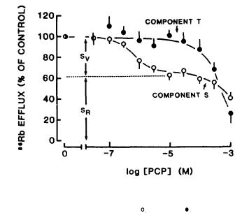
Pharmacological evidence indicates that components R, S, T, and C correspond to distinct classes of K channels (Bartschat and Blaustein 1985a; Bartschat and Blaustein 1985b). Component T is blocked by low concentrations of 4-aminopyridine (4-AP); component C is selectively blocked by micromolar quinine sulfate, but not by 4-AP. Components R and S are much less sensitive to both drugs.
EFFECTS OF PCP ON K CHANNELS IN SYNAPTOSOMES
To determine which of these K channels are inhibited by PCP, this drug was tested for its ability to block the various components of Rb efflux. In the experiment of figure 1, the effects of 10 µM
PCP (closed circles) and 100 µM PCP (closed |
squares) |
were |
examined |
|
in nominally Ca-free solutions. |
In 100 mM |
[K]o medium, 10 µM PCP |
||
d e p r e s s e d 86Rb efflux through S |
by about 35 percent, |
but |
had neg- |
|
ligible effect on component T. Increasing PCP to 100 µM had no additional effect on S, but blocked component T by about 45 percent. Although not shown here, component R and the Ca-dependent component, C, were virtually unchanged by 100 µM PCP.
The dose-response curves for the effects of PCP on components S and T are illustrated in figure 2. Note that low doses of PCP selectively blocked a portion (about one-third) of component S (SV); higher doses inhibited T, as well, with but little additional effect on S.
FIGURE 2. PCP dose-response curves |
for |
the |
inhibition of compo- |
n e n t s S |
( ) |
and |
T ( ) |
NOTE: The data points are the means of 4 determinations, ± SEW. SimiIar results were obtained in 3 other experlments.
40

EFFLUX COMPONENT S CAN BE DIVIDED INTO TWO FRACTIONS, SR AND SV, WITH DIFFERENT PROPERTIES
Before trying to interpret this partial inhibition of S by PCP, we need to consider the influence of membrane potential on the 8 6 Rb efflux. To obtain information about the relationship between component S and membrane potential, we measured S (increase in the slope of 86Rb efflux between 2 and 4 seconds) as a function of [K]o in the efflux solution, in the absence of drug. These data are shown in figure 3 (solid circles); the calculated depolarization, due to increasing [K]o, is given in the upper abscissa scale.
FIGURE 3. |
|
Effect |
of |
PCP |
on |
|
8 6Rb efflux |
component |
S |
|
|
|
|
|
|||||||
NOTE: The |
magnitude |
of |
component |
S |
at |
the indicated |
[K] |
was |
expressed |
as |
the |
|
|||||||||
slops |
of |
the |
least squares |
line |
fitting |
Rb |
efflox°between |
2 |
and |
4 |
seconds |
||||||||||
for |
control (•) |
and |
100 |
µM |
PCP-containing |
solution |
(o). |
|
The |
|
dotted |
line |
Is |
||||||||
the |
expected |
contrlbutlon |
of |
the |
"resting” |
K |
permeablllty |
(R) |
to |
component |
|||||||||||
S due |
to |
electrodiffusion |
effects (i.e., SR) |
when |
the |
synaptosomes |
are |
de- |
|||||||||||||
polarized |
to |
the level lndicated In the upper abscissa |
(also |
see |
Bartschat |
||||||||||||||||
and |
Elsusteln |
1985a). |
The |
data |
points |
are |
the means of |
4 |
determinatlons. |
||||||||||||
As discussed previously (Bartschat and Blaustein 1985a), depolarization of the terminals should increase the driving force for efflux of 86Rb through the "resting" K permeability (component R),
even if |
there is no increase in conductance (i.e., no voltage |
gat- |
||
ing of R). The magnitude of this calculated electrodiffusion |
com- |
|||
ponent |
of 86Rb efflux through the "resting" K conductance, denoted |
|||
as |
SR, |
is shown as the dotted line |
in figure 3 (also see figure 6c |
|
in |
Bartschat and Blaustein 1985a). |
The difference between this |
||
41
dotted line (fraction SR) and the solid line (component S) presumably represents the voltage-regulated fraction of component S, denoted as SV.
THE EFFECT OF PCP ON 86Rb EFFLUX THROUGH COMPONENT S
If two different types of K conductances contribute to component S, we might anticipate that some drugs will affect the two conductances differently. Indeed, the dose-response curves for inhibition of S by tetra-alkylamines and 4-AP appear to be biphasic (Bartschat and Blaustein 1985a), which is consistent with this prediction. The obviously biphasic dose-response curve for PCP inhibition of component S (figure 2, open circles) provides further evidence for this view.
Note that the magnitude of component S measured as a function of [Klo in the presence of 100 µM PCP (figure 3, open circles) corresponds very closely to the calculated magnitude of SR (figure 3, dotted line). These data clearly indicate that components SR and Sv are different. Component SV apparently is a manifestation of voltage-regulated, noninactivating K channels, and is blocked
selectively by low concentrations of PCP. Since the |
"resting" 86Rb |
||
efflux is unaffected by these concentrations of |
PCP |
(not shown), |
|
we would not expect PCP to affect |
86Rb efflux |
component SR if this |
|
flux is mediated by the resting K |
conductance, |
as described. |
|
The concentration of PCP that inhibits SV by 50 percent (IC50) is about 1 µM (figure 2, open circles). This is within the range of doses that produce confusional psychosis (0.1 to 1.0 mg/kg, corresponding to about 0.1 to 1 µM) (Burns and Lerner 1981). Higher concentrations of PCP induce convulsions and coma (Burns and Lerner 1981). Assuming that block of the K channels corresponding to SV does cause the behavioral deficit, it is important to consider the possibility that block of only a small fraction of these K channels may be sufficient to induce behavioral change.
THE EFFECTS OF PCP ANALOGUES ON COMPONENT Sv
If the behavioral activity of PCP is related to its block of presynaptic K channels (Albuquerque et al. 1981; Albuquerque et al. 1983; Blaustein and Ickowicz 1983), PCP-like analogues should block these same K channels with a rank order of potency that parallels their relative in vivo psychotomimetic activity. One of the most behaviorally potent PCP-like agents is TCP (1-[1-(2- thienyl)-cyclohexyl] piperidine) (Shannon 1983). Figure 4 illustrates the dose-response curves for the block of components S and T by this drug. The data indicate that TCP is a more potent blocker of Sv than is PCP (figure 2). TCP blocked component T only at high concentrations (>1O-5 M), and in this respect was approximately equivalent in potency to PCP (figure 2).
42

FIGURE 4. Dose-response curve illustrating the |
effects of |
the PCP |
a n a l o g u e T C P o n c o m p o n e n t s |
( ) and |
T (o) |
NOTE: The experiment was carried out as described in the figure 1 legend. The data points are the means of 6 determinations.
Figure 5 compares the ability of PCP and two of its congeners,
meta-amino-PCP (m-NH2-PCP) and |
meta-nitro-PCP (m-NO2-PCP), |
to |
|
block component S. |
All three drugs selectively blocked Sv, but |
||
with very different |
affinities. |
The rank order of potency |
for |
block of Sv by these three drugs and TCP (figure 4) was: TCP, m- NH2-PCP>PCP>m-NO2-PCP. This same sequence was observed when these agents were examined in various behavioral impairment paradigms that have been used to assess the psychotomimetic liability of these and related agents (Albuquerque et al. 1981; Shannon 1983; Aguayo et al. 1984). In contrast, all of these agents blocked T only at higher concentrations (IC5O's=100-500 µM; data not shown). The similarity between the relative potency of block of Sv by PCPlike drugs and the effects of these drugs in the behavioral impairment paradigms strongly supports the view that blockade of voltage-regulated, noninactivating K channels may be directly involved in the psychotomimetic actions of these drugs.
THE EFFECTS OF "SIGMA OPIATES" ON COMPONENT SV
Several "sigma opiates," which differ structurally from PCP, are known to induce a PCP-like toxic psychosis, and to displace PCP from rat brain membranes (Zukin 1982; Hampton et al. 1982;
43
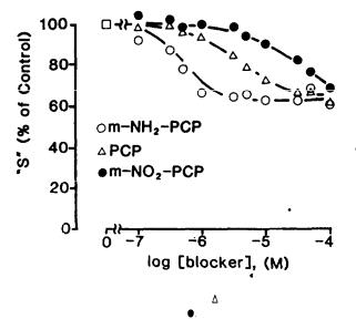
FIGURE 5. |
T h e e f f e c t s |
o f t h e P C P |
( |
), m-NH2-PCP (o), |
and m-NO2- |
|
|
P C P ( |
) |
on component S |
|
NOTE: The |
experiments were carried out as |
described in the figure 1 |
legend. The |
||
data |
points are the |
means of SiX |
determinations. |
|
|
Mendelsohn et al. 1984; Murray and Leid 1984). Of particular interest are several stereoisomer pairs of "sigma opiates," one isomer of which produces PCP-like behavioral effects and displaces bound 3H-PCP more potently than the other. These agents should be especially well suited for a test of the hypothesis mentioned in the preceding section, namely, that induction of the PCP-like behavioral deficit can be correlated with block of SV.
We examined the effects of three such stereoisomer pairs of "sigma opiates" on 86Rb efflux in synaptosomes in the presence of 10 µM naloxone (to avoid classical opiate effects). Our data show (Bartschat et al. 1985) that dexoxadrol blocks Sv at onethousandth the concentration of its stereoisomer, levoxadrol; (+)NANM (N-allyl-normetazocine or SKF-10,047) is tenfold more potent than (-)NANM; and (-)cyclazocine is twoto threefold more potent than (+)cyclarocine. This stereoselectivity for block of SV closely parallels the effects of these stereoisomers in behavioral and binding experiments (table 1). Furthermore, block of Sv occurs at nanoto micromolar concentrations of the more potent "sigma opiates"-- comparable to the concentrations at which PCP and its more potent congeners act.
44

TABLE |
1. |
C o m p a r i s o n o f |
a c t i o n s o f |
s t e r e o i s o m e r p a i r s |
o f " s i g m a |
|
|
|
|
|
o p i a t e s " |
|
|
|
|
|
|
|
|
|
|
|
|
|
|
Relative Potency |
for |
|
|
|
|
Block |
Displacement of |
PCP-Like |
Drug |
Pair |
|
of SV |
Bound 3H-PCP |
Behavior |
|
|
|
|
|
|
||
Dexoxadrol: |
|
|
|
>1,OOO:1a |
||
Levoxadrol |
~1,OOO:1 |
418: 1 |
||||
(+)NANM:(-)NANM |
|
~1O:1 |
6.4:1 |
3:1b |
||
(-)Cyclazocine: |
|
|
|
5:1C |
||
(+)Cyclazocine |
1-2:1 |
1.8:1 |
||||
|
|
|
|
|
||
SOURCES: |
aCone et al. 1984; |
bShannon 1982a; |
cShannon 1982b. |
|
||
IDENTIFICATION OF THE RAT BRAIN PCP RECEPTOR
The aforementioned results are consistent with the view that the rat brain PCP/"sigma opiate" high-affinity receptor is associated with the voltage-regulated, noninactivating K channels in the presynaptic terminals. Thus, we reasoned that the elucidation of the molecular composition of this PCP "receptor" might provide direct information about the subunit composition of these K channels. This could also prove to be a very useful first step in the effort to purify and characterize the channel protein. To label and identify the brain PCP receptor, we synthesized a tritiated photo affinity analogue of PCP, m-azido-3H-phencyclidine (3H-Az-PCP) (Haring and Kloog 1984). This photolabile ligand binds with high
affinity (K0.5 |
0.9 |
µM) to the rat brain PCP receptor (Sorensen |
and Blaustein |
1984) |
As illustrated by the fluorogram in figure |
6, when rat brain sinaptic membranes were incubated with 3H-Az-PCP and irradiated, several polypeptides incorporated the label. However, when excess unlabelled PCP was added before the samples were irradiated, incorporation of the label was markedly reduced in only two of the polypeptides, MR=80 kD (80,000 Daltons; P80) and 95 kD (P95). respectively. P95 was labelled more heavily than P80, and may, therefore, include the primary PCP binding site; however, we have not yet ruled out the possibility that P80 is a product of proteolytic cleavage (Sorensen and Blaustein 1985b).
Covalent labelling of these two polypeptides was also specifically blocked by other PCP analogues such as TCP, by some K channel
blockers |
(4-AP |
and |
tetrabutylammonium ions), and stereoselectively |
by certain PCP-like "sigma opiates" (dexoxadrol >> levoxadrol) |
|||
(not shown). |
The latter results parallel the ability of these |
||
ligands |
to displace |
3H-PCP from rat brain membranes and to block |
|
86Rb efflux component Sv in synaptosomes (Sorensen and Blaustein 1985a; Bartschat et al. 1985).
45

FIGURE 6. |
Fluorogram |
showing the |
covalent |
attachment of |
3 H-Az-PCP |
||||||||||||||||||||
|
|
|
t o r a t b r a i n s y n a p t i c m e m b r a n e p o l y p e p t i d e s |
|
|
||||||||||||||||||||
NOTE: Synaptic membranes |
were |
incubated |
in |
the |
|
dark |
for |
60 |
minutes at 0 °C in |
|
|||||||||||||||
either |
54 |
mM3Tris-HEPES (A) or 5 mM |
sodium |
phosphate |
(B) buffers at pH 7.0, |
||||||||||||||||||||
with 1.0 µM H-Az-PCP, without (-) or with (+) a five |
hundredfold excess |
of |
|||||||||||||||||||||||
unlabelled |
PCP. |
The |
samples |
were then |
Irradiated |
with |
366 |
nm |
UV |
light |
for |
||||||||||||||
15 |
minutes |
(irradiation at 254 nm UV light for 5 minutes |
gives |
similar |
re- |
||||||||||||||||||||
sults) |
to |
pnotolyze |
the |
Az-PCP. |
The membranes were |
solubilized |
in |
sodium |
|||||||||||||||||
dodecyl sulfate (SDS) dissociation buffer overnight. and subjected to SDS- |
|||||||||||||||||||||||||
polyacrylamide |
gel |
|
electrophoresis. |
The |
resulting |
geIs |
were |
|
impregnated |
||||||||||||||||
with |
“Fluore-Hance” |
(RPI, |
Mt. |
Prospect, |
III.) |
dried, |
and |
exposed |
to |
Kodak |
|||||||||||||||
X-Omat AR film for several weeks to obtain fluorographic |
|
patterns |
of |
the |
|||||||||||||||||||||
labelled polypeptides. Several polypeptldes that incorporate label are |
|
||||||||||||||||||||||||
lndlcated |
as PN |
where |
N |
is |
defined |
as |
the |
apparent |
molecular |
weight |
(kD) of |
||||||||||||||
the |
polypeptlde |
x |
10-3. |
|
Molecular |
weight |
standards |
are |
|
also |
Indicated. |
|
|||||||||||||
The acetylcholine receptor (AChR) of Torpedo electric organ is also a PCP "receptor." However, this nicotinic AChR has about one-tenth the affinity for PCP than that of the rat brain PCP re-
ceptor [K0.5  0.3 µM , versus
  4-6 µM for Torpedo (Heidmann et al. 1983; flaring et al. 1984)]. Moreover, the nicotinic AChR has
subunits of MR<66 kD, and these are the subunits that are specifically labelled with 3H-AZ-PCP in the Torpedo electroplax membranes (Heidmann et al. 1983; Haring and Kloog 1984; Haring et al. 1984). These data indicate that the nicotinic AChR-PCP receptor differs from the rat brain PCP receptor. Furthermore, our findings are
46
consistent with the view that the rat brain PCP receptor is the voltage-regulated, noninactivating K channel in the nerve terminals, and that this channel consists of at least two subunits of MR=80 kD and 95 kD.
HOW DOES PCP PRODUCE ITS BEHAVIORAL EFFECTS?
The striking correlation between behavioral potency and block of 86Rb efflux component SV, for PCP analogues and the stereoisomer pairs of "sigma opiates," provides strong circumstantial evidence that the K channel block may underlie the behavioral effects of these drugs. Block of K channels at the nerve tetmi-
nals should prolong the nerve action potential and thereby enhance Ca entry into the terminals. This would in turn, alter synaptic transmission by increasing Ca-dependent neurotransmitter release; virtually all neurotransmitter types might be affected, depending upon the distribution in the brain of these PCP-sensitive, volt- age-regulated K channels. Such disruption of synaptic transmission at central synapses could induce the disordered behavior that is characteristic of PCP intoxication. Thus, our observations may provide the physiological link between the binding of PCP to its high-affinity receptor in the brain and the ultimate behavioral effects of PCP intoxication.
It should be pointed out that PCP analogues also have a prominent postsynaptic action: they block the channels associated with the nicotinic cholinergic receptors (Albuquerque et al. 1981; Albuquerque et al. 1983). However, many of the analogues that display potent antinicotinic activity are behaviorally inactive (Albuquerque et al. 1983), whereas our data demonstrate that there is a direct relationship between block of certain presynaptic K channels and behavioral potency; the stereoselective effects of the "sigma opiates" are particularly striking in this regard
(table |
1). |
This supports the view that presynaptic K channels are |
the primary sites of action of these drugs in the brain. More- |
||
over, |
our data indicate that the voltage-regulated, noninactivat- |
|
ing K channels may be the high-affinity PCP binding sites, and |
||
that PCP may |
be a useful ligand to help identify and isolate these |
|
K channels. |
|
|
The behavioral effects of PCP have been associated with excessive release of a wide variety of neurotransmitters: in particular, a massive dopamine release may underlie some of the most prominent symptoms of PCP intoxication (Rappolt et al. 1980). Our results readily explain the genesis of such an effect, because activation of presynaptic K channels is one of the primary factors that influences Ca entry into nerve terminals and Ca-dependent transmitter release by limiting action-potential duration and regulating excitability.
47