

Drug Targeting Organ-Specific Strategies
.pdfReferences 155
[145]Van Zwieten PA, Kam KL, Pijl AJ, Hendriks MG, Beenen OH, Pfaffendorf M, Pharmacol. Res. 1996, 33, 95–105.
[146]Velasquez MT, Kimmel PL, Michaelis OE, FASEB J. 1990, 4, 2850–2859.
[147]Wapstra FH, Van Goor H, Navis G, De Jong PE, de Zeeuw D, Clin. Sci. 1996, 90, 393–401.
[148]Marinides GN, Groggel GC, Cohen AH, Border WA, Kidney Int. 1990, 37, 749–757.
[149]Rollason TP, Brewer DB, J. Pathol. 1981, 134, 39–56.
[150]Kluth DC, Rees AJ, J. Nephrol. 1999, 12, 66–75.
[151]Bagchus WM, Hoedemaeker PJ, Rozing J, Bakker WW, Lab. Invest. 1986, 55, 680–687.
[152]Jefferson JA, Johnson RJ, J. Nephrol. 1999, 12, 297–307.
[153]Wilson CB, Kidney Int. 1989, 35, 938–953.
[154]Jungquiera LC, Carneiro J, Coutopoulos AN, In: Basic Histology, pp. 354–371. Lange Medical Publications, Los Altos, 1975.
Drug Targeting Organ-Specific Strategies. Edited by G. Molema, D. K. F. Meijer Copyright © 2001 Wiley-VCH Verlag GmbH ISBNs: 3-527-29989-0 (Hardcover); 3-527-60006-X (Electronic)
6A Practical Approach in the Design of Colon-specific Drug Delivery Systems
Claudia S. Leopold
6.1 Introduction
Drug delivery to the large intestine has become attractive to researchers whose main interest is the treatment of colonic disorders and the delivery of peptide drugs to the colon. In contrast to colon-specific drug delivery, drug delivery to the small intestine can be easily achieved by using enteric coating polymers that are soluble in the neutral environment of the small intestine. The development and the design of colon-specific drug formulations represents a technological challenge as these dosage forms must pass through the upper gastrointestinal (GI) tract in intact form before delivering the drug to the colon.
Colon-specific drug delivery does not appear to make much sense at first because of the small area of absorption and the strong barrier properties of the colonic epithelium. However, the colon has some unique features, which make this organ attractive for site-specific drug delivery. On the one hand, the peptidase activity in the large intestine is significantly lower than that in the stomach and the small intestine and the colonic transit time is much longer than that of the upper GI tract. This allows the delivery of unstable peptide drugs and drugs with a low permeability to this lower intestinal region. On the other hand, the topical treatment of colonic disorders may lead to the reduction of both drug dose and side effects.
There are currently four strategies that are pursued to achieve colon specificity: first, by relying on the pH difference between the small and the large intestine (pH-controlled drug release); second, by exploiting the enzymatic activity of the colonic microflora (enzyme-con- trolled drug release); third, by relying on the relatively constant small intestinal transit time (time-controlled drug release) and fourth, by taking advantage of the increase in the luminal pressure in the colon due to strong peristaltic waves (pressure-controlled drug release). This chapter gives an overview of the delivery concepts in colon-specific drug delivery which are currently employed (see also Table 6.1).
6.2 Physiological Characteristics of the Colon
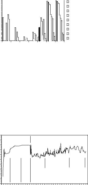
Drug delivery to the colon has become attractive to researchers interested in the delivery of peptide drugs to the large intestine and the topical treatment of colonic disorders. Because of the unique physiological characteristics of the large intestine, drug delivery to the colon can be achieved in different ways. One such feature is the colonic microflora (bacterial count: 1011–1012 cfu ml–1), which consist mainly of anaerobic or facultative anaerobic microorganisms [1] (Figure 6.1) that produce a variety of enzymes [2]. The ability of the colon to support

158 6 A Practical Approach in the Design of Colon-specific Drug Delivery Systems
Table 6.1. Overview of potential possibilities for achieving drug delivery to the colon.
Delivery method |
Principle |
Achieved with |
Examples |
|
|
|
|
pH-controlled |
Difference in pH between |
pH-dependent dissolution |
Enteric coatings, basic |
drug release |
the small and large |
of polymeric coatings |
polymers |
|
intestine |
|
|
|
|
pH-dependent |
Acrylic polymers |
|
|
polymer swelling |
|
|
|
of hydrogels |
|
|
|
pH-dependent drug |
Insulin + gelatin B |
|
|
release from drug/ion |
Olsalazine + anion |
|
|
exchange resin complexes |
exchange resin |
Enzyme-controlled |
Degradation of dosage |
Degradable pro-drugs |
Mono-, oligoor polymers |
drug release |
form components by the |
|
with degradable drug– |
|
enzymes of the colonic |
|
carrier bonds |
|
microflora |
|
|
|
|
Coating materials with deAzo polymers, polymers |
|
|
|
gradable bonds including |
with glycosidic bonds |
|
|
capsule shells |
|
|
|
Hydrogels and matrices |
Cross-linked guar, pectin, |
|
|
consisting of cross-linked, |
dextran, inulin, azo |
|
|
degradable polymers |
polymers |
|
|
Sustained release coating |
Ethylcellulose or Eudra- |
|
|
materials with degradable |
git® RS with galactoman- |
|
|
domains (pore formers) |
nans, β-cyclodextrin, |
|
|
|
glassy amylose, inulin |
Time-controlled |
Relatively constant transit |
Time-dependent |
Cellulose ethers, |
drug release |
time in the small intestine |
swellable polymers |
Eudragit® sustained- |
|
of about 3 h |
|
release coatings |
|
|
Slow build-up of an |
COER-24TM |
|
|
osmotic pressure in the |
|
|
|
dosage form |
|
|
|
Polymer layers with time- |
|
|
dependent erosion or |
|
|
dissolution |
Pressure-controlled |
Disintegration of the |
Thick coating consisting |
drug release |
dosage form in the colon |
of water-insoluble, non- |
|
by intra-luminal pressure |
swellable polymers |
|
resulting from strong |
|
|
peristaltic waves |
|
Cellulose ethers; Eudragit® E, chitosan (in combination with an acid in the dosage form)
Hard gelatin capsule with inner ethylcellulose coating
anaerobic bacterial flora is shown by its redox potential in the range of – 250 and – 480 mV [3,4]. Further characteristics are the slightly acidic environment in the proximal colon (pH 6.0–6.4), which results from the degradation of polyand oligosaccharides to short-chain fatty acids, and a neutral or slightly alkaline environment in the distal colon (pH 7.0–7.4) [5] (Figure 6.2). Moreover, as a result of the strong and prolonged propulsive motility in the distal colon that occurs once or twice a day, the luminal pressure and thus the potentially destructive forces increase temporarily in this lower part of the large intestine, where solid faeces are formed [6]. Drug absorption from the colon is affected by the small effective surface area available for absorption and the tightly packed colon epithelium; however, colonic tran-
6.3 Pathological Processes in the Colon |
159 |
log viable count / g content
11 |
|
|
|
|
|
|
|
|
Bacteroides |
|
|
|
|
|
|
|
|
||
10 |
|
|
|
|
|
|
|
|
Eubacteria |
|
|
|
|
|
|
|
|
||
9 |
|
|
|
|
|
|
|
|
Anaerobic streptococci |
|
|
|
|
|
|
|
|
||
8 |
|
|
|
|
|
|
|
|
|
|
|
|
|
|
|
|
|
Bifidobacteria |
|
|
|
|
|
|
|
|
|
||
7 |
|
|
|
|
|
|
|
|
|
|
|
|
|
|
|
|
|
Enterococci |
|
|
|
|
|
|
|
|
|
||
6 |
|
|
|
|
|
|
|
|
|
|
|
|
|
|
|
|
|
Coliform bacteria |
|
|
|
|
|
|
|
|
|
||
5 |
|
|
|
|
|
|
|
|
|
|
|
|
|
|
|
|
|
Lactobacilli |
|
|
|
|
|
|
|
|
|
||
|
|
|
|
|
|
|
|
|
4 
Veillonellae
3 
Clostridium perfringens
2 

oral cavity |
stomach |
duodenum |
jejunum |
ileum |
cecum |
rectum |
Figure 6.1. Bacterial flora of the human GI tract. Modified from reference [4].
sit time can last for up to 78 h, thereby allowing the absorption of drugs of even low permeability such as peptides.
6.3 Pathological Processes in the Colon
Ulcerative or inflammatory lesions may affect the physiology of the small and large intestine. Ulcer formation entails a circumscribed loss of tissue from the surface of an organ, which results from necrosis following cell destruction by chemicals and the like, or by restriction of the blood supply. Ulcers are among the most common and important lesions. Those that do not penetrate the muscularis mucosa are called erosions. Ulcerative conditions in humans must be differentiated from malignant ulcers, which are associated with neoplasia. Among
|
10 |
|
|
|
|
|
|
|
|
|
|
|
9 |
small intestine |
|
|
large intestine |
|
|
|
|||
|
8 |
|
|
|
|
|
|
|
|
|
|
|
7 |
|
|
|
|
|
|
|
|
|
|
|
6 |
proximal small intestine |
|
|
|
|
|
|
|
|
|
pH |
5 |
mid small intestine |
distal small intestine |
right |
|
mid |
|
|
left |
|
|
4 |
|
|
|
|
|||||||
colon |
|
colon |
|
|
colon |
|
|||||
3 |
|
|
|
|
|
|
|
||||
2 |
|
|
|
|
|
|
|
||||
1 |
|
|
|
|
|
|
|
||||
0 |
|
|
|
|
|
|
|
||||
|
|
|
|
|
|
|
|
|
|
|
|
|
0 |
2 |
4 |
6 |
8 |
10 |
12 |
14 |
16 |
18 |
20 |
time after ingestion of a radiotelemetry capsule [h]
Figure 6.2. pH profile in the GI tract of a healthy subject, measured with a radiotelemetry capsule. Modified from reference [5].
160 6 A Practical Approach in the Design of Colon-specific Drug Delivery Systems
the inflammatory bowel diseases of humans are regional enteritis, or Crohn’s disease, and chronic ulcerative colitis [7]. These diseases are primarily treated with mesalazine, various corticosteroids and immunosuppressants.
Crohn’s disease is granulomatous and in most cases it is a simultaneous disease of the ileum and colon.The primarily inflamed region is the distal ileum, and all intestinal layers are thickened. The mucosal surface is reddened, nodular, and cobblestone-like, with multiple linear ulcerations.The mucosal layer is thickened by inflammatory infiltrate, the submucosa and serosa by fibrosis, and the serosa by hypertrophy. Chronic ulcerative colitis is a systemic disease that starts at the rectum or the sigmoid colon and progresses proximally to involve the entire left side of the colon.The colonic crypts are the first sites of cell damage and death, and the disease primarily involves the mucosal layer of the intestine.
The aetiopathogenesis of inflammatory bowel disease is not yet known. Most authors agree that immunologic abnormalities in the local mucosa-associated immune system are of major importance. Under normal conditions this gut-associated immune system has to protect the host against invasion of potential pathogens or an inappropriate immune response to luminal antigens. Lymphocytes within the mucosal immune system differ in many respects from lymphocytes in other areas of the body.There are indications that the tissue-specific differentiation of mucosal T cells is disturbed in inflammatory bowel disease. An imbalance between helper and suppressor mechanisms in the intestinal mucosa could result in a sustained and overshooting inflammatory and immune reaction against antigens normally occurring in the intestinal lumen [8] (see Chapter 7 for a more detailed discussion on the pathophysiological processes in inflammatory bowel diseases). Further disease states of the large intestine that might be treated with colon-specific dosage forms in the future are diarrhoea, tropical sprue, coeliac disease, irritable bowel syndrome, and different types of cancer [7].
In those instances where a disease of the colon is to be treated locally through the use of a delivery system, testing in the appropriate animal model is extremely important. For example, the delivery of anti-inflammatory agents to the colon for treatment of inflammatory bowel disease must be evaluated in suitable animal models. A number of animal models for intestinal inflammation are available for the testing of colonic delivery systems. The methods employed include lymphatic obstruction, vascular changes, and neurogenic manipulation [9–11]. Intestinal inflammation in animals such as rodents may be produced by topical application or administration of irritant chemicals such as acetic acid, trinitrobenzenesulfonic acid, difluoromethyl ornithine, pepsin inhibitors, or degraded carrageenan [9,11]. Colon cancer may be induced by administration of carcinogens such as chanthrenes, aromatic amines, hydrazine derivatives, alkylnitrosamides, and aflatoxin [12]. In the future, transgenic animals will play an important role as models for various disease states.
6.4 Approaches to Colon-specific Drug Delivery
Four strategies are currently being pursued to achieve drug release specifically in the colon.
•The fact that the luminal pH of the healthy distal colon is slightly higher than that of the proximal small intestine has led to the development of oral dosage forms that are intended to release the drug at the colonic pH (pH-controlled drug release).
6.4 Approaches to Colon-specific Drug Delivery |
161 |
•Colonic microflora produce a variety of enzymes that are not present in the stomach or the small intestine and can therefore be used to deliver drugs to the colon after enzymatic cleavage of degradable formulation components or drug carrier bonds (enzyme-con- trolled drug release). It should be taken into consideration that because of the negative redox potential in the colon, enzymatic or chemical reduction reactions are favoured.
•The relatively constant transit time in the small intestine of approximately 3–4 h is another physiological characteristic which can be exploited to achieve colon specificity (timecontrolled drug release). After gastric emptying, a time-controlled drug delivery system is intended to release the drug after a predetermined lag phase.
•Another strategy relies on the strong peristaltic waves in the colon that lead to a temporarily increased luminal pressure (pressure-controlled drug release). Pressure-sensitive drug formulations release the drug as soon as a certain pressure limit is attained, i.e. destruction force is exceeded.
Using mostly anti-inflammatory model drugs or drugs that are absorbable in the colon, many colon-specific dosage forms have been developed in the past, including pro-drugs, cross-linked hydrogels, matrices and coated dosage forms. However, whereas the synthesis of pro-drugs is possible only if the drug has suitable functional groups that can be bound to a carrier molecule, biodegradable hydrogels and matrices are problematic insofar as polymer degradation rates and thus drug release are often too slow. Most colon-specific drug delivery systems belong to the group of coated dosage forms because of the flexibility in the design of the latter and the improved coating procedures that have been developed in the past.
With regard to peptide and protein absorption poor membrane permeability, enzymatic instability, and large molecular size are three factors that have remained major hurdles for peptide formulations. Absorption-enhancing agents that have been effective, at least in research environments with smaller drug candidates, have also shown some limited efficacy in small animal models with certain peptides. In most cases, however, effective formulations have only achieved fairly low peptide absorption (< 10%) and have also resulted in significant alterations in the normal cellular morphology of the gastrointestinal tract, at least on a transient basis [13]. Current data suggest that the successful development of oral peptide formulations remains a significant challenge.
6.4.1 pH-Controlled Drug Release
Many of the marketed dosage forms developed for colon-specific drug delivery, such as the enteric coated formulations Asacolitin®, Azulfidine®, Claversal®, Salofalk®, Colo-Pleon®, Entocort® and Budenofalk® rely on the physiological pH difference between the small and the distal large intestine. In healthy subjects this pH difference amounts to about 0.5 pH units [4,5] (Figure 6.2). However, it has been shown that this difference in pH between the small and the large intestine is too small to guarantee reliable drug release in the colonic region [14–16]. Moreover, in patients with inflammatory bowel disease the luminal colonic pH drops to values between 2.5 and 4.7 [17–19], a fact that has been attributed to a failure of bicarbonate secretion rather than excessive bacterial fermentation [18].
Enteric coating materials not only protect a dosage form from the acidic environment in the stomach and allow drug delivery to the small intestine, they may also pass through the
162 6 A Practical Approach in the Design of Colon-specific Drug Delivery Systems
small intestine and dissolve only in the colon, depending on their dissolution pH and the thickness of the coating applied. Many of the oral drug preparations for the treatment of inflammatory bowel disease available on the market (e.g. Asacolitin®, Claversal®, Salofalk®) are coated with enteric polymers such as Eudragit® L or S, i.e. methacrylic acid copolymers with different degrees of substitution, which show pH-dependent dissolution behaviour. These polymers are supposed to induce drug release as soon as the luminal pH in the GI tract exceeds values of 6 or 7. However, studies with Eudragit® S-coated tablets in healthy subjects have shown, that drug release in the colon is not sufficiently reproducible [14,15]. Other studies verify the reliability of this delivery method. One reason for these inconsistent results is the decrease in the luminal pH after passage through the ileocaecal valve as a result of bacterial fermentation of non-absorbable oligoand polysaccharides to short chain fatty acids. Only in the distal colon is a luminal pH of 7 attained, which differs only slightly from the average pH of the small intestine (6.5–6.8).
The OROS-CT™ delivery system (Oral Osmotic System for Colon Targeting) is an enteric formulation consisting of one drug compartment containing osmotically active excipients and a second compartment containing a swelling polymer (Figure 6.3). Both compartments are coated with a semi-permeable membrane and an outer enteric coating. After dissolution of the enteric coating the swelling polymer slowly pushes the liquid content of the osmotic compartment out of the micropore as a result of water penetration. This leads to combined pH-controlled and sustained drug release.
During an acute attack of inflammatory bowel disease the luminal pH of the large intestine which is normally 6.4–7.0, drops to values between 2.3 and 4.7 [17–19], which means that enteric coatings are unsuitable coating materials in this particular case. Coating materials that dissolve at a low pH or are degradable in an acidic environment may be used in such a case.Therefore, the basic polymers Eudragit® E, an aminoalkyl methacrylate copolymer, and polyvinylacetal diethylaminoacetate (AEA™) have been investigated in vitro as coating materials for oral dosage forms designed for the treatment of inflammatory bowel disease with dexamethasone as the model corticosteroid [20,21]. An in vivo study is planned.
OROS-CT ™ |
|
COER-24™ |
||
Micropore |
Drug + |
Micropore |
||
Osmotic agent |
||||
|
Polymer delay coat |
Drug compartment |
||
|
|
|||
|
|
|
||
|
|
|
|
|
|
|
|
|
|
|
|
|
|
|
|
|
|
|
|
|
|
|
|
|
|
|
|
|
|
|
|
|
|
|
|
|
|
|
|
|
|
|
|
|
|
|
|
|
|
|
|
|
|
|
|
|
|
|
|
|
|
|
|
|
|
|
|
|
|
|
|
|
|
|
|
|
|
|
|
|
|
|
|
|
|
|
|
|
|
|
|
|
|
|
|
|
|
|
|
|
|
|
|
|
|
|
|
|
|
|
|
|
|
|
|
|
|
|
|
|
|
|
|
|
|
|
|
|
|
|
|
|
|
|
|
|
|
|
|
|
|
|
|
|
|
|
|
|
|
|
|
|
|
|
|
|
|
|
|
|
|
|
|
|
|
|
|
|
|
|
|
|
|
|
|
|
|
|
|
|
|
|
|
|
|
|
|
|
|
|
|
|
|
|
|
|
|
|
|
|
|
|
|
|
|
|
|
|
|
|
|
|
|
|
|
|
|
|
|
|
|
|
|
|
|
|
|
|
|
|
|
|
|
|
|
|
|
|
|
|
|
|
|
|
|
|
|
|
|
|
|
|
|
|
|
|
|
|
|
|
|
|
|
|
|
|
|
|
|
|
|
|
|
|
|
|
|
|
|
|
|
|
|
|
|
|
|
|
|
|
|
|
|
|
|
|
|
|
|
|
|
|
|
|
|
|
|
|
|
|
|
|
|
|
|
|
|
|
|
|
|
|
|
|
|
|
|
|
|
Swelling agent |
Osmotic compartment |
||||||||||||||||||||||||||||||||||||||
Semipermeable |
|
Enteric coating |
|
|
|
|
|
|
|
|
|
Semipermeable membrane |
|||||||||||||||||||||||||||||||||||||||||||||||||||||||||||||
|
|
|
|
|
|
|
|
|
|
|
|
|
|
|
|
|
|
|
|
|
|
|
|
|
|
|
|
|
|
|
|
|
|
|
|
|
|
||||||||||||||||||||||||||||||||||||
membrane |
|
|
|
|
|
|
|
|
|
|
|
|
|
|
|
|
|
|
|
|
|
|
|
|
|
|
|
|
|
|
|
|
|
|
|
|
|
|
|||||||||||||||||||||||||||||||||||
|
|
|
|
|
|
|
|
|
|
|
|
|
|
|
|
|
|
|
|
|
|
|
|
|
|
|
|
|
|
|
|
|
|
|
|
|
|
|
|
|
|
|
|
|
|
|
|
|
|
|
|
|
|
|
|||||||||||||||||||
Figure 6.3. OROS-CTTM (Oral Osmotic System for Colon Targeting) and COER-24TM (Controlled Onset Extended Release) delivery systems.
6.4 Approaches to Colon-specific Drug Delivery |
163 |
pH-sensitive ion exchange systems represent another approach to how to achieve pH-con- trolled drug release in the colon. Drug ions, bound to an ion exchange resin, may be released pH-dependently into the large intestine, as in the case of insulin [22] or mesalazine [23]. In the latter case the drug is used in the form of its ionized pro-drug (olsalazine) and drug release occurs after microbial cleavage of the drug–carrier bonds. The ion exchange resin serves as polymer to prevent premature absorption of the pro-drug in the small intestine.
6.4.2 Enzyme-controlled Drug Release
Enzyme-controlled drug release takes advantage of the existence of enzyme-producing microorganisms in the colon (Figure 6.1). The colonic microflora produce a variety of enzymes, the activity of which may be exploited for colon-specific drug delivery.Among these enzymes are the azoreductase, various glycosidases, esterases and peptidases. Because of this physiological characteristic of the colon, biodegradable pro-drugs, coating materials, hydrogels and matrices have been developed (see below).
Pro-drugs are conjugates of drugs with carrier molecules mostly of inert nature. The microbial enzymes in the colon are responsible for the cleavage of the drug–carrier bond. A variety of pro-drugs have been synthesized, mainly azo compounds, glycosides, esters and amides [24].
Pro-drugs must not be cleaved by digestive enzymes of the upper GI tract and should not be susceptible to chemical hydrolysis. Moreover, pro-drug absorption in the small intestine should be negligible. Because of these requirements, the hydrophilicity, the molecular weight and the charge of the carrier molecules have to be regarded as critical parameters.
The azo pro-drug sulfasalazine (Azulfidine®, Colo-Pleon®), consisting of the drug mesalazine and the carrier molecule sulfapyridine, was the first pro-drug available for the treatment of inflammatory bowel disease. Because of side-effects caused by the pharmacologically active sulfapyridine carrier, other carrier molecules such as sulfanilic acid, p- aminobenzoic acid and its amino acid derivatives (benzalazine; ipsalazide; balsalazide, Colazide®) and mesalazine itself (olsalazine, Dipentum®) have been used in its place. In the case of glycoside pro-drugs, which were developed primarily for use with corticosteroids, monosaccharides have been intensively investigated as inert carrier molecules [25,26]. In addition, a variety of inert hydrophilic carriers of oligomeric as well as polymeric nature, some of them being enzymatically degradable themselves, have been used to prevent premature pro-drug absorption in the small intestine. Examples are β-cyclodextrin [27,28], dextran [29–34] and the polyanionic poly(L-aspartic acid) [35,36].
The Drug Delivery Index (DDI) allows a quantification of the reduction in the drug dose and the systemic exposure observed after drug release specifically to the colon [37]. It may be calculated using AUC (Area Under the plasma drug concentration–time Curve) data or drug concentrations in blood and colonic tissues under steady-state conditions:
|
AUC Tissue (Pro-drug) |
|
|
AUC Tissue (Drug) |
|
DDI (Pro-drug vs. Drug) = |
|
(6.1a) |
|
||
|
AUC Blood (Pro-drug) |
|
|
AUC Blood (Drug) |
|
164 6 A Practical Approach in the Design of Colon-specific Drug Delivery Systems
|
Css Tissue (Pro-drug) |
|
|
Css Tissue (Drug) |
|
DDI (Pro-drug vs. Drug) = |
|
(6.1b) |
|
||
|
Css Blood (Pro-drug) |
|
|
Css Blood (Drug) |
|
where Css is the steady-state drug concentration
The numerator of Eq. 6.1a describes to what extent drug concentrations are increased in the target tissue (colonic mucosa or muscle tissue) after pro-drug administration as opposed to drug administration. It may be regarded as the factor by which the pro-drug dose could be reduced in comparison to the drug dose. The denominator of Eq. 6.1a, which corresponds to the relative bioavailability of the drug released from the pro-drug, provides a measure of the reduction in the systemic exposure and thus the decrease of the systemic toxicity. In Eq. 6.1b the DDI is defined as the quotient of the tissue to blood concentration ratios after pro-drug and drug administration. With glucoside, glucuronide and dextran pro-drugs DDI values up to 9.7 have been reported in rats [25,38,39].
A disadvantage of the use of pro-drugs is the need for suitable functional groups such as amino-, hydroxyor carboxy groups in both drug and carrier molecules (see Chapter 11 for more details on chemical synthesis routes applied in drug targeting/delivery strategies). Sometimes spacer molecules are necessary to link the drug to the carrier molecule, which often leads to complicated drug release kinetics. Another disadvantage of the pro-drug approach is the fact that for the approval of any newly synthesized pro-drug a toxicity study is required by regulatory agencies.
The group of enzymatically degradable coating materials comprises film-forming azo polymers and polymers with glycosidic bonds as well as conventional sustained release coating materials based on acrylates or ethylcellulose with biodegradable pore formers. The films must not be soluble or degradable in the upper GI tract. They should only be degradable in the colon and their degradation products need to be toxicologically harmless.
Cross-linked azo polymers were the first coating materials that were investigated with regard to their enzymatic degradability in the colon using insulin as the model drug [40,41]. A problem observed with these polymers was the rather slow degradation rate, probably a result of the lipophilicity of these compounds. Sufficiently hydrophilic linear azo polymers and pH-sensitive terpolymers based on acrylates were found to be more suitable coating materials [42–45].
In general, several problems have to be considered if azo polymers are used as an enzymatically degradable component of a colon-specific dosage form [46]. As a result of the enzymatic reduction to primary aromatic amines in the colon, the toxicity of these polymers is a critical issue. Moreover, the rate of microbial reduction of the polymeric azo compounds is often too slow. The reduction reaction may stop at the level of the hydrazo compounds instead of leading to the amines, which may influence the drug release mechanism and rate. In addition, the reduction reaction does not necessarily depend on the presence of the azoreductase, but may be induced by the negative redox potential in the colon. The latter also applies to the degradation of pro-drugs or polymers with disulfide bonds, which is the result of a chemical reduction step with no enzymatic reaction involved [47].
6.4 Approaches to Colon-specific Drug Delivery |
165 |
Biodegradable polymer coatings with glycosidic bonds, that have been developed in the past, are mainly based on galactomannans [48–50], chitosan [51] or the high molecular weight dextran fatty acid ester lauroyl dextran [52]. A special enzyme-controlled delivery system with pH-induced drug release has been developed by Watanabe et al. [53]. Drug release from this enteric tablet formulation begins after microbial degradation of an outer disaccharide layer (lactulose) to short-chain fatty acids in the colon and subsequent dissolution of the underlying basic Eudragit® E film. The colon-specific dosage form is available on the market as Codes™ and can incorporate any drug suitable for colonic drug delivery.
Degradable matrix films, consisting of a sustained release coating material and a poorly water-soluble but degradable pore former, are used if the pore former itself does not form an homogeneous coating film. These pore formers guarantee drug release in the colon after microbial degradation and the formation of pores in the film. As pore formers several oligoand polysaccharides were investigated such as β-cyclodextrin [54], galactomannans [55], glassy amylose [56,57], pectin [58–60] and inulin [61]. A coated dosage form for colon-specif- ic drug delivery with a matrix film consisting of ethylcellulose and the pore former glassy amylose is available on the market as Colal™.
If a biodegradable polymer does not exhibit satisfactory film-forming properties, it may also be used as a compression coat requiring a compaction process onto a drug-containing core [62–64].
Further examples of enzymatically degradable ‘drug formulation wrappings’ are capsule shells made of the polysaccharides chitosan [65,66] or cross-linked dextran [67].
Biodegradable polymers that cannot be used as coating materials for colon-specific drug delivery because they are readily water soluble and/or do not form films, may be used in the form of hydrogels and matrices. Hydrogels, consisting of cross-linked polymers, have been developed based on acrylates, polyether-esters and polysaccharides. In the case of acrylateand polyether-ester-copolymers, azo-aromatic compounds have been used for cross-linking purposes and to guarantee degradability in the colon [68–70]. The higher the cross-linking density of these polymers, the lower their tendency to swell and the slower the degradation rate by azoreductase, ultimately resulting in slower drug release. Long chain azo-aromatic crosslinking compounds lead to faster polymer degradation and thus higher drug release rates. In general, the azo-aromatic cross-linking compounds that are used should have a rather small negative redox potential in order to ensure rapid degradation [71].
In the design of coatings, hydrogels and matrices based on azo polymers, the number of azo bonds in the polymers should not be too high, as this could lead to enzyme-saturated conditions slowing down the degradation process and thus drug release [72].
Polymer cross-linking leads to a decrease in the water solubility of many readily soluble polysaccharides, low water solubility being a requirement for colon-specific drug delivery. Dextrans [73,74], the mucopolysaccharide chondroitin sulfate [75,76], guar gum [77,78], pectin [79–82] and inulin [83–85] have all been investigated in cross-linked forms.Again, with a higher degree of cross-linking, the swelling properties of these polymers tend to be lower and this leads to a slower degradation rate and thus slower release of the drug. Poorly watersoluble drugs are usually released by an erosion-type mechanism.