

History of Modern Biotechnology II
.pdfThe Morphology of Filamentous Fungi |
29 |
the genetics of morphology where many enzymes, and their corresponding genes, are involved (see the introduction to Sects. 4 and 4.3.1). What is wanted is a kind of Holy Grail: a mould with the morphology of yeast and without loss of inherent production capacity.
The third reason is a screening problem. Mass screening for a better productivity is relatively easy, there are many assays and hundreds of thousands of new, mutated strains can be produced and tested per year for any given product. However, no assays that perform mass screening for a better morphology can handle so many mutant strains.
6
Conclusions and Prospects
1.Powerful new research tools have been developed, resulting in greater insight into the morphological details of fungi and an increase in the quantity of information and its quality.
2.Highly structured models exist for morphogenesis and for the relation between morphology and productivity. The application of these models in industry seems to be rather limited for a number of practical reasons.
3.The mechanistic postulates underlying these models vary from some that are firmly based on sound physiological principles to others that are in conflict with these principles.
4.Whether or not it is important that a model have a sound mechanistic background depends on the purpose of the model and whether or not it will be used for extrapolation.
5.The physiology of the morphogenesis of fungi is making progress, but the know-how of the genetics behind it is very limited. This is most unfortunate, because a sound genetic base is very important for the future development of fungi with a high inherent productivity combined with morphological properties that result in high rates of momentum, mass, and heat transport.
6.Mass screening and enrichment cultures for favourable morphologies may possibly fill the gap until the basic genetics for morphology has been developed sufficiently far.
7.The control of morphology should be based on insight into genetics, physiology and biochemical engineering ,and on real integration of those three areas, in particular among the people working in each of these areas on one project.
8.Other methods of overcoming the morphology problems might be:
a.Growing fungi on carrier particles in the form of pellets or as layers. This has been the subject of many studies, but here too the application in industry is limited.
b.The use of other microorganisms, such as yeasts or bacteria.
c.The use of moulds in solid state processes.
d.The use of plants [68, 69]. This point needs some explanation. Production of microbial enzymes, such as phytase, in plants has been proven to be feasible. Phytase is a very interesting enzyme for the manufacture of fodder for pigs and poultry, because it reduces the phosphate content of the
30 |
N.W.F. Kossen |
manure considerably. The enzyme was originally produced in Aspergillus. The expression of the phytase gene in plants can be made tissue-specific. Its expression in seeds results in a product with relatively high enzyme concentrations. The product is very stable, free-flowing and non-dusting This method has a broad and very interesting potential for applications.
9.To end with a special remark. The school of Trinci has been standing like a beacon in the landscape of morphology of fungi for a number of decades.
Acknowledgements. The author wishes to thank Dr. Sietsma, of the University of Groningen, for his positive criticism and his additions to the introduction of the physiology of the growth of fungi, and Dr. Krabben, presently a post-doctoral student at the Delft University of Technology, for numerous of discussions. The author remains fully responsible for any remaining faux pas.
Appendix
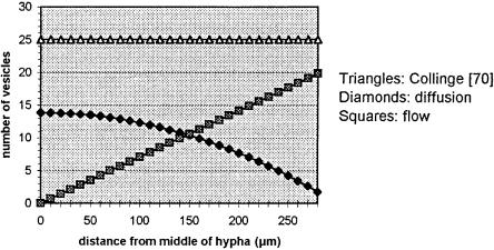
To show how little influence the kind of model can have on the outcome of a simulation, two very simple models for the growth of hyphae due to the transport of vesicles to the tip are compared. In one model, transport is based on diffusion; in the other model, transport is based on flow.
Diffusion Model. The assumptions are: transport of vesicles takes place by ordinary diffusion, and the vesicles are formed with 0th order kinetics. The rate of formation is given by Prosser and Trinci [11]: rl =1.5 vesicles mm–1 min–1. The value of rl is not important; the important point is that rl is constant. The hyphal element consists of one hypha. In the middle of the hypha the concentration gradient of the vesicles is zero; at its tip their concentration is zero, due to rapid uptake of vesicles by the wall of the apex.
The assumptions lead to the following differential equation and boundary conditions:
d2C |
4 |
· rl |
(7) |
–ID · 61 |
= 613 |
||
dx2 |
p · dh2 |
|
|
The boundary conditions are dCx/dx = 0 for x = 0 and C = 0 for x = L The solution of this equation is:
2 · rl
C = p062 (8) · dh · ID
Flow Model. The assumptions are: transport of vesicles takes place by constant flow. The rate of formation of vesicles is identical to that in the diffusion model. In the middle of the hypha the concentration is zero.
These assumptions lead to the following differential equation and boundary condition:
dC |
4 |
· rl |
(9) |
v · 5 |
= 91 |
||
dx |
p · dh2 |
|
|
The Morphology of Filamentous Fungi |
31 |
The boundary condition is C = 0 for x = 0.
The solution is:
4 · rl
C = p962 (10) · dh · v
For both equations, the flow at x = L is equal to rl · L That is as it should be, because this is the total amount of vesicles formed per second over the length L, and – in the steady state – it should be equal to the amount arriving at x = L. Because the rate of growth of the hypha is proportional to rl L, both models predict exactly the same rate of growth. Needless to say, this is independent of the value of ID or v!
These models can also be used to calculate the apparent velocity due to diffusion of the vesicles. If one calculates the diffusion coefficient ID with the Einstein equation and assumes that the observed transport velocity for a diffusion process can be calculated with
dC
vobserved · C = –ID 51 (11)
dx
where C and dC/dx can be calculated from Eq. (1), then the observed velocity is
of the same order of magnitude as that found by Prosser and Trinci [11], about 10 mm min–1.
It is clear that we cannot conclude which model is the correct one from a mechanistic point of view by comparing the simulated rates of growth or the transport velocities with the experimental values. However, if we look at the concentration gradients of vesicles, as observed by Collinge and Trinci [70], then the picture changes completely. In fact, the values of C (x) calculated
Fig. 7. Concentration of vesicles (number/mm3) as a function of the distance to the middle of the hypha in mm.
32 |
N.W.F. Kossen |
by both models conflict with the measurements of C (x) for Neurospora crassa where C (x) is almost constant at about 25 vesicles per mm3 until the apical compartment has been reached, where it increases sharply.
Both models presented above are gross oversimplifications of reality and are therefore not very realistic. However, the message is clear: without close observation of reality, the models for the growth of hyphae cannot be considered to be mechanistic in the physical/physiological sense.
References
1.Scheper T, Schügerl K (eds) (1998) Adv Biochem Biotechnol, vol 60, Springer, Berlin Heidelberg New York
2.Nielsen J, Villadsen J (1994) Bioreaction engineering principles. Plenum Press, New York
3.Metz B, Kossen NWF, van Suijdam JC (1979) Adv Biochem Eng Biotechnol 1:103
4.Megee RD, Kinorhita S, Fredrickson AG (1970) Biotechnol Bioeng 12 : 771
5.Paul GC, Thomas CR (1996) Biotechnol Bioeng 51: 558
6.Bellgardt KH (1998) Process models for production of b-lactam antibiotics. In: Scheper T, Schügerl K (eds) Adv Biochem Biotechnol 60 :153
7.Dion WM, Carilli A, Sermonti G, Chain EB (1954) Rend Ist Super Sanita 17 :187
8.Dion WM, Kaushal R (1959) Sel Sci Pap Ist Super Sanita 2 : 357
9.van Suijdam JC, Metz B (1981) Biotechnol Bioeng 23 :111
10.Fiddi C Trinci APJ (1976) J Gen Microbiol 97 :169
11.Prosser JI, Trinci APJ (1979) J Gen Microbiol 111:153
12.Metz B (1976) From pulp to pellet. Dissertation, Delft University of Technology
13.Metz B, de Bruin EW, van Suijdam JC (1981) Biotechnol Bioeng 23 :149
14.Adams HL, Thomas CR (1987) Biotechnol Bioeng 32 : 707
15.Paul GC, Thomas CR (1998) Characterisation of mycelial morphology using image analysis. In: Scheper T, Schügerl K (eds) Adv Biochem Biotechnol 60 :1
16.Paul GC, Kent CA, Thomas CR (1992) Trans I Chem Eng (Part C) 70 :13
17.Pons MN, Vivier H (1998) Beyond filamentous species. In: Scheper T, Schügerl K (eds) Adv Biochem Biotechnol 60 : 61
18.Krabben P, Nielsen J (1998) Modelling the mycelium morphology of penicillium species in submerged cultures. In: Scheper T, Schügerl K (eds) Adv Biochem Biotechnol 60 :125
19.Spohr A, Dam-Mikkelsen C, Carlsen M, Nielsen J, Villadsen J (1998) Biotechnol Bioeng 58 : 541
20.Schügerl K, Gerlach SR, Siedenberg D (1998) Adv Biochem Biotechnol 60 :195
21.Nielsen J, Johansen CL, Villadsen J (1994) J Biotechnol 38 : 51
22.Thiele EW (1939) Ind Eng Chem 31: 916
23.Rvesbech NP, Jørgensen BB, Brix O (1981) Limnol Oceanogr 26(4) : 717
24.Hooijmans CM (1990) Diffusion coupled with bioconversion in immobilized systems. Dissertation, Delft University of Technology
25.Cronenberg CCH, Ottengraf SPP, van den Heuvel IC, Pottel F, Sziele D, Schügerl K, Bellgardt KH (1994) Bioproc Eng 10 : 209
26.Cronenberg CCH (1994) Biochemical engineering on a micro-scale:biofilms investigated with needle type glucose sensors. Dissertation, University of Amsterdam
27.Gooday GW, Lloyd D, Trinci APJ (1980) 13th Symp Soc Gen Microbiol, p 207
28.Scott WA, Tatum EL (1970) Proc Nat Acad Sci USA 66 : 515
29.Schlegel HG (1993) General microbiology. Cambridge University Press, Cambridge, p 170
30.Howard RJ, Aist JR (1980) J Cell Biol 87 : 55
31.Regalado CM, Sleeman BD, Ritz K (1997) Philos Trans R Soc Lond 352 :1963
32.Cabib E, Roberts R, Bowers B (1982) Annu Rev Biochem 51: 763
33.Drgonová J, Drgon T, Tanaka K, Kollár R, Guang-Chao Chen, Ford RA, Chan CSM, Takai Y, Cabib E (1996) Science 272 : 277
The Morphology of Filamentous Fungi |
33 |
34.Kamada Y, Qadota H, Python CP, Anraku Y, Ohya Y, Levin DE (1996) J Biol Chem 271: 9193
35.Yamochi W,Tanaka K, Nonaka H, Maeda A, Musha T, Takai Y (1994) J Cell Biol 125 :1077
36.Wessels JGH (1993) Advances in microbial physiology 34 :147
37.Fredrickson AG, Tsuchia HM (1963) AIChE J 9 : 459
38.Randolph AD (1964) Can J Chem Eng 280
39.Randolph AD, Larson MA (1971) Theory of particulate processes. Academic Press, New York, p 41
40.Nielsen J (1993) Biotechn Bioeng 41: 715
41.Ainsley M, Ward AC, Wright AR (1990) Biotechnol Bioeng 35 : 820
42.Bergter F (1978) Z Allg Mikrobiol 18 :143
43.Trinci APJ (1970) Arch Microbiol 73 : 353
44.Trinci APJ [1970] Trans Br Mycol Soc 55 :17
45.Plomley NJB (1959) Aust J Biol Sci 12 : 53
46.Caldwell IY, Trinci APJ (1973) Arch Microbiol 88 :1
47.Emerson S (1950) J Bacteriol 60 : 221
48.Nielsen J, Krabben P (1995) Biotechnol Bioeng 46 : 588
49.Hinze JO (1975) Turbulence. McGraw Hill, New York, p 221
50.van Suijdam JC (1980) Mycelial pellet suspensions. Dissertation, Delft Univerity of Technology
51.van Suijdam JC, Metz B (1981) J Ferm Technol 59 : 329
52.Ayazi Shamlou P, Makagiansar HY, Ison HY, Lilly MD, Thomas CR (1994) Chem Eng Sci 49 : 2621
53.Yang H, King R, Reichl U, Gilles ED (1992) Biotechnol Bioeng 39 : 49
54.King R (1998) Mathematical modeling of the morphology of streptomyces species. In: Scheper T, Schügerl K (eds) Adv in Biochem Eng Biotechnol 60. Springer, Berlin Heidelberg New York, p 95
55.May PN (1974) Stability and complexity in model ecosystems. Princeton University Press, Princeton, NJ
56.Nielsen J (1992) In: Scheper T, Schügerl K (eds) Adv Biochem Eng Biotechnol 46. Springer, Berlin Heidelberg New York, p 187
57.Topiwala HH (1973) Methods Microbiol 8 : 35
58.Popper KR (1946) Lecture, Signific. Congress Bussum, Holland (unpublished)
59.Wei J (1975) Chemtech Feb: 128
60.Kossen NWF (1993) Scale-up strategy in fermentation. In: Mortensen U, Norman HJ (eds) Proceedings of the International Symposium on Bioreactor Performance, Helsingør
61.Brock TD, Madigan MT (1991) Biology of microorganisms. Prentice Hall, Englewood Cliffs, NJ, p 18
62.Nonaka H, Tanaka K, Hirano H, Fujiwara T, Kohno H, Umikawa M, Mino A, Takai Y (1995) EMBO J 4 : 5931
63.Brody S, Tatum EL (1966) Proc Nat Acad Sci USA 290
64.Lettinga G (1973) Agricultural University Wageningen, personal communication
65.Olsvik E, Tucker KG, Thomas CR, Kristiansen B (1993) Biotechnol Bioeng 42 :1046
66.Bongenaar JJTM, Kossen NWF, Metz B, Meijboom FW (1973) Biotechnol Bioeng 15 : 201
67.Allen DG, Robinson CW (1990) Chem Eng Sci 45 : 37
68.van Ooijen AJJ, Rietveld K, Hoekema A, Pen J, Sijmons PC, Teunis C, Verwoerd TC, Quax WJ (1996) US patent 5, 543, 576
69.van Ooijen AJJ, Rietveld K, Hoekema A, Pen J, Sijmons PC, Teunis C, Verwoerd TC 1997) US patent 5, 593, 963
70.Collinge AJ, Trinci APJ (1974) Arch Microbiol 99 : 353
Received December 1998

Antibiotica Research in Jena from Penicillin and Nourseothricin to Interferon
Harald Bocker, Wolfgang A. Knorre
Hans-Knöll-Institute for Natural Products Research, Beutenbergstraße 11, 07745 Jena,
Germany
Fax: +49 3641 656800
Milestones of antibiotics research and biotechnology in Jena/Thuringia are: 1938 – Hans Knöll established a strain collection of microorganisms; 1942 – production of penicillin on laboratory scale by Hans Knöll; since 1945 – development of industrial production processes for penicillin and streptomycin; 1952 – production of BCG-vaccine; since 1956 – development of biotechnical processes in the Institute of Microbiology and Experimental Therapy for actinomycin C, oxytetracyclin, erythromycin, paromomycin, turimycin, griseofulvin, nystatin, and nourseothricin, and in the 1980s for streptokinase, staphylokinase, and interferons. After the German unification the Hans-Knöll-Institute for Natural Products Research was founded.
Keywords. Bioprocess development, Penicillin, Streptomycin, BCG-vaccine, Nourseothricin, Lysin, Streptokinase, Staphylokinase, Interferons.
In 1937 the well-known glass factory Jenaer Glaswerk Schott & Gen. started cooperation with the young physician Dr. Hans Knöll, living in Frankfurt (Main), in order to check their all-glass bacterial filters. These filters were produced for the first time in 1935 according to an invention of Dr. Paul Prausnitz, Head of the Department for Design and Manufacture of Apparatus, in this glassworks. Knöll had already dealt with the problems of tuberculosis and chemotherapeutics. He subsequently became known for the filtration of bacteria. On behalf of this company, Knöll developed an accurate measuring procedure for checking such filters. Being very interested in this, the Schottfactory offered him the opportunity to establish and manage a bacteriological laboratory in the glassworks. Knöll started this job on 1st November 1938, not being aware at this time that his work would become of special importance both for him and for future biotechnology activities in Jena.
Knöll established a still existing collection of defined strains of different microorganisms as a basis for filter checking, and further works in the fields of microbiology, chemotherapy, and cell biology. His activities in identifying new methods in phase contrast and fluorescence microscopy led to cooperation with the precision-mechanical-optical factory Carl Zeiss in Jena.
Through reference to the literature, Knöll’s attention was drawn to penicillin, which had been discovered by Fleming in 1928 and which had been isolated by
a research team in Great Britain in 1939. He started experiments to obtain this new antibiotic in mold cultures of Penicillium notatum.After a short time crude
samples of this antibiotic were obtained. Originally Knöll intended to test penicillin for its effectiveness against cancer cells. However, the immense importance of penicillin in the fight against several human bacterial infectious
Advances in Biochemical Engineering/
Biotechnology, Vol. 70
Managing Editor: Th. Scheper
© Springer-Verlag Berlin Heidelberg 2000
36 |
H. Bocker and W.A. Knorre |
diseases soon became known. Penicillin-producing mold strains were sought in the environment and were cultivated in flat glass flasks such as Fernbach flasks. The use of Schott-borosilicatglass proved to be advantageous. It was found that the use of cheaper glass-types spoiled the synthesis of this antibiotic because of its arsenic content. Because of this, and in spite of wartime, an effective exchange – via foreign countries – of information about penicillin took place between Schott-glass and Jena.
Soon afterwards, penicillin wound powder was available from Jena on the laboratory scale. In late 1942, for the first time, it was applied to man. A factory worker, who had a suppurating injury to his hand, was cured successfully by application of this penicillin produced in the Bacteriological Laboratory.
In 1944, this successful work, together with support from the Carl Zeiss factory, led to the transformation of the Bacteriological Laboratory, whose staff had increased from 4 to 15 employees, into the Institute of Microbiology (Schott-Zeiss-Institute), supported by the founding firms of Schott and Zeiss.
At the end of the Second World War in June 1945, the US Army administration at that time in Jena intended to transfer the Institute of Microbiology to the western part of Germany. However, this intention was not realized as in July 1945 the occupation by the Soviet Army began. This new military administration ordered an immediate expansion in penicillin production. Cultivation of the producing mold had been intensified, while stage fermentors made from glass, aluminum, and steel, respectively, were installed in an empty factory building belonging to the firm Carl Zeiss. Railway tank wagons were also modified into fermentors. A great deal of effort was put in to erecting a production plant for penicillin attached to the Institute.
As a result of the rapid increase in the size of the operation, the fermentation section of the Institute of Microbiology was named Jenapharm in 1947. To improve the unsatisfactory supply of medicines, other medicaments and drugs, such as vitamins, analgesics, and transfusion solutions were incorporated into its production program. The number of employees grew to more than 800. Finally, in 1950 the Institute of Microbiology became an independent nationally owned factory, the VEB Jenapharm (VEB means a state-owned company). In addition to penicillin, the VEB Jenapharm produced another antibiotic, streptomycin, which was used to fight tuberculosis, incidence of this illness having increased considerably as a result of war.
A few months after the foundation of the German Democratic Republic (GDR), as a measure in the fight against tuberculosis, the Ministry of Health ordered Knöll to start the immediate production of vaccines according to the methods of Calmette and Guérin as a prerequisite for the introduction of BCGvaccination. The first research building on the Beutenberg Hill in Jena was therefore erected as a production unit (see Fig. 1), where from 1952 onwards the BCG-vaccine was produced for the whole of the GDR.
In 1953, Knöll left VEB Jenapharm to become director of the newly founded Institute of Microbiology and Experimental Therapy (IMET), which was built in accordance with his ideas, also on the Beutenberg Hill. This institute was taken over by the German Academy of Sciences (later Academy of Sciences of

Antibiotica Research in Jena from Penicillin and Nourseothricin to Interferon |
37 |
Fig. 1. Hans Knöll (1913–1988, founder of the Institute of Microbiology and Experimental Therapy on the Beutenberg and the pharmaceutical industry in Jena) left of Werner Eggenrath (Prime Minister of Thuringia) at the topping-out ceremony of the Microbiology Institute under construction in 1951. Knöll was awarded the National Prize of the German Democratic Republic. His Institute became a refuge for the politically displaced. Today the name of Hans Knöll is synonymous with Jena as are the names of Carl Zeiss and Otto Schott
the GDR) in 1956 and was transformed into the Central Institute of Microbiology and Experimental Therapy (ZIMET). Prof. Knöll was director of the ZIMET up to 1976. During this time the personnel at the institute increased to about 1000 workers.
The task of ZIMET was to work on therapeutics, particularly on microbial agents for use in human and veterinary medicine, and later it took over additional technical tasks. The structure of this institute incorporated all the research requirements necessary under the same roof in order to reach a high level of self-sufficiency. ZIMET was composed of the divisions Antibiotic Research, Biotechnology, Experimental Therapy, Medical Microbiology, Methods and Theory, Molecular Biology and Microbial Genetics, Steroid Research, Environmental Microbiology, and Scientific Engineering.
To maintain a continuous line of investigations from screening and increasing efficiency to testing the isolates and purified final products on animals, it was necessary to install qualified microbial and chemical laboratories as well as an efficient experimental breeding system, technical media preparing groups, and different workshops.
38 |
H. Bocker and W.A. Knorre |
|
Antibiotics research in all its complexity and relevant applications became |
an essential task of this institute. In the 1950s further improvement in the production of penicillin and streptomycin, in cooperation with VEB Jenapharm, was its main objective. Later on the research potential was systematically developed, including corresponding fields of basic research.
For many years, both closely cooperating divisions “Antibiotic Research” and “Biotechnology” were mainly involved in the elaboration of specific bioprocess methods and down-stream processing by contract with the pharmaceutical industry of the GDR. Further shared research works were the search for producing microbial strains, and the developing of technical instructions for the biosynthetic production and chemical isolation of various antibiotics and other substances for therapeutic and technical purposes, respectively.
The behavior of microbial production strains in shaking flasks and laboratory fermentors was investigated to optimize process conditions and the composition of media on the basis of process kinetic analyses, as well as to elucidate the importance of certain medium compounds for special types of biosynthesis. For such investigations a new biometric screening method, based on the (2n+1)-spectrum, was developed and used with high efficiency.
In the 1960s basic investigations into growth and product-formation kinetics, as well as metabolic regulation in microorganisms, were started, aiming at scientific progress in the optimization of production methods obtained from models. On model systems, in a number of instances, the bistability of specific product formations of microbial processes was demonstrated successfully. Methods for optimal control of biotechnological processes were developed on the basis of mathematical descriptions of growth, metabolism, and product formation in microorganisms as well as by computer simulation of kinetic models.
In a pilot plant with reactors up to 3 m3 net capacity the biotechnical methods were further adapted to the conditions of particular industrial production. Subsequently, the biosynthetically formed substances were obtained by selected known down-stream processes. Moreover, large numbers of small quantities of microbial agents for experimental purposes were produced there.
A widely recognized way to rationalize the multistage procedures of screening and selection, respectively, was opened by introducing six types of selection machine developed by an automation team of the ZIMET. This socalled Autoselect System includes machines to deliver small quantities of agar, to inoculate colonies, to dilute samples, to punch test plates, to pour sample solutions into the punched holes, and to measure the diameters of inhibition zones on the test plates by an optoelectronic method. Thus, by means of the Autoselect Systems, the number of colonies and samples tested could be considerably increased, and the accuracy of working steps and measurements was remarkably improved. The data were evaluated by computer. The optoelectronic measuring device was adopted by the production program of the factory VEB Carl Zeiss Jena.
The search for new antibiotics producing microorganisms had been a permanent task of the ZIMET, particularly of the Antibiotic Research division. Up to the 1980s a collection with more than 20,000 taxonomically identified, freeze-dried strains with defined antibiotic activity had been established.
Antibiotica Research in Jena from Penicillin and Nourseothricin to Interferon |
39 |
New production methods were developed and transferred to industrial production for antibacterial antibiotics, such as actinomycin C, streptomycin, oxytetracyclin, erythromycin, paromomycin, and turimycin, as well as for the antifungal antibiotics griseofulvin and nystatin and the nutritive antibiotic nourseothricin, which was required in animal nutrition. Remarkable results, which were used in the industrial production of nourseothricin, were obtained from basic investigations on the regulating effect of phosphate on secondary metabolism. It was detected, that a regulated feeding with phosphate, liquefied starch, and ammonia, while realizing a phosphorus limitation and simultaneously sufficient concentrations of the other components in the medium, can increase the biosynthetic production of this antibiotic in stirred and intensively aerated bioreactors up to yields of 50 g/l nourseothricin. Furthermore, an economic mode of precipitating this agent, basing on its adsorption to bentonite, was invented and applied in industrial production.
Taking multivalent advantages of growth media, an example was set in the industrial streptomycin processes. By a specific change in down-stream processing, a proteolytic enzyme complex could be obtained in addition to the main product, streptomycin. This by-product, after concentrating in its aqueous solution, was used on the industrial scale in the VEB Filmkombinat ORWO Wolfen (Saxony-Anhalt) for the de-gelatination of photographic films in order to recover silver residues. Furthermore, the important VEB Lederfabrik Weida (Thuringia) used this product as a softening-enzyme in the leather-tanning process.
A new method of processing was adapted and tested successfully in the con- tamination-free production of L-lysine yielding an important food supplement in animal nutrition. In order to guarantee the required high oxygen transfer rate, deep-stream pilot reactors (0,45 m3 capacity) were used. In cooperation with the Research Center of Biotechnology Berlin the developed processing was transferred to industrial application. In the VEB Gärungschemie Dessau (Saxon-Anhalt) non-contaminated production was realized constantly with yields of 80–90 g/l in a production time of 70 h, using a technical deep-stream bioreactor-device with a capacity of 8 m3, fitted out with a continuously acting sterilization-unit for nutrient media. Such a high yield of L-lysine from a bioprocess without contamination was outstanding at that time.
In the early 1980s the ZIMET was concentrating its efforts on the further extension of biotechnology, particularly in the important field of the development of microbial agents from genetically engineered microorganisms. In this connection, genetic engineering protein technology and process development for rDNA products has been promoted.
The genes coding for the plasminogen activators streptokinase and staphylokinase were cloned and sequenced. Expression studies in a variety of hosts led to the construction expression vectors based on staphylokinase signals. Several bacterial strains producing significant amounts of human interferons alpha 1, alpha 2, and gamma have been constructed on the basis of these vectors. In order to maximize the production of interferons in the E. coli cells a high cell density bioprocess for the industrial application was developed in 1985. In 1987, after only two years, the first lots (about 1010 IU) of purified human inter-
- #
- #
- #15.08.20134.04 Mб17Hastie T., Tibshirani R., Friedman J. - The Elements of Statistical Learning Data Mining, Inference and Prediction (2002)(en).djvu
- #
- #
- #
- #
- #
- #
- #
- #15.08.201315.44 Mб27Hudlicky M, Pavlath A.E. (eds.) - Chemistry of Organic Fluorine Compounds 2[c] A critical Review (1995)(en).djvu