

Frantisek Svec - Capillary Electrochromatography
.pdfShort Monolithic Columns as Stationary Phases for Biochromatography |
61 |
For this special case, the following expression for the critical distance, X0 , at which the quasi-steady state is obtained:
X0 = l · u · Cc . |
(7) |
87Z · B |
|
In Eq. (7), l is an auxiliary parameter, u is the linear velocity of the mobile phase, Cc is critical concentration of the displacing salt, Z is the effective charge on the solute ion divided by the charge on the mobile phase ion and B is the gradient steepness.
The results obtained by Tennikova et al. [55] for ion-exchange chromatography, together with the results previously presented by Belenkii et al. [53] for reversed phase chromatography, form a theoretical basis for HPMC, which is now more conveniently called chromatography on short monolithic columns (SMC). According to the authors, the quasi-steady state is realized at an X0 less than L, thus, it might occur at steep gradients or on long columns.As the aim of any separation is to obtain good resolution, it is not important in which state the separation is performed. The main conclusion was that it is possible to use ultra-short columns for the separation of proteins and other molecules that traditionally have been separated using steep gradients,with appropriate resolution and at low gradient times. Furthermore, because of the short separation layer length, the pressure drop on short continuous beds is much lower (typically less than 5 MPa) and extremely high flow rates can be applied. This, together with the improved mass transfer characteristics, may lead to the possibility of performing very fast separations, which are extremely important for large and labile therapeutic macromolecules.
Very similar results were presented by Coffman et al. [2]. They introduced a yet different approach to the prediction of the efficiency of large molecule (proteins) separations on very short columns. Their approach is based on the fact that since short columns yield non-Gaussian effluent distributions, measuring the degree of binary separation using conventional chromatographic resolution is inadequate. Instead, they proposed the fractional purification Pi of component i, defined as:
Yi |
. |
(8) |
Pi = |
||
Y91i +Yj |
|
|
Where Yi represents the fractional mass yield of i in the i-rich product.
The purity of the final product in a single-stage (ultra-short) column is defined through the separation factor calculated as:
ui |
= |
ln [1–Pi] |
(9) |
4uj |
. |
||
|
961ln Pi |
|
As further shown by the authors, for e.g., a fifty-plate column, a separation factor of about 1.5 is needed to achieve a 99% purity. This value of the separation factor is in the range of many practical protein separations. Therefore, the use of a fifty-plate column can achieve high purity indicating that the tens of thousands of plates found in many conventional chromatography columns are not necessary
62 |
A. Strancar et al. |
for most protein separations. Short columns can be efficiently used for the separation of proteins and probably for other large molecules as well, especially in reversed phase chromatography, where extremely large values of separation factors are not uncommon for some protein separations.
2.3
Resolution and Efficiency in SMC-Chromatography
Finally, let us briefly speculate about the efficiency of SMC to separate different (bio)molecules. The usual measure for the efficiency of conventional HPLC column is the so-called height equivalent of a theoretical plate (HETP) [56], which is simply the ratio between the column length, L, and the number of theoretical plates, N. Normally, the lower the HETP value, the more efficient the column is (larger N at the same column length). Columns with a large efficiency have HETP values in the range from 8 µm to 14 µm. For the typical column lengths (e.g. 10–25 cm) this HETP value translates into approximately 12000 to 30000 theoretical plates, N,per column. In ion-exchange chromatography of large molecules like proteins and DNA the HETP are much larger and can even reach 100–200 mm depending on the column type. These HETP values still translate into relatively high number of theoretical plates, N, ranging from 1000 to 2500. In the case of SMC with a “length” of only a few mm, the number of theoretical plates is much lower, ranging only from 10 to 50. As has been shown [57], this number is still high enough to allow an isocratic separation of different oligonucleotides. This was explained by a large difference in the Z factor between individual nucleotides owing to the increased chain length and, consequently, to the increased charge density. If the difference in Z factors of two molecules is large enough, the resolution achievable even on a short column can be satisfactory. This is especially true for large (bio)molecules, which differ significantly in the Z factor [59]. The reason lies in their high molecular mass and, consequently, a high heterogeneity of charged groups on the molecule surface. Due to these pronounced differences, the binding characteristics between individual molecules are quite different and an efficient separation can be achieved by selective elution using a linear gradient. A more detailed and comprehensive description of the processes governing differential elution of large biomolecules on the SMC can be found in a recent review paper published by Tennikova and Freitag [43].
3
Preparation of SMC and Scale-Up Strategies
3.1
Synthesis of the SMC
Most of the SMC described in this chapter are prepared by free radical polymerization of a mixture of glycidyl methacrylate (providing functional groups), ethylene dimethacrylate (as a cross-linking reagent), 2,2´-azobisisobutyronitrile (as an initiator) and a porogenic solvent (cyclohexanol and dodecanol) in barrels of polypropylene syringes, as published elsewhere [61, 62], yielding glycidyl
Short Monolithic Columns as Stationary Phases for Biochromatography |
63 |
methacrylate-co-ethylene dimethacrylate (GMA-EDMA) monoliths. Another method uses a free radical polymerization of the mixture of styrene and divinylbenzene (the latter as cross-linking reagent) using 2,2´-azobisisobutyroni- trile as an initiator and a porogenic solvent (dodecanol and toluene) to ensure adequate porosity.After the polymerization, the block of polymer formed in disk or tube shape, is mounted in a specially designed housing allowing good sample distribution and low dead volume. Then the disk or tube is washed with methanol, a methanol-water mixture (50:50) and distilled water to remove porogenes and residual monomers from the porous polymer.After this the monolithic bed is ready for further derivatization or ligand immobilization if desired. GMAEDMA monoliths have active epoxide groups which can easily be further modified using various chemistries, e.g. diethyl amine, propane sulfone for ion exchange chromatography, butyl for hydrophobic interaction chromatography or any desired protein ligand for affinity chromatography.
The preparation of monolithic columns is in many cases considered an easy and straightforward process, especially when compared to the tedious and timeconsuming preparation of monosized spherical particles and subsequent packing of conventional columns. In principle, this is true for the in-situ preparation of methacrylate and styrene-divinylbenzene based HPLC columns, or capillary micro HPLC and CEC columns [63].According to various authors, the procedure consists of simply filling the column or capillary with a liquid monomer mixture (that also contains an organic solvent and an initiator), sealing the column/capillary at both ends and triggering the polymerization procedure by placing the column in an appropriate water bath. This procedure is very easily done and it removes the need for tedious slurry column packing. However, according to our own experience,this method of monolithic column preparation only works in the case of micro or small-scale (up to a few ml in volume) monolithic columns. The preparation of large volume monolithic columns with a well-defined and homogeneous structure still represents a considerable challenge to manufacturers. In contrast to the scale up of particle columns, containing particles that range in size from a few micrometers up to 100 micrometers, which is obtained by packing these very small particles in larger columns, large-scale monolithic columns are obtained by producing a large block of a polymer cast in a proper cartridge (monolith holder). The main problems that occur during this process are connected to the heat release and heat dissipation (gel-effect) during polymerization.
3.2
Preparation of Large Scale SMC
The production of conventional stationary phases in the form of porous polymer particle is based on suspension polymerization. Namely,the polymerization is allowed to proceed in a solvent under vigorous stirring that assures obtaining particles of the desired diameter. Since the particle size is typically in the range of a few micrometers,no problems with heat transfer are encountered. In contrast,the preparation of monoliths requires a so-called ‘bulk’ polymerization. A polymer mixture consisting of monomers and porogenic solvent is mixed with an initiator. As the temperature is increased, the initiator decomposes and oligomer nu-
64 |
A. Strancar et al. |
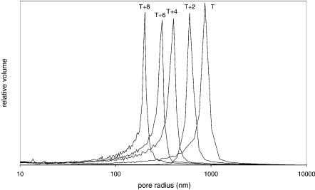
clei start to form. The solubility of the polymers in the reaction mixture decreases during growth and at some point they start to precipitate. Thermodynamically speaking, the monomers are better solvents for the polymer than the porogenes. Consequently, the precipitated nuclei are swollen with the monomers. Since the monomer concentration is higher than in the surrounding solution, the polymerization in the nuclei is kinetically preferred. In the absence of mixing and due to their higher density, insoluble nuclei sediment and accumulate at the bottom of the mould. Initially, they form a very loose structure, which is highly porous. During the course of the polymerization, nuclei continue to grow and crosslink until the final structure is achieved. As it can be deduced from the above description, the pore size distribution of the polymer depends on the chemical composition, but also the polymerization temperature. In particular, the temperature defines the degradation rate of the initiator and,therefore,also the number of nuclei formed in a given time. Since the amount of the monomers is constant, the lower number of nuclei formed at lower temperatures within a defined volume corresponds to a larger size and thus, to larger pores between the clusters of growing nuclei. In contrast, at higher polymerization temperatures, where the initiator decomposition is much faster, the number of growing nuclei is much larger. Therefore, the pores formed will be smaller. A dramatic effect of the polymerization temperature is demonstrated in Fig. 1. As can be seen, a change of only 8 °C shifts the average pore radius from 400 to 850 nm and completely changes the flow characteristics of such a monolith. Therefore, the polymerization temperature is a powerful tool for the control of pore formation.
Fig. 1. Effect of the polymerization temperature on the pore size distribution. At the highest temperature (T+8) the average pore radius is 400 nm while at the lowest T the pores are much larger with an average pore radius of 850 nm

Short Monolithic Columns as Stationary Phases for Biochromatography |
65 |
The polymerization of a methacrylate-based monolith is an exothermic process. Therefore, during the course of the reaction heat is released. If no mixing takes place and if the size of the mould is in a range of a centimeter or more, the released heat cannot be dissipated fast enough.As a consequence, an increase of the temperature inside the reaction mixture occurs as shown in Fig. 2. The temperature increase during polymerization is over 77 °C in a mould of 5 cm diameter. The effect of this temperature increase during the polymerization was carefully studied by Peters et al. [64]. They initially performed experiments in a 26 mm mould using AIBN as an initiator. The extremely fast reaction led to a monolith with a badly scarred structure due to the nitrogen released during the reaction. In subsequent experiments, benzoyl peroxide was used instead as an initiator. During the polymerization in a 26-mm mould an increase of temperature of only 7 °C across the radius of the column was recorded and no influence on the pore size distribution across the radius as well as along the height was found. On the other hand, when a mould of 50 mm was used, a temperature increase of 113 °C was observed and a 25 °C temperature differential was recorded across the radius of the column. Pore size distribution measurements revealed that the pores in the middle of the polymer were larger than on the outer part resulting in pore size distribution inhomogeneity. Obviously, the preparation of large volume monoliths is limited by the exothermic nature of the polymerization and the fact that the temperature exerts a pronounced influence on the pore size distribution. To avoid these problems, Peters et al. [64] suggested perform-
Fig. 2. Temperature profile in the middle of a 5 cm cylindrical mould during the polymerization of a GMA-EDMA monolith. The increase of the temperature by 77 °C significantly influences the structure of the GMA-EDMA monolith
66 |
A. Strancar et al. |
ing the polymerization at a slow reaction rate. This is accomplished by gradually adding the reaction mixture to the mould. To investigate this, the authors fed the reaction mixture into the mould at a rate of 20 ml/h for 12 h to ensure slow polymerization. They found only a slight temperature increase of 10 °C and a much more uniform pore size distribution. The problem, which might arise with such a procedure, is the conditioning of the reaction mixture. The initiator is either continuously added and dissolved into a thermostatted reaction mixture or the reaction mixture must be significantly colder to prevent polymerization over such a long period of time. That the conditions in a gradual addition of the polymerization mixture were not the same as in the previously presented experiments can be concluded from the comparison of the pores of the monolith prepared in a conventional way (batch mode) in a 26 mm mould and with a gradual feeding (fed-batch mode) in a 50 mm mould.Although the temperature increase was similar in both cases, a pore diameter of the highest peak of the pore size distribution ranges from 1.50 to 1.56 mm in the former case and 1.66 to 1.76 mm in the latter case (in the upper part of the later monolith, the pores were even larger, i.e. up to 2 mm).
Due to the problems with scale up by increasing the diameter, an alternative approach has been proposed, which consists of preparing long monolithic rods with small diameter. Indeed, there are several publications describing GMAEDMA monolithic columns with a length of up to 300 mm [29]. As with conventional columns, an increased length results in increased backpressure. Since the monolithic structure is the most advantageous for fast separation of large molecules, which is predominantly based on a gradient elution, the column length should not improve the resolution significantly. In fact, as was discussed in Sect. 2.2, longer columns might even result in additional band spreading, thus lowering resolution. Therefore, to take advantage of the monolithic structure on a large scale, the most suitable design seems to be a tube shaped monolith. The first such monolith used in a radial chromatography mode was designed by Strancar et al. [46] in 1997. It was a 22 ml tubular monolithic GMA-EDMA column used for the purification of plasma proteins. The backpressure was significantly lower than the rod-shaped monolithic columns of similar volume.
Still the production of larger volume tubular shaped monoliths was hampered by the problem of providing the required uniform pore size distribution. To overcome this problem, a new approach has recently been proposed by Podgornik et al. [47]. Instead of gradually adding the polymerized mixture to form a single large volume monolith, this approach is based on the preparation of monoliths of a precisely defined shape. This process avoids temperature increases during polymerization, thus preventing uneven pore size distribution. To accomplish this in practice,a mathematical model based on the heat balance during the polymerization process was developed. To simplify the model, the authors assumed that the heat released per unit volume (S) is constant during the polymerization and uniformly released over the entire volume. Furthermore, they assumed that the thermal conductivity l is constant and that the system is in thermal equilibrium. These assumptions were justified since only the determination of the maximal temperature increase is of interest and all other parameters can be considered to be in steady state during this process. For a more precise mathematical

Short Monolithic Columns as Stationary Phases for Biochromatography |
67 |
model predicting the temperature profile during the course of a polymerization and analysis of the reaction rate is required. Recently, the experiment has been conducted that indicates that this polymerization system follows a first order reaction kinetics [65]. However, using the previously described assumptions, the following equation for determination of the maximal temperature increase inside the reaction mixture can be derived:
|
|
|
|
|
4 |
|
|
4 |
|
|
||
|
|
|
|
|
|
|
|
|
|
2 |
|
|
|
S |
|
|
r2 |
–r2 |
|
|
1 – r0 |
|
|
||
|
|
|
2 |
1 |
0 |
|
|
4 |
|
|
||
Tmax =T0 + |
4 l |
· r1 + |
|
r1 |
· |
ln |
1 |
–1 . |
(10) |
|||
|
r1 |
|||||||||||
|
5 |
|
453 |
|
|
444 |
|
|
||||
|
|
|
|
2 ln r |
0 |
|
|
2 ln r |
0 |
|
|
|
|
|
|
|
|
|
|
|
|
|
|
||
Based on this equation one can predict the temperature increase to be expected for a defined annulus thickness as shown in Fig. 3.With the above-described approach one can in addition construct a monolithic annulus of a desired radius but limited thickness. By preparing a series of annuluses where the outer diameter of the smaller monolith is equal to the inner diameter of a larger one, a large volume monolithic unit can be constructed by forming a so called “tube in a tube”system,as shown in Fig. 4. In this way,a monolithic unit of the required volume and uniform pore size distribution can be prepared. Furthermore, the voids between the annuluses can be filled with the reaction mixture and polymerization is allowed to proceed for a second time. Since the voids are very thin, no increase in temperature during the course of the reaction is expected.
Fig. 3. Effect of the annulus thickness on the maximal temperature increase during the polymerization of a GMA-EDMA monolith. Inner annulus radius is 10 mm; calculation is based on Eq. (10). (Reprinted with permission from Podgornik A, Barut M, Strancar A, Josic D, Koloini T (2000) Anal Chem 72:5693)

68 |
A. Strancar et al. |
Fig. 4. Construction of a large volume GMA-EDMA monolithic unit. The monolithic unit (4) consists of three monolithic annuluses (1, 2 and 3). Total thickness of the unit 4 is a sum of the thickness of the monolithic annuluses 1, 2 and 3. (Reprinted with permission from Podgornik A, Barut M, Strancar A, Josic D, Koloini T (2000) Anal Chem 72:5693)
Fig. 5. Effect of the flow rate on the separation efficiency. Separation of a protein mixture at six different flow rates (40, 80, 120, 160, 200 and 240 ml/min) normalized to the elution volume. Conditions: Column: 80 ml CIM® DEAE Tube Monolithic Column; Mobile phase: buffer A: 20 mM Tris-HCl buffer, pH 7.4; buffer B: 20 mM Tris-HCl buffer +1 M NaCl, pH 7.4; Gradient: 0–100% buffer B in 200 ml; Sample: 2 mg/ml of myoglobin (peak 1), 6 mg/ml of conalbumin (peak 2) and 8 mg/ml of soybean trypsin inhibitor (peak 3) dissolved in buffer A; Injection volume: 1 ml; Detection: UV at 280 nm. (Reprinted with permission from Podgornik A, Barut M, Strancar A, Josic D, Koloini T (2000) Anal Chem 72:5693)

Short Monolithic Columns as Stationary Phases for Biochromatography |
69 |
This approach was verified by the construction of an 80 ml tubular monolithic column. The monolithic column was characterized by low backpressures even at high flow rates (below 2.5 MPa at the flow rate of 250 ml/min). One interesting feature, which should be highlighted at this point, is that, in contrast to conventional radial columns of large diameter and small bed thickness, the bed in this case had an outer diameter that was 35 mm while the inner diameter was only 1.5 mm. Because of that, the linear velocity of the mobile phase increases more then 23 times from the outer to the inner surface of the column. In the case of conventional porous particle supports, such changes in the linear velocity would generally result in a pronounced deterioration of the column efficiency. However, the characteristics of the monoliths were found to be flow independent, therefore the change in linear velocity should not have any influence either on the resolution or on the binding capacity. This was proved by the separation of a protein mixture as well as by measuring the dynamic binding capacity determined at different flow-rates. As shown in Fig. 5, the curves obtained overlap nicely at different flow rates. As the authors calculated, this unit can purify around 15 g of protein per hour.
4
Characteristics and Application of SMC in the Liquid Chromatography of Biomolecules
4.1
Characteristics of the SMC
Most of the SMC presented in this chapter are highly cross-linked porous rigid monolithic polyglycidylmethacrylate-co-ethyleneglycoldimethacrylate or styrene-divinilybenzene polymers produced by BIA Separations under the trade name of Convective Interaction Media (CIM). Following the idea of a short chromatographic layer,the smaller units are produced in the form of disks (see Fig. 6) and the larger units in the form of tubes (see Fig. 4).
Both units are engineered to ensure well-defined, narrow pore-size distributions, excellent separation power and exceptional chemical stability and flow characteristics. To ensure scalability, the smaller and bigger units are of the same
Fig. 6. Some of the smaller commercially available SMC – the CIM® Disk Monolithic Columns from BIA Separations d.o.o., Ljubljana, Slovenia

70 |
A. Strancar et al. |
Fig. 7. Semi-Preparative Anion Exchange Purification of a 16-mer Oligodeoxynucleotide on a CIM® DEAE Disk Monolithic Column. Conditions: Column: 0.34 ml CIM® DEAE Disk (3¥12 mm ID); Instrumentation: Gradient HPLC system with extra low dead volume mixing chamber; Sample: 16mer oligodeoxynucleotide from the reaction mixture – bold line, standards of 1, 2, 3, 4, 5, 6, 7, 9, 10, 11, 12, 14, 15, 16mer – thin line; Injection Volume: 20 mL; Mobile Phase: Buffer A: 20 mM Tris-HCl, pH 8.5; Buffer B: Buffer A+ 1 M NaCl; Gradient: as shown in the Figure; Flow Rate: 4 ml/min; Detection: UV at 260 nm
Fig. 8. Fast semi-industrial scale separation of a protein mixture using an 80 ml CIM® DEAE Tubular Monolithic Column. Conditions: Column: 80 ml CIM® DEAE Tubular Monolithic Column; Mobile phase: Buffer A: 20 mM Tris-HCl buffer, pH 7.4; Buffer B: 20 mM Tris-HCl buffer +1 M NaCl, pH 7.4; Gradient: 0–100% Buffer B in 30 s; Sample: 2 mg/ml of myoglobin (peak 1), 6 mg/ml of conalbumin (peak 2) and 8 mg/ml of soybean trypsin inhibitor (peak 3) dissolved in buffer A; Flow Rate: 400 ml/min; Injection volume: 1 ml; Detection: UV at 280 nm