
Dewick P.M. Medicinal natural products VCH-Wiley, Weinheim, 2002 / booktext@id88013692placeboie
.pdf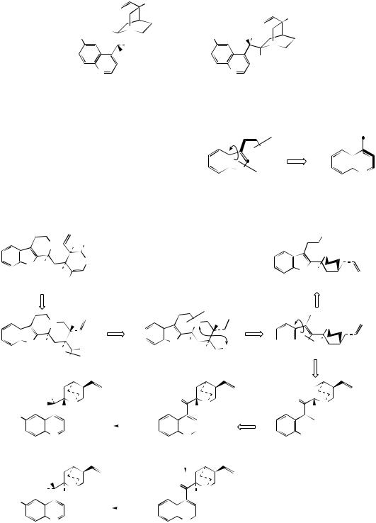
ALKALOIDS DERIVED FROM TRYPTOPHAN |
361 |
|||
|
H |
|
|
|
|
|
|
|
H |
H S |
N |
R |
H OH |
|
R |
8 |
|
||
R |
9 OH |
|
R |
N |
|
H |
|
S |
|
|
|
H |
|
|
|
|
|
|
|
N |
|
|
N |
|
R = OMe, (–)-quinine |
|
R = OMe, (+)-quinidine |
||
R = H, (–)-cinchonidine |
|
R = H, (+)-cinchonine |
||
Figure 6.88
and cinchonine (Figure 6.88), long prized for their antimalarial properties. These structures are remarkable in that the indole nucleus is no longer present, having been rearranged into a quinoline system (Figure 6.89). The relationship was suspected quite early on, however, since the indole derivative cinchonamine (Figure 6.90) was known
|
|
NR2 |
|
|
N |
|
N |
|
|
||
|
H |
|
|
|
|
|
|
indole alkaloid |
quinoline alkaloid |
||
Figure 6.89
|
|
NH H OGlc |
N |
H |
O |
|
||
H |
|
H |
CO2Me
strictosidine
cleavage of C–N bond (via iminium) then formation of new C–N bond (again via iminium)
|
|
N |
H |
|
|
|
|
|
N |
H |
|
N H |
|
CHO |
|
N |
H |
CHO |
|||
|
|
|
||||||||
|
|
|
||||||||
|
H |
H |
|
H |
H |
|||||
|
|
MeO2C |
hydrolysis and |
|
corynantheal |
|
||||
|
|
|
|
decarboxylation |
|
|
|
|
|
|
|
|
H |
|
|
|
|
O |
8 |
|
|
|
HO |
N |
|
|
|
N |
|
|||
|
|
|
|
|
|
|
||||
R |
H |
|
|
|
|
|
H |
|
||
|
NADPH |
|
|
|||||||
|
|
|
|
|
|
|||||
|
|
N |
|
|
|
|
|
|
N |
|
|
|
|
|
|
|
|
|
|
||
|
|
|
|
|
|
|
|
|
||
|
R = H, cinchonidine |
|
|
|
cinchoninone |
|
||||
|
R = OMe, quinine |
|
|
epimerization at |
|
|
||||
|
|
|
|
|
||||||
|
|
|
|
|
|
|
|
|||
|
|
|
|
|
|
C-8 via enol |
|
|
||
|
|
|
|
|
|
|
|
|||
OH
N
N H
H cinchonamine
 CHO
CHO
N

 N H
N H
Hcleavage of indole C–N bond
O
N
H
CHO
NH2
|
|
H |
|
|
O |
|
HO |
N |
|
|
|||
|
|
|
N |
|||
R |
H |
|
|
|
H |
|
|
NADPH |
|||||
|
|
N |
|
|
|
N |
|
|
|
|
|
||
|
|
|
|
|
||
R = H, cinchonine |
|
|
|
|
||
R = OMe, quinidine |
|
|
|
|
||
Figure 6.90

362 |
ALKALOIDS |
to co-occur with these quinoline alkaloids. An outline of the pathway from the Corynanthe-type indole alkaloids to cinchonidine is shown in Figure 6.90. The conversion is dependent on the reversible processes by which amines plus aldehydes or ketones, imines (Schiff bases), and their reduction products are related in nature. Suitable modification of strictosidine leads to an aldehyde (compare the early reactions in the ajmalicine pathway (Figure 6.76)). Hydrolysis/decarboxylation would initially remove one carbon from the iridoid portion and produce corynantheal. An intermediate of the cinchonamine type would then result if the tryptamine side-chain were cleaved adjacent to the nitrogen, and if this nitrogen were then bonded to the acetaldehyde function. Ring opening in the indole heterocyclic ring
could generate new amine and keto functions. The new heterocycle would then be formed by combining this amine with the aldehyde produced in the tryptamine side-chain cleavage. Finally, reduction of the ketone gives cinchonidine or cinchonine. Hydroxylation and methylation at some stage allows biosynthesis of quinine and quinidine. Quinine and quinidine, or cinchonidine and cinchonine, are pairs of diastereoisomers, which have opposite chiralities at two centres (Figure 6.88). Stereospecific reduction of the carbonyl in cinchoninone can control the stereochemistry adjacent to the quinoline ring (C-9). The stereochemistry at the second centre (C-8) is also determined during the reduction step, presumably via the enol form of cinchoninone (Figure 6.90).
Cinchona
Cinchona bark is the dried bark from the stem and root of species of Cinchona (Rubiaceae), which are large trees indigenous to South America. Trees are cultivated in many parts of the world, including Bolivia, Guatemala, India, Indonesia, Zaire, Tanzania, and Kenya. About a dozen different Cinchona species have been used as commercial sources, but the great variation in alkaloid content, and the range of alkaloids present, has favoured cultivation of three main species, together with varieties, hybrids, and grafts. Cinchona succirubra provides what is called ‘red’ bark (alkaloid content 5–7%), C. ledgeriana gives ‘brown’ bark (alkaloid content 5–14%), and C. calisaya ‘yellow’ bark with an alkaloid content of 4–7%. Selected hybrids can yield up to 17% total alkaloids. Bark is stripped from trees which are about 8–12 years old, the trees being totally uprooted by tractor for the process.
A considerable number of alkaloids have been characterized in cinchona bark, four of which account for some 30–60% of the alkaloid content. These are quinine, quinidine, cinchonidine, and cinchonine, quinoline-containing structures representing two pairs of diastereoisomers (Figure 6.88). Quinine and quinidine have opposite configurations at two centres. Cinchonidine and cinchonine are demethoxy analogues, but unfortunately use of the -id- syllable in the nomenclature does not reflect a particular stereochemistry. Quinine is usually the major component (half to two-thirds total alkaloid content) but the proportions of the four alkaloids vary according to species or hybrid. The alkaloids are often present in the bark in salt combination with quinic acid (see page 122) or a tannin material called cinchotannic acid. Cinchotannic acid decomposes due to enzymic oxidation during processing of the bark to yield a red pigment, which is particularly prominent in the ‘red’ bark.
Cinchona and its alkaloids, particularly quinine, have been used for many years in the treatment of malaria, a disease caused by protozoa, of which the most troublesome is Plasmodium falciparum. The beneficial effects of cinchona bark were first discovered in South America in the 1630s, and the bark was then brought to Europe by Jesuit missionaries. Religious intolerance initially restricted its universal acceptance, despite the widespread occurrence of malaria in Europe and elsewhere. The name cinchona is a mis-spelling derived from Chinchon. In an often quoted tale, now historically disproved, the Spanish Countess of Chinchon, wife of the viceroy of Peru, was reputedly cured of malaria by the bark. For
(Continues)
ALKALOIDS DERIVED FROM TRYPTOPHAN |
363 |
(Continued )
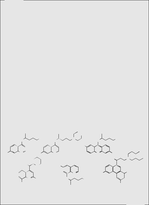
many years, the bark was obtained from South America, but cultivation was eventually established by the English in India, and by the Dutch in Java, until just before the Second World War, when almost all the world’s supply came from Java. When this source was cut off by Japan in the Second World War, a range of synthetic antimalarial drugs was hastily produced as an alternative to quinine. Many of these compounds were based on the quinine structure. Of the wide range of compounds produced, chloroquine, primaquine, and mefloquine (Figure 6.91) are important antimalarials. Primaquine is exceptional in having an 8-aminoquinoline structure, whereas chloroquine and mefloquine retain the 4- substituted quinoline as in quinine. The acridine derivative mepacrine (Figure 6.91), though not now used for malaria treatment, is of value in other protozoal infections. Halofantrine (Figure 6.91) dispenses with the heterocyclic ring system completely, and is based on phenanthrene. At one time, synthetic antimalarials had almost entirely superseded natural quinine, but the emergence of Plasmodium falciparum strains resistant to the synthetic drugs, especially the widely used prophylactic chloroquine, has resulted in reintroduction of quinine. Mefloquine is currently active against chloroquine-resistant strains, but, whilst ten times as active as quinine, does produce gastrointestinal upsets and dizziness, and can trigger psychological problems such as depression, panic, or psychosis in some patients. The ability of P. falciparum to develop resistance to modern drugs means malaria still remains a huge health problem, and is probably the major single cause of deaths in the modern world. Chloroquine and its derivative hydroxychloroquine (Figure 6.91), although antimalarials, are also used to suppress the disease process in rheumatoid arthritis.
Quinine (Figure 6.88), administered as free base or salts, continues to be used for treatment of multidrug-resistant malaria, though it is not suitable for prophylaxis. The specific mechanism of action is not thoroughly understood, though it is believed to prevent polymerization of toxic haemoglobin breakdown products formed by the parasite (see artemisinin, page 200). Vastly larger amounts of the alkaloid are consumed in beverages, including vermouth and tonic water. It is amusing to realize that gin was originally added to quinine to make the bitter antimalarial more palatable. Typically, the quinine dosage was up to 600 mg three times a
|
|
|
|
|
|
|
OH |
|
|
|
|
|
RS |
NEt2 |
|
RS |
N |
|
RS |
NEt2 |
|
|
|
HN |
|
|
|
|||||
|
|
|
|
HN |
|
|
HN |
|
|
|
|
|
|
|
|
|
|
MeO |
|
|
|
Cl |
|
N |
Cl |
|
N |
|
|
N |
Cl |
|
|
|
|
|
|
||||||
|
|
|
|
|
||||||
|
chloroquine |
|
hydroxychloroquine |
|
|
mepacrine |
HO RS |
N |
||
|
|
|
|
|
|
|
|
|
||
|
|
HO RS |
SR |
|
MeO |
|
|
|
|
|
|
|
N |
|
|
|
|
|
|
||
|
|
|
|
|
|
|
|
|
||
|
|
|
|
|
|
|
|
|
|
|
|
|
|
H |
|
|
|
|
|
|
|
Cl
|
|
HN |
N |
F3C |
|
|
|
|
|
||||
|
N |
RS |
|
|
|
|
|
CF3 |
|
NH2 |
|||
CF3 |
|
|
||||
|
|
Cl |
||||
mefloquine |
primaquine |
halofantrine |
||||
Figure 6.91
(Continues)
364 |
ALKALOIDS |
(Continued )
day. Quinine in tonic water is now the mixer added to gin, though the amounts of quinine used (about 80 mg l−1) are well below that providing antimalarial protection. Quinine also has a skeletal muscle relaxant effect with a mild curare-like action. It thus finds use in the prevention and treatment of nocturnal leg cramp, a painful condition affecting many individuals, especially the elderly.
Quinidine (Figure 6.88) is the principal cinchona alkaloid used therapeutically, and is administered to treat cardiac arrhymias. It inhibits fibrillation, the uncoordinated contraction of muscle fibres in the heart. It is rapidly absorbed by the gastrointestinal tract and overdose can be hazardous, leading to diastolic arrest.
Quinidine, cinchonine, and cinchonidine also have antimalarial properties, but these alkaloids are not as effective as quinine. The cardiac effect makes quinidine unsuitable as an antimalarial. However, mixtures of total Cinchona alkaloids, even though low in quinine content, are acceptable antimalarial agents. This mixture, termed totaquine, has served as a substitute for quinine during shortages. Quinine-related alkaloids, especially quinidine, are also found in the bark of Remija pendunculata (Rubiaceae).
|
N |
|
|
|
N |
|
|
|
|
N |
N |
|
H |
|
|
|
|
β-carboline alkaloid |
pyrroloquinoline alkaloid |
|
(pyridoindole) |
|
|
Figure 6.92
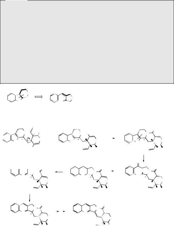
Camptothecin (Figure 6.93) from Camptotheca acuminata (Nyssaceae) is a further example of a quinoline-containing structure that is actually derived by modification of an indole system. The main rearrangement process is that the original β- carboline 6–5–6 ring system becomes a 6–6–5 pyrroloquinoline by ring expansion of the indole
|
|
NH H OGlc |
|
|
|
ester hydrolysis, |
||
|
|
|
|
NH CO2Me |
lactam formation |
|||
|
|
O |
≡ |
|
H |
|
|
|
|
|
|
|
|
|
|||
N |
H |
|
N H |
O |
||||
|
||||||||
H |
|
|
|
|
|
|||
H |
|
|
|
H |
OGlc |
|||
|
|
CO2Me |
|
|
H |
|||
strictosidine |
|
|
|
|
|
|
||
reduction of carbonyl then dehydration to give pyridine ring


 O N
O N
N
H
O
H
deoxypumiloside |
H OGlc |
|
O |
aldol-type |
||||
|
condensation |
|||||
|
|
|
|
|||
|
|
|
|
O |
||
|
|
|
|
|||
|
|
|
|
N |
|
|
|
|
|
|
|
||
|
N |
|
|
|
||
|
H H |
O |
||||
|
|
|
|
|||
|
|
|
|
H |
||
pumiloside |
H OGlc |
|||||
|
|
O |
|
|
|
N |
|
|
|
H |
O |
N |
H |
|
|
|
|
||
H |
|
|
OGlc |
strictosamide |
H |
||
|
|
||
|
|
oxidation of double |
|
O |
|
bond to yield two |
|
|
carbonyls |
|
|
|
|
|
|
|
|
O |
|
|
O N |
|
|
N |
|
|
|
H |
H |
|
O |
|
|
H |
|
|
|
|
|
|
|
H |
OGlc |
allylic isomerization
|
O |
|
|
|
O |
|
||
|
N |
|
|
|
N |
|
||
N |
|
|
|
|
|
N |
|
|
|
|
|
|
|
|
|
||
H |
O |
|
|
|
|
O |
||
|
|
|
|
|
||||
|
H OGlc |
|
camptothecin |
HO |
O |
|||
|
|
|
|
|
|
|
||
Figure 6.93

ALKALOIDS DERIVED FROM TRYPTOPHAN |
365 |
heterocycle (Figure 6.92). In camptothecin, the iridoid portion from strictosidine is effectively still intact, the original ester function being utilized in forming an amide linkage to the secondary amine. This occurs relatively early, in that strictosamide is an intermediate. Pumiloside
(also isolated from C. acuminata) and deoxypumiloside are potential intermediates. Steps beyond are not yet defined, but involve relatively straightforward oxidation and reduction processes (Figure 6.93).
Pyrroloindole Alkaloids
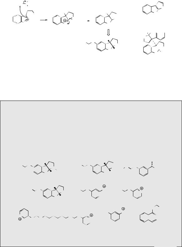
Both C-2 and C-3 of the indole ring can be regarded as nucleophilic, but reactions involving C-2 appear to be the most common in alkaloid biosynthesis. There are examples where the nucleophilic character of C-3 is exploited, however, and the rare pyrroloindole skeleton typified by physostigmine (eserine) (Figure 6.95) is a likely case. A suggested pathway to physostigmine is by C-3 methylation of tryptamine, followed by ring
Camptothecin
Camptothecin (Figure 6.93) and derivatives are obtained from the Chinese tree Camptotheca acuminata (Nyssaceae). Seeds yield about 0.3% camptothecin, bark about 0.2%, and leaves up to 0.4%. Camptotheca acuminata is found only in Tibet and West China, but other sources of camptothecin such as Nothapodytes foetida (formerly Mappia foetida) (Icacinaceae), Merilliodendron megacarpum (Icacinaceae), Pyrenacantha klaineana
(Icacinaceae), Ophiorrhiza mungos (Rubiaceae), and Ervatmia heyneana (Apocynaceae) have been discovered. In limited clinical trials camptothecin showed broad-spectrum anticancer activity, but toxicity and poor solubility were problems. The natural 10hydroxycamptothecin (about 0.05% in the bark of C. acuminata) is more active than camptothecin, and is used in China against cancers of the neck and head. Synthetic analogues 9-aminocamptothecin (Figure 6.94) and the water-soluble derivatives topotecan and irinotecan (Figure 6.94) showed good responses in a number of cancers; topotecan and irinotecan are now available for the treatment of ovarian cancer and colorectal cancer, respectively. Irinotecan is a carbamate pro-drug of 10-hydroxy-7-ethylcamptothecin, and is converted into the active drug by liver enzymes. These agents act by inhibition of the enzyme topoisomerase I, which is involved in DNA replication and reassembly, by binding to and stabilizing a covalent DNA–topoisomerase complex (see page 137). Camptothecin has also been shown to have potentially useful activity against pathogenic protozoa such as Trypanosoma brucei and Leishmania donovani, which cause sleeping sickness and leishmaniasis respectively. Again, this is due to topoisomerase I inhibition.
|
NH2 |
|
NMe2 |
N |
|
|
|
|
|
|
|
|
|
|
|
||
|
9 |
O |
HO |
O |
N O |
O |
||
10 |
|
|||||||
|
|
N |
|
N |
|
|
|
N |
|
|
|
O |
|||||
|
|
N |
|
N |
|
N |
||
|
|
|
|
|
|
|||
|
|
|
O |
O |
|
|
O |
|
|
|
OH O |
OH O |
|
|
OH O |
||
|
|
9-aminocamptothecin |
|
topotecan |
|
|
|
irinotecan |
Figure 6.94
366 |
ALKALOIDS |
Ad
Me S
R
N H
tryptamine
C-alkylation at C-3 due to nucleophilic character
Me
NH2
nucleophilic attack on to iminium ion
|
|
|
|
Me |
|
||
NH2 |
|
|
|
|
|
|
NH |
|
|
|
|
|
|||
N |
|
|
|
|
N |
|
|
H |
|
|
|
|
H |
|
|
MeHN |
|
|
|
Me |
|
||
|
|
O |
|
||||
|
|
|
|
|
|
|
NMe |
|
|
O |
|
|
N |
H |
|
|
|
|
|
|
|
||
|
|
|
|
|
|
Me |
|
|
|
|
|
physostigmine |
|
||
|
|
|
|
(eserine) |
|
||
Figure 6.95
|
3 |
|
|
4 |
|
3a |
2 |
|
|
|
|
5 |
|
|
NH |
|
|
|
|
6 |
|
8a |
1 |
|
|||
|
8 N |
|
|
|
|
|
|
7 |
|
H |
|
pyrroloindole
H
H N

MeN
 NMe
NMe
N H H
chimonanthine
formation involving attack of the primary amine function on to the iminium ion (Figure 6.95). Further substitution is then necessary. Dimers with this ring system are also known, e.g. chimonanthine
(Figure 6.95) from Chimonanthus fragrans (Calycanthaceae), the point of coupling being C-3 of the indole, and an analogous radical reaction may be proposed. Physostigmine is found in seeds of
Physostigma
Physostigma venenosum (Leguminosae/Fabaceae) is a perennial woody climbing plant found on the banks of streams in West Africa. The seeds are known as Calabar beans (from Calabar, now part of Nigeria) and have an interesting history in the native culture as an ordeal poison. The accused was forced to swallow a potion of the ground seeds, and if the mixture was subsequently vomited, he/she was judged innocent and set free. If the poison took effect, the prisoner suffered progressive paralysis and died from cardiac and respiratory failure. It is said that slow consumption allows the poison to take effect, whilst emesis is induced by a rapid ingestion of the dose.
MeHN O |
|
Me |
|
|
|
MeHN |
|
|
O |
Me |
|
|
|
Et |
|
|
|||||||
|
|
|
|
|
|
|
|
|
|
|
|
|
|
|
|
|
|
|
|
||||
|
|
|
|
N |
CONHMe |
|
|
O |
Me |
|
N O |
NMe2 |
|||||||||||
|
|
|
|
|
|
|
|
|
|
|
|
|
|||||||||||
|
|
|
|
|
|
|
|
|
|
|
|
|
|
|
|
|
|
|
|
||||
O |
|
|
N |
|
H |
|
|
O |
|
N |
H |
|
|
|
|
|
|
|
|
|
|||
|
|
|
|
|
|
|
|
|
|
|
|
|
|
O |
|
|
|
||||||
|
|
|
|
Me |
|
|
|
|
|
|
|
|
Me |
|
|
|
|
|
|
|
|
|
|
|
|
|
eseramine |
|
|
|
|
|
|
|
physovenine |
|
|
|
|
|
|
rivastigmine |
|
|
|||
Me(CH2)6HN O |
Me |
|
Me2N |
|
|
O |
|
NMe3 |
Me2N |
|
|
O |
|
|
|||||||||
|
|
|
|
|
|
|
|
|
|
||||||||||||||
|
|
|
|
|
|
|
NMe |
|
|
|
|
|
|
|
|
|
|
|
|
|
NMe |
|
|
|
|
|
|
|
|
|
|
|
|
|
|
|
|
|
|
|
|
|
|
|
|
||
|
|
O |
|
|
N H |
|
O |
|
|
|
|
O |
|
O |
|||||||||
|
|
|
eptastigmine |
Me |
|
|
|
neostigmine |
|
|
pyridostigmine |
||||||||||||
|
|
|
|
|
|
|
|
|
|
|
|||||||||||||
|
|
|
|
|
|
|
|
|
|
|
|
|
|
|
|
|
|
|
|
|
|
|
|
O NHMe
MeN |
|
O |
Me |
|
|
HO |
|
NMe2Et |
|
|
||||
|
|
|
|
|
|
|||||||||
|
|
|
|
|
|
|
|
|
|
|
|
|||
|
|
|
|
|
N |
|
O |
|
|
|
|
|||
|
|
O N |
|
|
|
|
NMe |
|
|
|
|
|||
|
|
|
|
|
|
|
|
|
|
|
|
|
|
|
|
|
|
|
Me |
|
|
O |
|
edrophonium |
carbaryl |
||||
|
|
|
|
|
distigmine |
|
|
|
|
|
||||
Figure 6.96
(Continues)

ALKALOIDS DERIVED FROM TRYPTOPHAN |
367 |
(Continued )
|
|
|
|
|
|
|
oxidation to N-oxide |
Me |
|
||||||||
|
|
|
|
Me |
|
|
|
|
|
|
|
|
|
|
|||
MeHN |
O |
|
|
|
|
|
|
|
MeHN |
O |
Me |
||||||
|
|
|
|
|
|
|
NMe |
O |
|
|
|
|
|
N |
|||
|
|
|
|
|
|
|
|
|
|
||||||||
|
|
|
|
|
|
|
|
|
|
|
|
|
|
|
|
|
O |
O |
|
|
|
N |
H |
|
O |
|
N H |
||||||||
|
|
|
|
|
|
|
|
|
|
|
|||||||
|
|
|
|
|
Me |
|
|
|
|
|
|
|
|
|
|
Me |
|
|
|
physostigmine |
|
|
|
|
|
|
|
|
|
physostigmine N-oxide |
|
||||
hydrolysis of |
|
|
H2O |
|
oxidation to quinol, and then |
|
|||||||||||
|
|
|
|
||||||||||||||
carbamate |
|
|
|
|
|||||||||||||
|
|
|
|
|
|||||||||||||
|
|
|
|
to ortho-quinone |
|
|
|
||||||||||
|
|
|
Me |
|
Me |
|
|||||||||||
|
HO |
|
|
|
|
O |
|
||||||||||
|
|
|
|
|
|
|
|
|
|
|
|||||||
|
|
|
|
|
|
NMe |
|
O |
|
NMe |
|
||||||
|
|
|
|
|
|
|
|
|
|
|
|
|
|
|
|||
|
|
|
|
|
N |
H |
|
|
|
O |
N H |
|
|||||
|
|
|
|
|
|
|
|
|
|||||||||
|
|
|
|
|
Me |
|
|
|
|
|
|
|
|
|
Me |
|
|
|
|
|
eseroline |
|
|
|
|
|
|
|
|
|
rubreserine |
|
|||
MeHN O |
Me |
NMe |
||
|
|
|||
|
|
|
|
O |
|
|
|
|
|
O |
|
N |
H |
|
|
|
|
||
|
|
|
Me |
|
|
|
geneserine |
|
|
Figure 6.97
The seeds contain several alkaloids (alkaloid content about 1.5%), the major one (up to 0.3%) being physostigmine (eserine) (Figure 6.95). The unusual pyrroloindole ring system is also present in some of the minor alkaloids, e.g. eseramine (Figure 6.96), whilst physovenine (Figure 6.96) contains an undoubtedly related furanoindole system. Another alkaloid, geneserine (Figure 6.97), is an artefact produced by oxidation of physostigmine, incorporating oxygen into the ring system, probably by formation of an N-oxide and ring expansion. Solutions of physostigmine are not particularly stable in the presence of air and light, especially under alkaline conditions, oxidizing to a red quinone, rubeserine (Figure 6.97).
Physostigmine (eserine) is a reversible inhibitor of cholinesterase, preventing normal destruction of acetylcholine and thus enhancing cholinergic activity. Its major use is as a miotic, to contract the pupil of the eye, often to combat the effect of mydriatics such as atropine (see page 297). It also reduces intraocular pressure in the eye by increasing outflow of the aqueous humour, and is a valuable treatment for glaucoma, often in combination with pilocarpine (see page 380). Because it prolongs the effect of endogenous acetylcholine, physostigmine can be used as an antidote to anticholinergic poisons such as hyoscyamine/atropine (see page 297), and it also reverses the effects of competitive muscle relaxants such as curare, tubocurarine, atracurium, etc (see page 324). Anticholinesterase drugs are also of value in the treatment of Alzheimer’s disease, which is characterized by a dramatic decrease in functionality of the central cholinergic system. Use of acetylcholinesterase inhibitors can result in significant memory enhancement in patients, and analogues of physostigmine are presently in use (e.g. rivastigmine) or in advanced clinical trials (e.g. eptastigmine (Figure 6.96)). These analogues have a longer duration of action and less toxicity than physostigmine.
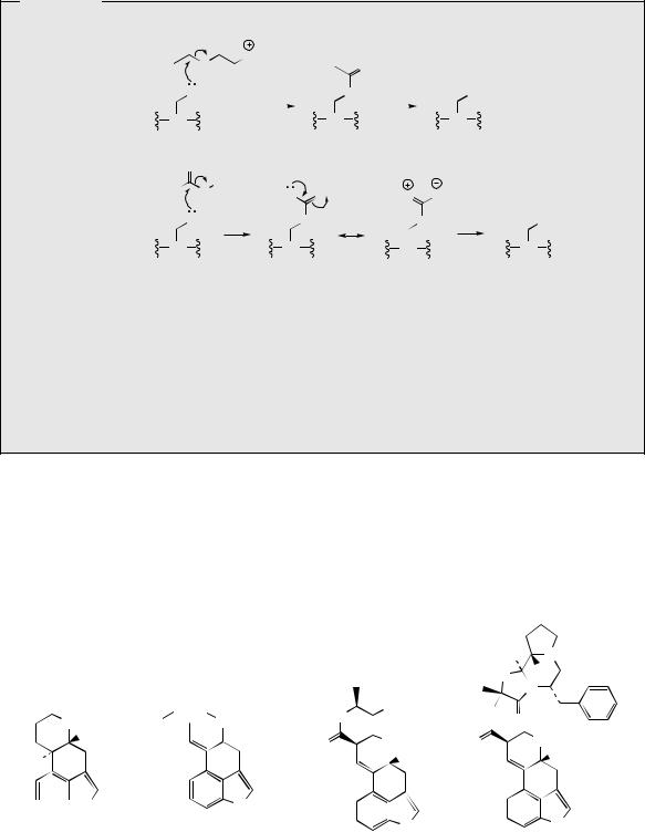
The biological activity of physostigmine resides primarily in the carbamate portion, which is transferred to the hydroxyl group of an active site serine in cholinesterase (Figure 6.98). The enzyme is only slowly regenerated by hydrolysis of this group, since resonance contributions reduce the reactivity of the carbonyl in the amide relative to the ester. Accordingly, cholinesterase becomes temporarily inactivated. Synthetic analogues of physostigmine which have been developed retain the carbamate residue, an aromatic ring to achieve binding and to provide a good leaving group, whilst ensuring water-solubility through possession of a quaternary ammonium system. Neostigmine, pyridostigmine, and distigmine (Figure 6.96) are examples of synthetic anticholinesterase drugs used primarily for enhancing neuromuscular transmission in the rare autoimmune condition myasthenia
(Continues)
368 ALKALOIDS
(Continued )
O |
|
|
|
|
|
|
|
|
|
O |
NMe3 |
O |
|
||||
|
|
|||||||
|
acetylcholine |
|
||||||
|
|
|
fast |
|
||||
|
OH |
|
|
O |
|
OH |
||
|
|
|
hydrolysis |
|||||
|
|
|
|
|
|
|
|
|
|
Ser |
Ser |
|
|
cholinesterase |
|
|
phenoxide system |
O |
|
|
|
Ar |
||
provides good |
MeHN O |
||
leaving group |
MeHN O |
||
|
|||
|
|
||
|
OH |
O |
|
|
Ser |
Ser |
|
|
cholinesterase |
|
Ser
resonance stabilization decreases carbonyl character
and slows rate of hydrolysis
MeHN O
slow |
OH |
O hydrolysis |
|
Ser |
Ser |
Figure 6.98
gravis, in which muscle weakness is caused by faulty transmission of nerve impulses. Edrophonium is a short-acting competitive blocker of the acetylcholinesterase active site, which is used to help diagnose myasthenia gravis. A number of carbamate insecticides, e.g. carbaryl (Figure 6.96), also depend on inhibition of cholinesterase for their action, insect acetylcholinesterase being more susceptible to such agents than the mammalian enzyme. Physostigmine displays little insecticidal action because of its poor lipid solubility.
Physostigma venenosum (Leguminosae/Fabaceae) and has played an important role in pharmacology because of its anticholinesterase activity. The inherent activity is in fact derived from the carbamate side-chain rather than the heterocyclic ring system, and this has led to a range of synthetic materials being developed.
Ergot Alkaloids
Ergot is a fungal disease commonly found on many wild and cultivated grasses, and is caused by species of Claviceps. The disease is eventually characterized by the formation of hard, seedlike ‘ergots’ instead of normal seeds, these structures,
|
|
7 |
6 |
|
8 |
|
|
NH |
|
9 |
|
10 |
|
H |
|
5 |
4 |
||
|
H |
|
||
|
11 |
|
3 |
|
|
|
|
||
12
2
13 
 N
N
14 H
ergoline
R
O
 NMe
NMe
 H
H
N
H
R = OH, (+)-lysergic acid R = NH2, ergine
|
|
|
|
HO |
N |
|
||
|
|
|
|
O |
H |
|
O |
|
|
|
|
|
|
||||
|
|
|
|
|
||||
|
|
|
|
N |
|
|||
|
|
|
|
|
|
|
||
|
|
OH |
HN |
|
|
|
|
|
HN |
|
O |
|
|
|
|||
|
|
|
|
|
|
|
|
|
O |
|
NMe |
O |
|
|
NMe |
|
|
|
|
H |
|
|
|
H |
|
|
|
|
N |
|
|
|
N |
|
|
|
|
|
|
|
|
|||
|
|
|
|
|
|
|||
|
ergometrine H |
|
|
|
H |
|
||
|
|
ergotamine |
|
|||||
Figure 6.99
ALKALOIDS DERIVED FROM TRYPTOPHAN |
369 |
called sclerotia, forming the resting stage of the |
|
NH2 |
HO2C |
|
fungus. The poisonous properties of ergots in grain, |
|
CO2H |
|
NMe |
|
|
H |
||
especially rye, for human or animal consump- |
C5 |
|
|
|
|
|
|
|
|
tion have long been recognized, and the causative |
|
|
|
|
agents are known to be a group of indole alka- |
|
N |
|
|
loids, referred to collectively as the ergot alkaloids |
|
|
N |
|
or ergolines (Figure 6.99). Under natural condi- |
|
H |
|
H |
|
Trp |
D-(+)-lysergic acid |
||
tions the alkaloids are elaborated by a combination |
|
|
|
|
of fungal and plant metabolism, but they can |
|
Figure 6.100 |
||
 OPP
OPP
|
|
CO2H |
|
|
N-methylation |
|
|
CO2H |
||||||
|
|
|
|
|
|
|||||||||
|
|
|
|
CO2H |
|
|
|
|
|
|
||||
|
|
|
|
|
|
|
|
|
|
|||||
|
|
|
|
|
|
|
|
|
|
|||||
|
|
|
|
|
|
|
|
|
|
|||||
|
|
NH2 |
|
|
NH2 |
|
SAM |
|
|
NHMe |
||||
|
N |
|
|
|
|
|
N |
|
|
|
|
|
N |
|
|
C-alkylation at position 4 |
|
|
|
|
|
||||||||
|
H |
which is nucleophilic due to |
H |
|
|
|
|
|
H |
|||||
|
L-Trp |
indole nitrogen |
|
|
4-dimethylallyl- |
|
|
|
|
|
|
O |
||
|
|
|
|
|
|
|
|
|
||||||
|
|
|
|
|
|
|
|
|
|
|||||
|
|
|
|
|
|
|
L-Trp |
|
|
|
|
|
|
|
|
|
|
|
|
|
|
|
|
|
|
|
|
|
|
O |
|
|
|
|
|
1,4-elimination of |
H |
|
||||||
epoxidation |
|
|
|
|||||||||||
H2O to give diene |
|
|
|
|||||||||||
|
|
H |
|
|||||||||||
|
|
|
|
|
|
|
|
|||||||
|
|
CO2H |
|
|
CO2H |
|
|
|
|
OH |
CO2H |
|||
|
|
|
|
|
|
|
|
|||||||
|
|
NHMe |
O |
NHMe |
|
|
|
|
|
|
NHMe |
|||
|
N |
|
|
|
|
|
N |
|
|
|
|
|
N |
|
|
|
|
|
|
|
|
|
|
|
|
||||
|
|
|
|
|
|
|
|
|
|
|
||||
|
H |
|
|
|
|
|
H |
|
|
|
|
|
H |
|
|
≡ |
|
|
|
|
|
|
Schiff base formation followed by reduction; a |
||||||
|
|
|
|
|
|
|
|
|||||||
H |
|
|
|
|
|
|
cis–trans isomerization |
is necessary to suitably |
||||||
|
|
|
|
|
|
|
|
position the amine and aldehyde groups |
||||||
O |
O |
|
|
|
|
|
|
OHC |
|
|
|
|
|
|
|
|
|
|
|
|
||||
|
|
|
NHMe |
|
|
|
|
|
|
NHMe |
|
|
|
|
|
|
|
NMe |
||||||
|
MeHN |
|
|
|
HO |
|
|
|
|
|
|
|
|
|
|
|
|
|
|
|
||||
|
|
|
H |
|
H |
|
|
|
|
|
|
|
H |
|
|
|
|
|
|
|
|
H |
||
|
|
|
O |
|
|
|
|
|
|
|
|
|
|
|
|
|
|
|
|
|||||
|
|
|
|
|
|
|
|
|
|
|
|
|
|
|
|
|
|
|
|
|
|
|||
|
|
|
– CO2 |
H |
NADPH |
H |
|
|
|
|
|
H |
||||||||||||
|
|
|
|
|
|
|
|
|
|
|
||||||||||||||
|
N |
|
|
|
|
|
N |
|
|
|
|
|
|
|
N |
|
|
|
|
|
|
|
|
N |
|
|
opening of epoxide |
|
|
|
|
|
|
|
|
|
|
|
|
|
|
|
|
||||||
|
|
|
|
|
|
|
|
|
|
|
|
|
|
|
|
|
|
|||||||
|
|
|
|
|
|
|
|
|
|
|
|
|
|
|
|
|
|
|||||||
|
|
|
|
|
|
|
|
|
|
|
|
|
|
|
|
|
|
|||||||
|
H |
|
|
H |
|
|
|
|
|
|
|
H |
|
|
|
|
|
|
|
|
H |
|||
|
|
allows ring closure and |
|
|
|
|
|
|
|
|
|
|
|
|
|
|
|
|
||||||
|
|
|
chanoclavine-I |
|
|
|
|
|
chanoclavine-I |
|
|
|
|
|
agroclavine |
|||||||||
|
|
|
decarboxylation |
|
|
|
|
|
|
|
|
|
aldehyde |
|
|
|
|
|
|
|
|
|
||
|
|
|
|
|
|
|
allylic isomerization; gives |
|
|
|
|
|
|
|
|
|
|
|
O2 |
|||||
|
|
|
|
|
|
|
|
|
|
|
|
|
|
|
|
|
|
|||||||
|
|
|
|
|
|
|
conjugation with aromatic |
|
|
|
|
|
|
|
|
|
|
|
NADPH |
|||||
|
|
|
|
|
|
|
|
|
|
|
|
|
|
|
|
|
|
|||||||
|
|
|
|
|
HO2C |
|
ring system |
|
|
|
|
HO2C |
|
|
|
|
|
|
|
|
|
|
|
|
|
|
|
|
|
|
NMe |
|
|
|
|
|
|
NMe |
|
|
|
HO |
|
|
|
NMe |
|||
|
|
|
|
|
H |
|
|
|
|
|
|
|
|
|
|
|
|
|
|
|||||
|
|
|
|
|
|
H |
|
|
|
|
|
|
|
H |
|
|
|
|
|
|
|
|
H |
|
|
|
|
|
|
|
|
|
|
|
|
|
|
H |
|
|
O |
H |
|||||||
|
|
|
|
|
|
|
|
|
|
|
|
|
|
|
|
|
|
|||||||
|
|
|
|
|
|
|
N |
|
|
|
|
|
|
|
N |
|
|
|
|
|
|
|
|
N |
|
|
|
|
|
|
|
|
|
|
|
|
|
|
|
|
|
|
|
|
|
|
|||
|
|
|
|
|
|
|
|
|
|
|
|
|
|
|
|
|
|
|
|
|
|
|||
|
|
|
|
|
|
|
|
|
|
|
|
|
|
|
|
|
|
|
|
|
|
|||
|
|
|
|
|
|
|
|
|
|
|
|
|
|
|
|
|
|
|
|
|
|
|||
|
|
|
|
|
|
|
H |
|
|
|
|
|
|
|
H |
|
|
|
|
|
|
|
|
H |
|
|
|
|
|
D-(+)-lysergic acid |
|
|
|
|
|
paspalic acid |
|
|
|
|
|
elymoclavine |
|||||||
Figure 6.101
370 |
ALKALOIDS |
be synthesized in cultures of suitable Claviceps species. Ergoline alkaloids have also been found in fungi belonging to genera Aspergillus, Rhizopus, and Penicillium, as well as Claviceps, and simple examples are also found in some plants of the Convolvulaceae such as Ipomoea and Rivea (morning glories) . Despite their toxicity, some of these alkaloids have valuable pharmacological activities and are used clinically on a routine
basis. |
|
|
Medicinally |
useful |
alkaloids are derivatives |
of (+)-lysergic |
acid |
(see Figure 6.99), which |
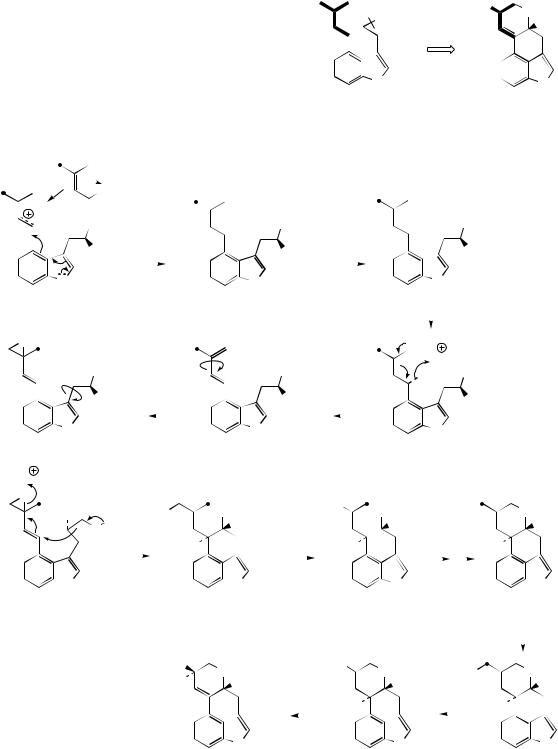
is typically bound as an amide with an amino alcohol as in ergometrine, or with a small polypeptide structure as in ergotamine. The building blocks for lysergic acid are tryptophan (less the carboxyl group) and an isoprene unit (Figure 6.100). Alkylation of tryptophan with dimethylallyl diphosphate gives 4-dimethylallyl-L- tryptophan, which then undergoes N-methylation (Figure 6.101). Formation of the tetracyclic ring system of lysergic acid is known to proceed through chanoclavine-I and agroclavine, though the mechanistic details are far from clear. Labelling studies have established that the
double bond in the dimethylallyl substituent must become a single bond on two separate occasions, allowing rotation to occur as new rings are established. This gives the appearance of cis–trans isomerizations as 4-dimethylallyl- L-tryptophan is transformed into chanoclavine- I, and as chanoclavine-I aldehyde cyclizes to agroclavine (Figure 6.101). A suggested sequence to account for the first of these is shown. In the later stages, agroclavine is hydroxylated to elymoclavine, further oxidation of the primary alcohol occurs giving paspalic acid, and lysergic acid then results from a spontaneous allylic isomerization.
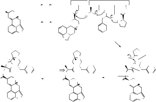
Simple derivatives of lysergic acid require the formation of amides; for example, ergine (Figure 6.99) in Rivea and Ipomoea species is lysergic acid amide, whilst ergometrine from Claviceps purpurea is the amide with 2-aminopro- panol. The more complex structures containing peptide fragments, e.g. ergotamine (Figure 6.99), are formed by sequentially adding amino acid residues to thioester-bound lysergic acid, giving a linear lysergyl–tripeptide covalently attached to the enzyme complex (Figure 6.102). Peptide formation
|
|
L-Ala, L-Phe, L-Pro |
ATP |
EnzSH |
|
|
|
|
|
|
Enzyme |
|
|
||||||||||||
|
|
|
|
|
|
|
|
|
|
|
|
|
|
|
|
|
|||||||||
HO2C |
|
|
|
|
|
|
|
|
|
|
O |
S |
|
|
|||||||||||
|
|
|
NMe |
|
|
|
|
|
|
|
|
|
|
|
|
|
|
|
|||||||
|
|
|
|
|
|
|
|
|
|
|
|
|
|
|
|
|
|
|
|||||||
|
|
|
|
H |
|
|
|
|
|
|
|
|
|
|
|
|
|
|
|
|
1 |
H2N |
|||
|
|
|
|
|
|
|
ATP |
EnzSH |
|
|
|
|
|
|
|
|
|
||||||||
|
|
|
|
|
|
|
|
|
|
|
|
|
|
|
|
L-Ala |
|||||||||
|
|
|
|
|
|
|
|
|
|
|
|
|
|
|
|
|
|
|
|
|
|
|
|||
|
|
|
|
|
|
|
|
|
|
|
|
|
|
|
|
|
|
|
|
|
|
NMe |
|
|
|
|
|
|
|
|
|
|
|
|
|
|
|
|
|
|
|
|
|
|
|
|
|
|
|
||
|
|
|
|
N |
|
activation via AMP |
|
|
|
|
|
|
H |
|
|
||||||||||
|
|
|
|
|
|
|
|
|
|
|
|
|
|||||||||||||
|
|
|
|
|
|
|
|
|
|
|
|
|
|||||||||||||
|
|
|
|
|
|
|
|
|
|
|
|
|
|
|
|||||||||||
|
|
|
|
|
esters, |
|
|
|
|
|
|
|
|
|
|
|
|
|
|
||||||
|
|
|
|
H |
|
|
|
|
|
|
|
|
|
|
|
|
|
|
|
||||||
D-(+)-lysergic acid |
then attachment to |
N |
|
|
|
|
|
|
|
|
|||||||||||||||
|
|
|
|
|
|
|
enzyme |
|
|
|
|
|
|
|
|
|
|
|
|
||||||
|
|
|
|
|
|
|
|
|
|
|
H |
|
|
|
|
|
|
|
|
||||||
|
|
|
|
|
|
|
(see Figure 7.15) |
|
|
|
|
|
|
|
|
|
|
||||||||
|
HO |
N |
|
|
|
hydroxylation then |
|
|
|
|
N |
|
|
||||||||||||
|
|
|
|
formation of |
|
|
|
|
|
|
|
|
|||||||||||||
|
O |
H |
|
O |
|
|
|
|
O |
|
|
|
H |
|
O |
||||||||||
|
|
|
|
hemiketal-like |
|
|
|
|
|
|
|||||||||||||||
|
|
|
|
|
|
|
|
|
|
||||||||||||||||
|
N |
|
|
|
|
|
|
|
|
|
N |
|
|
||||||||||||
|
|
|
|
|
|
|
product |
|
|
|
|
|
|
|
|
||||||||||
|
|
|
|
|
|
|
|
|
|
|
|
|
|
||||||||||||
HN |
|
|
|
|
|
|
|
|
|
|
|
|
|
|
O |
|
|
|
|
|
|
|
|
|
|
|
|
O |
|
|
|
|
|
|
|
|
|
|
|
|
HN |
|
|
|
|
O |
|
|
|||
|
|
|
|
|
|
|
|
|
|
|
|
|
|
|
|
|
|
|
|
|
|||||
O |
|
|
|
NMe |
|
|
|
|
|
|
|
|
|
|
O |
|
|
|
|
NMe |
|
|
|||
|
|
|
|
|
|
|
|
|
|
|
|
|
|
|
|
|
|
|
|||||||
|
|
|
|
H |
|
|
|
|
|
|
|
|
|
|
|
|
|
|
|
|
|||||
|
|
|
|
|
|
|
|
|
|
|
|
|
|
|
|
|
|
|
|
H |
|
|
|||
|
|
|
|
|
|
|
|
|
|
|
|
|
|
|
|
|
|
|
|
|
|
|
|
||
|
|
|
|
N |
|
|
|
|
|
|
|
|
|
|
|
|
|
|
|
|
N |
|
|
||
|
|
|
|
|
|
|
|
|
|
|
|
|
|
|
|
|
|
|
|
|
|
||||
|
|
|
|
|
|
|
|
|
|
|
|
|
|
|
|
|
|
|
|
|
|
||||
|
|
|
|
|
|
|
|
|
|
|
|
|
|
|
|
|
|
|
|
|
|
||||
|
|
|
|
|
|
|
|
|
|
|
|
|
|
|
|
|
|
|
|
|
|
||||
|
|
|
|
H |
|
|
|
|
|
|
|
|
|
|
|
|
|
|
|
|
|
|
|||
|
|
|
|
|
|
|
|
|
|
|
|
|
|
|
|
|
|
|
|
H |
|
|
|||
|
ergotamine |
|
|
|
|
|
|
|
|
|
|
|
|
|
|
|
|
|
|
||||||
|
Enzyme |
|
|
|
|
|
|
|
|
|
|
|
|||
|
|
|
|
|
|
|
|
|
the enzyme is known to |
||||||
|
S NH2 |
|
O |
S |
|
be comprised of two |
|||||||||
|
2 |
|
|
|
S |
|
|
subunits which bind |
|||||||
|
|
|
|
|
|
|
|||||||||
O |
|
|
H |
|
|
substrates as indicated |
|||||||||
|
|
|
|
||||||||||||
|
|
|
|
|
|
|
|
|
|
|
|||||
|
|
|
O |
3 |
N |
|
|
|
|
|
|
|
|
|
|
|
|
|
|
|
|
|
|
|
|
|
|
|
|||
|
|
|
|
|
|
|
L-Pro |
|
|
|
|
|
|
||
|
|
L-Phe |
|
|
sequential formation |
|
|
||||||||
|
|
|
|
|
|
|
|
|
|||||||
|
|
|
|
|
|
|
of tripeptide |
|
|
||||||
|
|
|
|
|
|
|
|
|
|
|
L-Pro |
|
|
||
|
lactam formation |
|
EnzS |
|
|
N |
|
|
|||||||
|
and release from |
|
|
|
|
|
|
O |
L-Phe |
||||||
|
|
|
|
|
|
||||||||||
|
enzyme |
|
|
|
O |
|
|
||||||||
|
|
|
|
|
|
|
|
||||||||
|
|
|
|
|
L-Ala |
|
HN |
|
|
|
|
|
|
||
|
|
|
|
|
|
|
|
|
|
|
|||||
|
|
|
|
|
|
|
|
|
|
|
|
|
|||
|
|
|
|
|
|
|
HN |
|
O |
|
|
|
|
|
|
|
|
|
|
|
|
|
O |
|
|
NMe |
|
|
|||
|
|
|
|
|
|
|
|
|
|
|
|||||
|
|
|
|
|
|
|
|
|
|
H |
|
|
|||
N
H
Figure 6.102
