

Atlas of breast surgery
.pdf86 |
7.2 |
Placement of Definitive Prosthesis |
tied. Once the prosthesis or expander is fitted into the muscular pocket, the sutures are tied down.Also, dissection of the muscular pocket should continue to the inferior extent of the pectoralis major muscle.
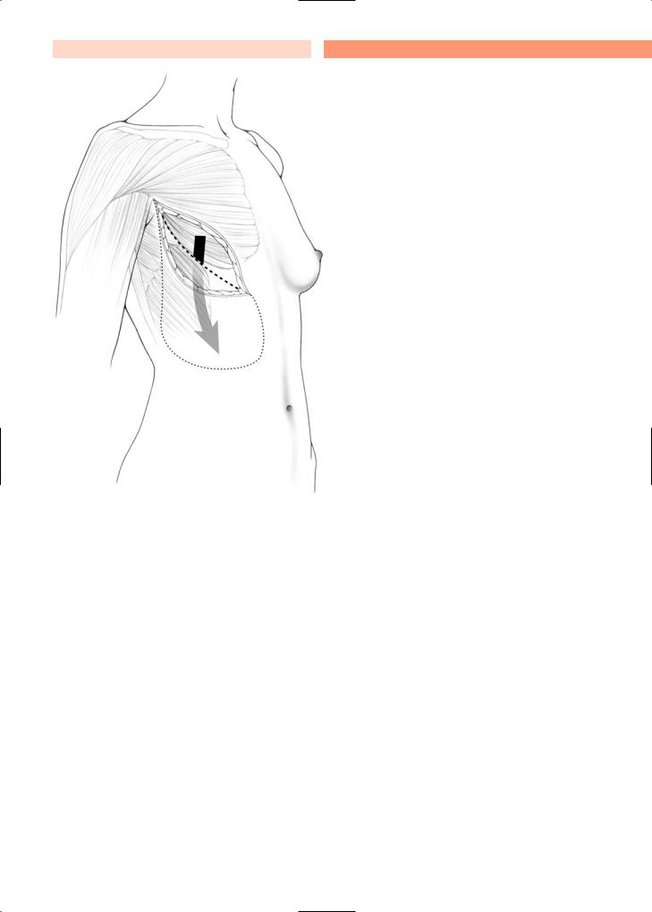
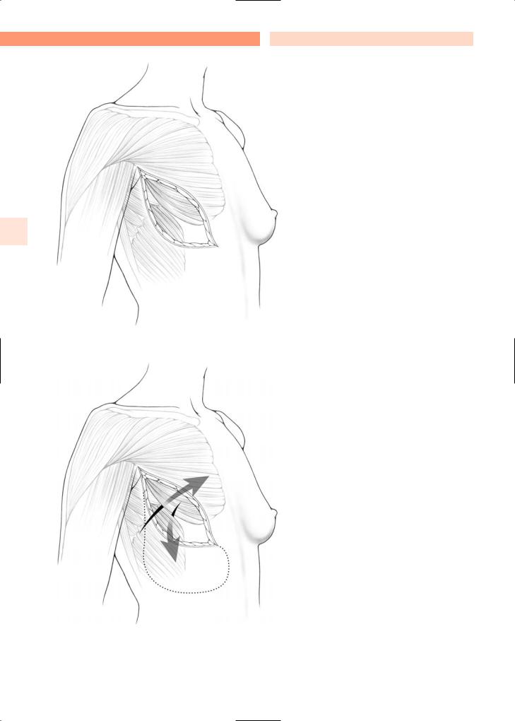
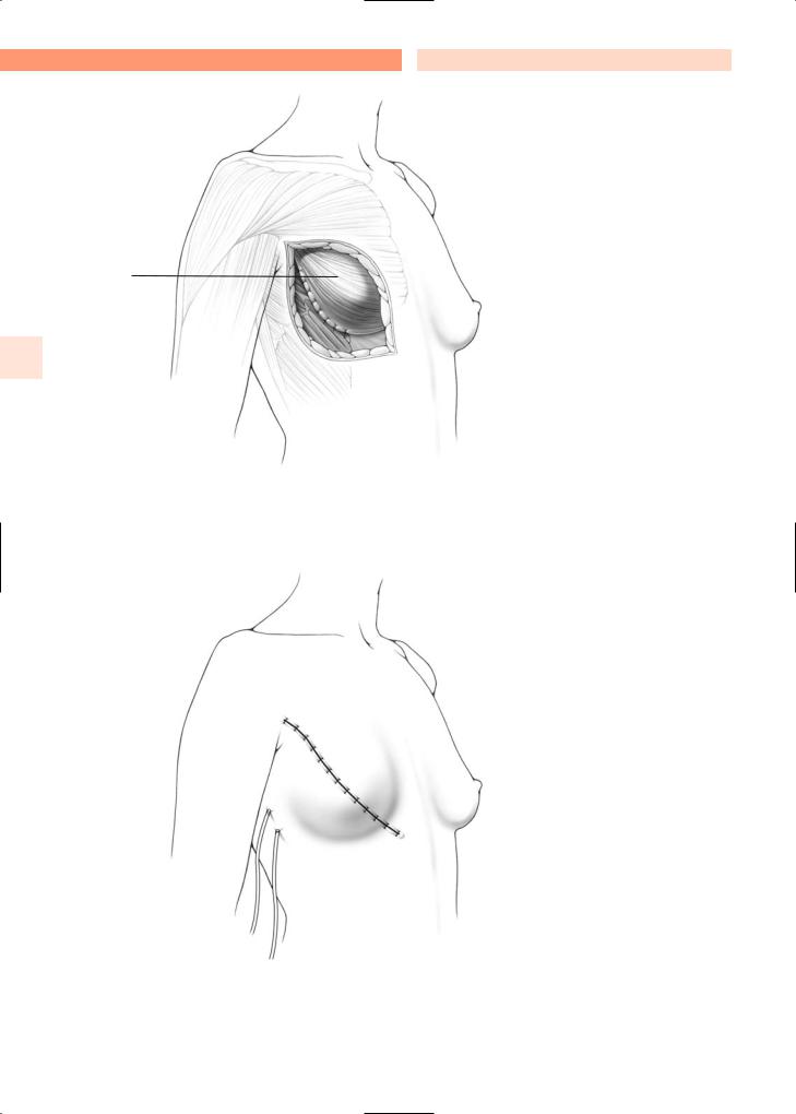
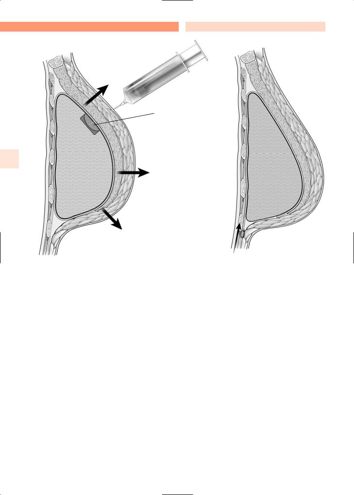
As illustrated, immediate breast reconstruction following mastectomy can be achieved with placement of a definitive prosthesis within the muscular pocket (Fig. 7.5a). This pocket is comprised of the pectoralis and serratus anterior muscles. Once the prosthesis is placed in the muscular pocket,the pock-
et should be closed with absorbable stitches. However, if the pectoralis muscle is wide enough, and if the lateral aspect of the skin is well supplied with blood, the muscular pocket can be left open laterally after placement of the prosthesis. Two Jackson–Pratt drains are generally left in the muscular pocket and brought out laterally after closure of the wound (Fig. 7.5b). These drains are hooked to bulb suction, and removed postoperatively once drainage is minimal (about 30 ml/day from each drain).
7
a
Fig. 7.1a, b. Immediate breast reconstruction using the “suspension technique”. A new mammary fold is created to bring tissue up before putting in a prosthesis
Plastic and Reconstructive Breast Surgery |
Chapter 7 |
87 |
b
Fig. 7.1a, b. Immediate breast reconstruction using the “suspension technique”. A new mammary fold is created to bring tissue up before putting in a prosthesis
88 |
7.2 |
Placement of Definitive Prosthesis |
7
Fig. 7.2. Immediate reconstruction after Patey mastectomy
Fig. 7.3. Serratus muscle is brought up and pectoralis muscle brought down to create a pocket for a prosthesis

Plastic and Reconstructive Breast Surgery |
Chapter 7 |
89 |
a
b
c
a
b
c
Fig. 7.4. Limits of the subpectoral undermining
90 |
7.2 |
Placement of Definitive Prosthesis |
Prosthesis |
|
underneath |
• |
muscle |
7
a
b
Fig. 7.5a, b. Immediate breast reconstruction, with placement of the prosthesis in the muscular pocket
Plastic and Reconstructive Breast Surgery
7.3 Breast Reconstruction with Expander
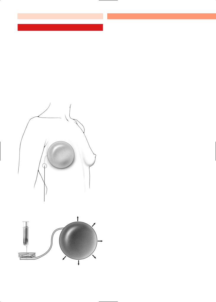
A muscular pocket is created, as described previously for placement of a definitive prosthesis. The pocket is comprised of the pectoralis major and serratus anterior muscles. The expander is placed in the muscular pocket and the catheter and port are tunneled and brought to a position under the skin in the axilla (Fig. 7.6).
Alternatively, in some cases, the port is located on the surface of the implant itself (Fig. 7.7). When the port is located on the sheet of the implant itself, a magnetic device can help to localize the port and thereby guide the inflation.
Chapter 7 |
91 |
The expander is inflated by passing a 22-gauge needle through the port and injecting saline solution through it. This is done once or twice each week until the expander is inflated such that the area of the nipple projection is at the same level bilaterally. This will provide a degree of ptosis. After suitable expansion, the expander is replaced by a permanent prosthesis.
Figures 7.6 and 7.7a, b illustrate the principle of cutaneous expansion, which is necessary to obtain a degree of ptosis. The ptosis is achieved with gradual inflation of the prosthesis by injecting saline solution through the port once or twice each week over a period of several weeks. An over-expansion is recommended by many surgeons, in order to gain the additional tissue required for a more natural ptosis.
•
Subcutaneous port
Fig. 7.6. Breast reconstruction with expander
92 |
7.3 |
Breast Reconstruction with Expander |
•
•
7
Port inside prosthesis
Metal shield for protection
Fig. 7.7a. Cutaneous expander |
Fig. 7.7b. Position of the definitive prothe- |
|
sis. After definitive substitution with a de- |
|
finitive prothesis same stiches in the sulcus |
|
improve the natural ptosis |
Plastic and Reconstructive Breast Surgery |
Chapter 7 |
93 |
7.4Hypothesis to Explain Capsular Contracture
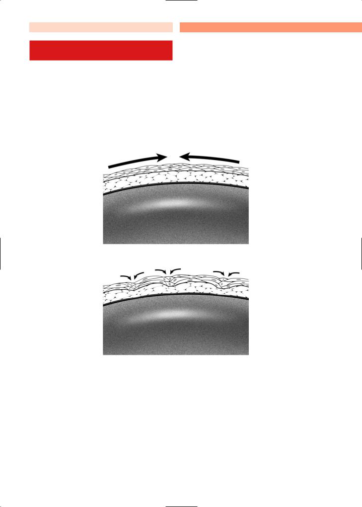
Specialized fibroblasts that have the ability to contract will migrate to the area around breast implants and expanders. These contractile fibroblasts, known as myofibroblasts, are responsible for wound contraction, and seem also to be responsible for capsular contraction. After a wound heals, myofibroblasts generally disappear. However, myofibroblasts persist in capsules that form around breast prostheses, and
this might explain the mechanism for capsular contracture [2].
As shown in the accompanying illustrations, when an implant is placed in the breast, it becomes lined with contractile fibroblasts (Fig. 7.8). Thus, the implant can be viewed as a three-dimensional wound. The contractile fibroblasts are responsible for initial contracture, but pressure from the implant prevents further contracture. Firmer implants should therefore be less prone to capsular contracture than softer implants. If the implant ruptures, this may result in a worse contracture.
Fig. 7.8. A hypothesis to explain capsular contracture

94 |
7.5 |
Suspension Technique |
7.5Suspension Technique (Advancement Abdominal Flap)
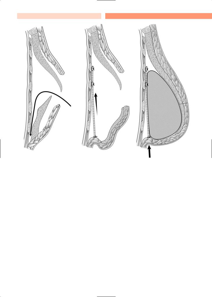
If a permanent prosthesis is to be placed following a mastectomy, the cosmetic appearance can often be improved with the “suspension technique.” First, a wide area of abdominal tissue is undermined inferiorly, just along the anterior surface of the rectus abdominis muscle (Fig. 7.9a). In this manner, the skin and subcutaneous tissue along the inferior aspect of the mastectomy wound are mobilized. The head of the operating table should then be raised, placing the
patient in a semi-sitting position. The abdominal flap that has been mobilized is then pulled up and maintained with a triangular nonabsorbable mesh that is fixed at the level of the future inframammary fold. The mesh is brought up superiorly, posterior to the pectoralis major muscle, as shown (Figs. 7.9b, 7.10), and the superior aspect of the mesh is attached to the costal cartilage with two nonabsorbable stitches (3–0 Prolene), as depicted in the illustration (Fig. 7.9b). The prosthesis is placed anterior to the mesh and posterior to the pectoralis muscle as shown (Fig. 7.10). This “suspension technique,” or abdominal flap advancement, was first described by Rietjens [3].
7 |
Nylon or titan mesh placed and |
fixed at inframammary fold |
•
a |
b |
Fig. 7.9a, b. Immediate breast reconstruction, using the “suspension technique II”
Plastic and Reconstructive Breast Surgery |
Chapter 7 |
95 |
a |
b |
c |
Fig. 7.10. a Immediate breast reconstruction using the “suspension technique” (profile view). b With mesh. c Suspension technique with mesh and subpectoral prothesis