

Pocket Atlas of Sectional Anatomy, Volume 2 Thorax, Abdomen and Pelvis
.pdfaxial 23
1 |
2 |
3 |
4 |
5 |
6 |
7 |
8 |
9 |
10 |
11 |
12 |
13 |
14 |
15 |
16 |
17 |
18 |
|
|
|
|
|
|
|
|
|
|
|
|
|
|
|
|
|
|
|
|
|
|
|
|
|
|
|
|
|
|
|
|
|
|
|
|
|
19 |
20 |
21 |
22 |
23 |
24 |
25 |
26 |
27 |
28 |
29 |
30 |
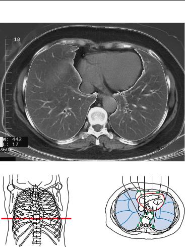
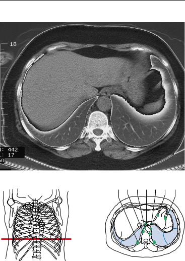
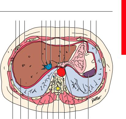
1. |
Latissimus dorsi muscle |
|
|
|
19. |
Rib |
|
|
|
|||
2. |
Serratus anterior muscle |
|
|
|
20. |
Intercostal muscle |
||||||
3. |
Right lung |
|
|
|
|
|
21. |
Azygos vein |
|
|||
4. |
Right atrium |
|
|
|
|
|
22. |
Erector spinae muscle |
||||
5. |
Internal thoracic artery and vein |
|
23. |
Trapezius muscle |
||||||||
6. |
Right coronary artery |
|
|
|
|
24. |
Spinal cord |
|
||||
7. |
Right atrioventricular (tricuspid) |
|
25. |
Spinous process |
||||||||
|
valve |
|
|
|
|
|
|
26. |
Esophagus |
|
||
8. |
Sternum (body) |
|
|
|
|
|
27. |
Sympathetic trunk |
||||
9. |
Right ventricle |
|
|
|
|
|
28. |
Thoracic duct |
||||
10. |
Interventricular septum |
|
|
|
29. |
Descending aorta |
||||||
11. |
Left atrium |
|
|
|
|
|
30. |
Left lung |
|
|
||
12. |
Rib (costal cartilage) |
|
|
|
|
31. |
Paramammary lymph nodes |
|||||
13. |
Left coronary artery (interventricu- |
|
32. |
Paravertebral lymph nodes |
||||||||
|
lar branch) |
|
|
|
|
|
33. |
Lateral pericardial lymph nodes |
||||
14. |
Left ventricle |
|
|
|
|
|
34. |
Parasternal lymph nodes |
||||
15. |
Coronary sinus |
|
|
|
|
|
35. |
Prepericardial lymph nodes |
||||
16. |
Left coronary artery (circumflex |
|
36. |
Juxtaesophageal lymph nodes |
||||||||
|
branch) |
|
|
|
|
|
|
37. |
Para-aortal lymph nodes |
|||
17. |
Phrenic nerve and pericardium |
|
|
38. |
Intercostal lymph nodes |
|||||||
18. |
Myocardium |
|
|
|
|
|
|
|
|
|
|
|
Moeller, Pocket Atlas of Sectional Anatomy, Vol. 2 © 2001 Thieme All rights reserved. Usage subject to terms and conditions of license.
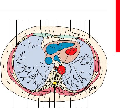
24 CT of the Thorax
30 31 32 33 34 35 36
Right Lung
4.Lateral segment of middle lobe
5.Medial segment of middle lobe
7.Medial basal (cardiac) segment of lower lobe
8.Anterior basal segment of lower lobe
9.Lateral basal segment of lower lobe
10Posterior basal segment of lower lobe
4 |
5 |
|
5 |
|
|
||
|
|
|
|
8 |
7 |
|
8 |
|
|
||
9 |
|
7 |
9 |
10 |
|
||
|
10 |
|
|
|
|
|
——= Borders of lung segments
——= Pericardium
Left Lung
5. Inferior lingular segment
7.Medial basal (cardiac) segment of lower lobe
8.Anterior basal segment of lower lobe
9.Lateral basal segment of lower lobe
10.Posterior basal segment of lower lobe
Moeller, Pocket Atlas of Sectional Anatomy, Vol. 2 © 2001 Thieme All rights reserved. Usage subject to terms and conditions of license.
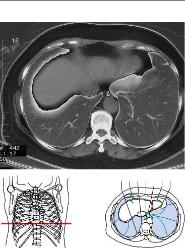
axial 25
1 |
2 |
3 |
4 |
5 |
6 |
7 |
8 |
9 |
10 |
11 |
12 |
13 |
14 |
15 |
16 |
|
|
|
|
|
|
|
|
|
|
|
|
|
|
|
|
|
|
|
|
|
|
|
|
|
|
|
|
|
|
|
|
|
17 |
18 |
19 |
20 |
21 |
22 |
23 |
24 |
25 |
26 |
27 |
28 |
29 |
1. |
Latissimus dorsi muscle |
|
|
|
18. |
Sympathetic trunk |
|||||||
2. |
Intercostal muscle |
|
|
|
|
19. |
Thoracic vertebra |
|
|||||
3. |
Rib |
|
|
|
|
|
20. |
Trapezius muscle |
|
||||
4. |
Right lung |
|
|
|
|
|
21. |
Erector spinae muscle |
|||||
5. |
Inferior vena cava |
|
|
|
|
22. |
Esophagus |
|
|
||||
6. |
Internal thoracic artery and vein |
23. |
Spinal cord |
|
|
||||||||
7. |
Right atrium |
|
|
|
|
|
24. |
Azygos vein |
|
|
|||
8. |
Xiphoid process |
|
|
|
|
25. |
Thoracic duct |
|
|
||||
9. |
Right coronary artery |
|
|
|
26. |
Vagus nerve |
|
|
|||||
10. |
Right atrioventricular (tricuspid) |
27. |
Descending aorta |
|
|||||||||
|
valve |
|
|
|
|
|
28. |
Left lung |
|
|
|
||
11. |
Coronary sinus |
|
|
|
|
|
29. |
Myocardium of the left ventricle |
|||||
12. |
Rib (costal cartilage) |
|
|
|
30. |
Paravertebral lymph nodes |
|||||||
13. |
Right ventricle |
|
|
|
|
|
31. |
Parasternal lymph nodes |
|||||
14. |
Circumflex branch of left coronary |
32. |
Prepericardial lymph nodes |
||||||||||
|
artery |
|
|
|
|
|
33. |
Juxtaesophageal lymph nodes |
|||||
15. |
Left ventricle |
|
|
|
|
|
34. |
Para-aortal lymph nodes |
|||||
16. |
Phrenic nerve and pericardium |
|
35. |
Intercostal lymph nodes |
|||||||||
17. |
Serratus anterior muscle |
|
|
36. |
Lateral pericardial lymph nodes |
||||||||
Moeller, Pocket Atlas of Sectional Anatomy, Vol. 2 © 2001 Thieme All rights reserved. Usage subject to terms and conditions of license.
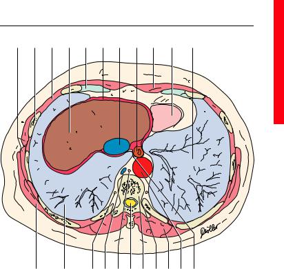
26 CT of the Thorax
Right Lung
5. Medial segment of middle lobe
8.Anterior basal segment of lower lobe
9.Lateral basal segment of lower lobe
10.Posterior basal segment of lower lobe
23 24 25 26 27 28 29
5 |
5 |
7 |
8 |
8 |
|
910 
 10 9
10 9
——= Borders of lung segments
——= Pericardium
Left Lung
5. Inferior lingular segment
7.Medial basal (cardiac) segment of lower lobe
8.Anterior basal segment of lower lobe
9.Lateral basal segment of lower lobe
10.Posterior basal segment of lower lobe
Moeller, Pocket Atlas of Sectional Anatomy, Vol. 2 © 2001 Thieme All rights reserved. Usage subject to terms and conditions of license.
axial 27
1 |
2 |
3 |
4 |
5 |
6 |
7 |
8 |
9 |
10 |
11 |
|
|
|
|
|
|
|
|
|
|
|
|
|
|
|
|
|
|
|
|
|
|
|
12 |
13 |
14 |
15 |
16 |
17 |
18 |
19 |
20 |
21 |
22 |
|
1. |
Serratus anterior muscle |
|
|
16. |
Thoracic duct |
|
||||||
2. |
Rib |
|
|
|
|
17. |
Spinal cord |
|
||||
3. |
Intercostal muscle |
|
|
|
18. |
Thoracic vertebra |
||||||
4. |
Liver |
|
|
|
|
19. |
Costovertebral joint |
|||||
5. |
Rib (costal cartilage) |
|
|
|
20. |
Erector spinae muscle |
||||||
6. |
Diaphragm |
|
|
|
|
21. |
Descending aorta |
|||||
7. |
Inferior vena cava |
|
|
|
22. |
Vagus nerve |
|
|||||
8. |
Esophagus |
|
|
|
|
23. |
Intercostal lymph nodes |
|||||
9. |
Rectus abdominis muscle |
|
|
24. |
Parasternal lymph nodes |
|||||||
10. |
Base of heart |
|
|
|
|
25. |
Paravertebral lymph nodes |
|||||
11. |
Left lung |
|
|
|
|
26. |
Para-aortal lymph nodes |
|||||
12. |
Latissimus dorsi muscle |
|
|
|
27. |
Superior phrenic lymph nodes |
||||||
13. |
Right lung |
|
|
|
|
28. |
Prepericardial lymph nodes |
|||||
14. |
Sympathetic trunk |
|
|
|
29. |
Lateral pericardial lymph nodes |
||||||
15. |
Azygos vein |
|
|
|
|
|
|
|
|
|
|
|
Moeller, Pocket Atlas of Sectional Anatomy, Vol. 2 © 2001 Thieme All rights reserved. Usage subject to terms and conditions of license.
28 CT of the Thorax
26 27 28 29 30 31 32
Right Lung
9.Lateral basal segment of lower lobe
10.Posterior basal segment of lower lobe
5 |
8 |
9 |
|
10 |
9 |
10 |
|
—— = Borders of lung segments
Left Lung
5. Inferior lingular segment
8.Anterior basal segment of lower lobe
9.Lateral basal segment of lower lobe
10.Posterior basal segment of lower lobe
Moeller, Pocket Atlas of Sectional Anatomy, Vol. 2 © 2001 Thieme All rights reserved. Usage subject to terms and conditions of license.
axial 29
1 |
2 |
3 |
4 |
5 |
6 |
7 |
8 |
9 |
10 |
11 |
12 |
3 |
13 |
|
|
|
|
|
|
|
|
|
|
|
|
|
|
|
|
|
|
|
|
|
|
|
|
|
|
|
|
|
14 |
15 |
16 |
17 |
18 |
19 |
20 |
21 |
22 |
23 |
24 |
25 |
|
1. |
Intercostal muscle |
|
|
|
|
17. |
Sympathetic trunk |
||||||
2. |
Rib |
|
|
|
|
|
|
18. |
Erector spinae muscle |
||||
3. |
External oblique muscle |
|
|
|
19. |
Azygos vein |
|
||||||
4. |
Right hepatic vein |
|
|
|
|
20. |
Thoracic duct |
|
|||||
5. |
Inferior vena cava |
|
|
|
|
21. |
Spinal cord |
|
|||||
6. |
Left hepatic vein |
|
|
|
|
22. |
Thoracic vertebra |
||||||
7. |
Diaphragm |
|
|
|
|
|
23. |
Descending aorta |
|||||
8. |
Vagus nerve |
|
|
|
|
|
24. |
Posterior vagal trunk |
|||||
9. |
Esophagus |
|
|
|
|
|
25. |
Spleen |
|
||||
10. |
Liver (left lobe) |
|
|
|
|
26. |
Intercostal lymph nodes |
||||||
11. |
Rectus abdominis muscle |
|
|
27. |
Paravertebral lymph nodes |
||||||||
12. |
Stomach |
|
|
|
|
|
|
28. |
Para-aortal lymph nodes |
||||
13. |
Left lung |
|
|
|
|
|
|
29. |
Superior phrenic lymph nodes |
||||
14. |
Latissimus dorsi muscle |
|
|
|
30. |
Inferior phrenic lymph nodes |
|||||||
15. |
Liver (right lobe) |
|
|
|
|
31. |
Right gastric lymph nodes |
||||||
16. |
Right lung |
|
|
|
|
|
32. |
Left gastroomental lymph nodes |
|||||
Moeller, Pocket Atlas of Sectional Anatomy, Vol. 2 © 2001 Thieme All rights reserved. Usage subject to terms and conditions of license.
30 MRI of the Thorax
34 |
|
35 |
|
36 |
1 |
37 |
|
|
2 |
|
3 |
|
|
38 |
|
6 |
|
|
|
||
|
|
|
|
39 |
|
|
|
|
5 |
|
10 |
40 |
|
|
|
|
|
|
—— = Border of lung segments —— = Pericardium
Right Lung
1. Apical segment of upper lobe
2. Posterior segment of upper lobe
3. Anterior segment of upper lobe
5. Medial segment of middle lobe
6. Superior segment of lower lobe
10. Posterior basal segment of lower lobe
Moeller, Pocket Atlas of Sectional Anatomy, Vol. 2 © 2001 Thieme All rights reserved. Usage subject to terms and conditions of license.
|
|
|
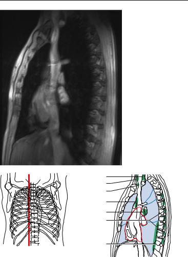
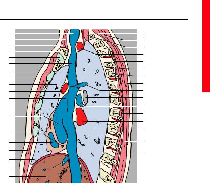
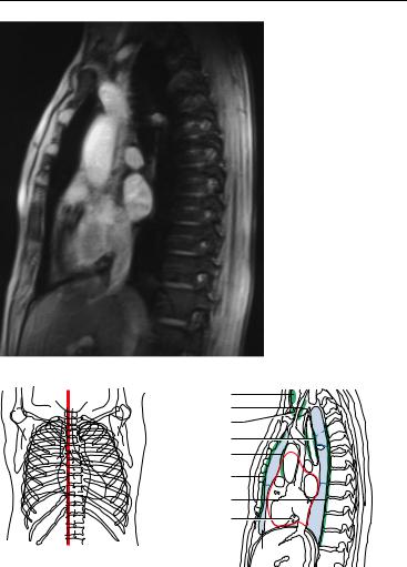
sagittal |
31 |
1 |
|
|
19 |
|
2 |
|
|
20 |
|
3 |
|
|
21 |
|
4 |
|
|
|
|
|
|
22 |
|
|
5 |
|
|
|
|
|
|
23 |
|
|
6 |
|
|
|
|
|
|
24 |
|
|
7 |
|
|
|
|
8 |
|
|
25 |
|
9 |
|
|
26 |
|
10 |
|
|
27 |
|
11 |
|
|
28 |
|
|
|
|
29 |
|
12 |
|
|
30 |
|
|
|
31 |
|
|
|
|
|
|
|
13 |
|
|
9 |
|
|
|
|
|
|
14 |
|
|
32 |
|
15 |
|
|
33 |
|
16 |
|
|
|
|
17 |
|
|
|
|
18 |
|
|
|
|
1. |
Jugular vein |
21. |
Longus colli muscle |
|
2. |
Sternothyroid muscle |
22. |
Subclavian artery |
|
3. |
Sternocleidomastoid muscle |
23. |
Rib |
|
4. |
Clavicle |
24. |
Posterior intercostal artery |
|
5. |
Sternoclavicular joint |
25. |
Vertebra |
|
6. |
Superior vena cava |
26. |
Trapezius muscle |
|
7. |
Sternum |
27. |
Azygos vein |
|
8. |
Pectoralis major muscle |
28. |
Right main stem bronchus |
|
9. |
Right lung |
29. |
Right pulmonary artery |
|
10. |
Ascending aorta |
30. |
Intercostal muscle |
|
11. |
Right auricle |
31. |
Pulmonary vein |
|
12. |
Right atrium |
32. |
Erector spinae muscle |
|
13. |
Right coronary artery |
33. |
Inferior vena cava |
|
14. |
Right ventricle |
34. |
Deep cervical lymph nodes |
|
15. |
Rectus abdominis muscle |
35. |
Superficial cervical lymph nodes |
|
16. |
Diaphragm |
36. |
Supraclavicular lymph nodes |
|
17. |
Hepatic veins |
37. |
Anterior mediastinal lymph nodes |
|
18. |
Liver |
38. |
Bronchopulmonary lymph nodes |
|
19. |
Semispinalis capitis muscle |
39. |
Posterior intercostal lymph nodes |
|
20. |
Splenius cervicis and capitis mus- |
40. |
Paravertebral lymph nodes |
|
|
cle |
|
|
|
Moeller, Pocket Atlas of Sectional Anatomy, Vol. 2 © 2001 Thieme All rights reserved. Usage subject to terms and conditions of license.
32 MRI of the Thorax
40 |
|
|
41 |
|
|
42 |
|
1 |
43 |
|
2 |
44 |
3 |
6 |
|
||
45 |
|
|
|
|
|
46 |
|
|
47 |
|
10 |
|
5 |
|
—— = Borders of lung segments —— = Pericardium
(Segments of the lungs, see page 2)
1. |
Sternocleidomastoid muscle |
6. |
Sternoclavicular joint |
2. |
Sternohyoid, sternothyroid and |
7. |
Brachiocephalic trunk |
|
omohyoid muscle |
8. |
Sternum |
3. |
Jugular vein |
9. |
Superior vena cava |
4. |
Subclavian artery |
10. |
Right lung |
5. |
Clavicle |
11. |
Ascending aorta |
Moeller, Pocket Atlas of Sectional Anatomy, Vol. 2 © 2001 Thieme All rights reserved. Usage subject to terms and conditions of license.