

Lymphocyte Recirculation and Homing |
4 |
What the USMLE Requires You To Know
•The structure and function ofthe secondary lymphoid organs
•The areas in which B and T lymphocytes localize in the peripheral lymphoid organs
•The role of chemokines and adhesion molecules in lymphocyte trafficking
Lymphocytes of the B- and T-cell lineages that have completed their selection in the bone marrow and thymus respectively are now mature, naive lymphocytes ready to begin their role in the surveillance of the body against invaders. These mature, naive lymphocytes willbegin the process of recirculation through the body, which is essen tial for ensuring that the limited number of cells with receptors for a specific antigen is enabled to search for that antigen throughout the body. Naive cells preferentially recirculate through the peripheral (secondary) lymphoid organs, the lymph nodes, spleen, and mucosal-associated lymphoid tissue (MALT) to maximize the chances of encounter with foreign antigen and thereby initiate specific immune responses.
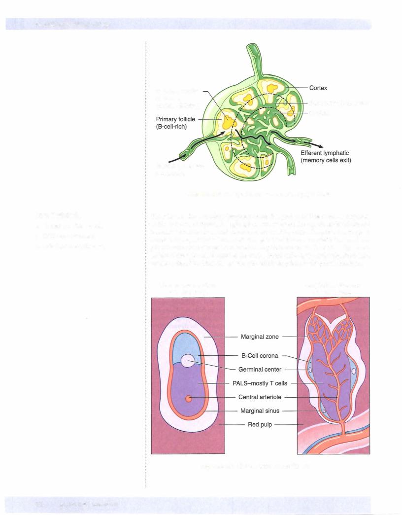
Lymph nodes are the small nodular aggregates of secondary lymphoid tissue found along the lymphatic channels of the body and are designed to initiate immune re sponses to tissue-borne antigens. Each lymph node is surrounded by a fibrous cap sule that is punctured by afferent lymphatics, which bring lymphinto the subcapsu lar sinus. The fluid percolates through an outer cortex area that contains aggregates of cells called follicles. Thelymph then passes into the inner medulla and the medul lary sinus before leaving the node through the hilum in an efferent lymphatic ves sel. Ultimately, lymph from throughout the body is collected into the thoracic duct, which empties into the vena cava and returns it to the blood.
In a Nutshell
Peripheral (Secondary) Lymphoid organs
•Lymph nodes, spleen, and MALT
•Sites offoreign antigen exposure
In
•Lymph nodes filtertissue fluids.
•Outer cortex contains follicles (B-cell areas).
•Paracortex is a T-cell area.
•Inner medulla contains macrophages.
MEDICAL 33
Chapter It •
Naive lymphocytes enter the lymph nodes from the blood through high endothelial venules (HEVs). This migration involves a multistep sequence of interactions be tween the lymphocyte and adhesion molecules on the endothelium. The initial low affinity interaction is mediated by homing receptors, called L-selectins, present on the lymphocytes, which bind to addressins present on the HEVs.
Naive T cells migrate into the paracortical areas under the influence ofa chemokine gradient. Naive B cells are guided along a chemokine gradient into the lymphoid follicles.
Less isknown aboutlymphocyte recirculation through the spleen. NaiveT cells home to the periarteriolar lymphoid sheaths (PALS), whereas naive B cells create lym phoid follicles outside ofthe T cell area.
The rate of recirculation of naive lymphocytes through the body by this system is quite high. It is estimated that each lymphocyte passes through each lymph node in the body once a day and through the spleen once every two days on average. This highrate oftrafficking maximizes the possibilitythat theverysmall number ofcells havinga specificantigenreceptorhave themaximumpossibilityofencounteringthat antigen when it is present.
Chapter Summary
•Mature naive lymphocytes recirculate through the peripheral lymphoid organs (lymph nodes and spleen) to search for antigen.
•Lymph nodes are designed to filter antigens from the tissue fluids.
•Lymph enters through afferent lymphatics and percolates through an outer cortex and inner medulla before leaving through the efferent lymphatic in the hilus.
•The spleen is designed to filterantigens from blood. Blood enters through a single splenic artery, which branches into arterioles that become surrounded by cuffs of lymphocytes (periarteriolar lymphoid sheaths) with follicles and macro phages nearby.
•Lymphocytes leave the bloodstream to enter lymph nodes through high endothe lial venules.
•L-selectins on lymphocytes bind to addressins on the high endothelial venules, and chemokine receptors mediate the homing of specific cells to specific areas.
•T lymphocytes migrate into the lymph node paracortical areas.
•B lymphocytes migrate into the lymph node follicles.
•In the spleen, T lymphocytes are attracted to the periarteriolar lymphoid sheaths.
•B cells entering the spleen form a corona outside the T cell area.
•Every lymphocyte passes through every lymph node once per day and through the spleen once everytwo days on average.
Lymphocyte Recirculation and Homing
In a Nutshell
•Naive lymphocytes leave the blood through HEVs.
•L-selectins on lymphocytes bind to ad dressins on HEVs.
•Naive T cells home to the lymph node paracortex or the splenic PALS.
•Naive B cells create a corona outside the T cell area.
MEDICAL 35

Section I • Immunology
Review Questions
1.A lymph node biopsy ofa 6-year-old boy shows markedly decreased numbers oflymphocytes in the paracortical areas. Analysis ofhis peripheral blood leu kocytes is likelyto shownormalto elevated numbers ofcells expressing surface
2.
(A)CD2
(B)CD3
(C)CD4
(D)CDS
(E)CD19
A 65-year-old woman was involved in an automobile accident that necessitated the removal ofher spleen. To which ofthe following pathogens would she have the most increased susceptibility?
(A)Babesia microti
(B)Bordetella pertussis
(C) Corynebacterium diphtheriae
(D)Enteroaggregative Escherichia coli
(E)Human papilloma virus
3.A 4-year-old boy is referred to a specialist forthe diagnosis ofa possible immu nologic problem. The child has extremely elevated white blood cell counts, with a profound lymphocytosis. A biopsy performed on a cervical lymph node reveals extreme hypocellularity in both cortical and paracortical areas. Absence ofwhich ofthe followingleukocytesurface molecules couldresult in this clinical picture?
4.
(A)Addressins
(B)Chemokines
(C) Immunoglobulin family cell adhesion molecules
(D)Integrins
(E)L-selectins
A 6-year-old child is taken to his pediatrician because the parents are alarmed about an indurated fluctuant mass on the posterior aspect of his neck. The mass is nontender and shows no signs ofinflammation. The child is examined carefully, and no other masses are found. The pediatrician decides to submit a biopsy ofthis area to a pathologist. The pathologist reports back that the mass is a lymph node with markedly increased numbers of cells in the cortical area. Fluorescent antisera to which ofthe cell surface markers is most likely to bind to cells in this area?
(A)CD2
(B)CD3
(C)CD4
(D)CD16
(E)CD19
36 MEDICAL
Chapter If • Lymphocyte Recirculation and Homing
5.A radioactive tracer dye is injected subcutaneously into the forearm of an experimental subject. What is the first area of the first draining lymph node that would develop significant radioactivity?
(A)Cortex
(B)Medulla
(C)Paracortex
(D)Primary follicle
(E)Subcapsular sinus
Answers and Explanations
I.The correct answeris E.The paracortex of a lymph node is a T-cell--dependent area. If this area is lacking cellularity, then the patient has a deficiency of T lymphocytes. B-lymphocyte numbers could be normal or even elevated. The only B-cell marker on this list is CD19, the marker which is used clinically to enumerate B cells in the body.
CD2 (choice A), also known as LFA-2, is an adhesion molecule found on T cells, thymocytes, and NKcells. In a person with a T-cell deficiency, therewould be decreased numbers of cells bearing this marker.
CD3 (choice B) is found on all T cells. It is also called the "pan-T" cell marker. In a person with a T-cell deficiency, there would be decreased numbers of cells bearing this marker.
CD4 (choice C) is found on all helper T lymphocytes. In a person with a T-cell deficiency, there would be decreased numbers of cells bearing this marker.
CD8 (choice D) is found on all cytotoxic T lymphocytes. In a person with a T-cell deficiency, therewould be decreased numbers ofcells bearing this marker.
2.The correct answer is A. The spleen is the secondary lymphoid organ that is responsible for primary surveillance against blood-borne antigens. Babesia microti is an intraerythrocytic parasite ofhumans, transmitted by the same vec tor tick as Lyme disease. Red blood cells (and their parasites) are filtered by the spleen, so splenectomy is a predisposing factor in development of serious disease with this parasite.
Bordetella pertussis (choice B) is a mucosal surface pathogen that attaches to the upper airways. Although its toxin becomes blood-borne, the organism itself is confined to the respiratory tree.
Corynebacterium diphtheriae (choice C) is a mucosal surface pathogen that attaches to the upper airways. Although its toxin becomes blood-borne, the organism itself is confined to the respiratory tree.
Enteroaggregative Escherichia coli (choice D) is an organism that causes diar rhea by producing abiofilm-likeaggregation oforganisms on the surface ofthe colonic mucosa, which impedes absorption. It is not likely to be a blood-borne pathogen.
Human papilloma virus (choice E) produces localized infections in epithelial cells where it is transferred by human-to-human or human-to-fomite contact. It is not likely to be a blood-borne pathogen.
3.The correct answer is E. L-selectins are the molecules found on the surfaces of lymphocytes that mediate their binding to the high endothelial venules of lymph nodes. This is the means by which lymphocytes enter lymph nodes; without these molecules, they would be unable to leave the blood. Thus, they would rise in the blood to extreme levels and be absent from their appropriate areas in the secondary lymphoid organs.
MEDICAL 37

Section I • Immunology
Addressins (choice A) are the molecules complementaryto L-selectins, which are found on endothelial cells. Although their absence could also cause similar signs, the question asks about a leukocyte surface molecule, and addressins are not found on leukocytes.
Chemokines (choice B) are not cell surface molecules, but do play a role in the homing oflymphocytes to specific regions ofthe secondary lymphoid organs. An absence of chemokine receptors on leukocytes could have caused similar signs,butchemokines themselves are solublesubstances, notsurfacemolecules.
Immunoglobulin family cell adhesion molecules (choice C), or IgCAMs, are adhesion molecules found on various cells ofthe immune system. They medi ate cell-to-cell interactions, aswell as bindingto the extracellular matrix. Their ligands are the integrins. If these molecules were absent, cell-cell interactions would be diminished,butmovementoflymphocytes out ofthe circulation and into the secondary lymphoid organs would not be affected.
Integrins (choiceD) are the ligands for the IgCAMs. They are involved in cell to-cell interactions, as well as binding to the extracellular matrix. Ifthese mol ecules were absent, cell-cell interactions would be diminished, but movement oflymphocytes out ofthe circulation and into the secondary lymphoid organs would not be affected.
4.The correct answer is E. The cortex of lymph nodes is a B-lymphocyte area. Thus, cells in this area would stain with fluorescent antibodies against CD19, the molecule that serves as a portion oftheB-cell signal transduction complex. This molecule would be found on all B cells, but would be absent from T cells, macrophages, and NK cells.
CD2 (choiceA) is aT-cellmarker.T cellswillbe found in the paracortical areas oflymph nodes.
CD3 (choice B) is a T-cell marker. It is the signal transduction complex ofthe T cell and will be found on all T cells. T cells will be found in the paracortical areas oflymph nodes.
CD4 (choice C) is a marker forhelperT cells.Thesecellswouldbe found inthe paracortical areas oflymph nodes.
CD16 (choice D) is the Fe receptor for IgG antibodies. It would be found on naturalkiller and phagocytic cells, which would not be numerous in the cortex ofthe lymph nodes. Phagocytic cells typicallyare found in the medullarycords.
5.The correct answer is E. Lymph nodes are designed to filter tissue fluids. Fluids enteringthelymph nodes do so through the afferent lymphatics and are released into the subcapsular sinus. From there, fluids percolate through the cortex, into the medulla,through the medullarycords, and finallyexit through the efferent lymphatics in the hilum.
The cortex (choice A) of the lymph node is directly beneath the subcapsular sinus. It would be the second region of the lymph node to be exposed to the radioactive tracer. The cortex is a B-lymphocyte-rich area.
The medulla (choice B) ofthe lymph node is rich in macrophages. Itwould not receive the radioactive fluid until it had passed through the cortex and paracor tex.
The paracortex (choiceC) ofthe lymph node is a T-cell area. It lies betweenthe cortex and the medulla and thus would receive the radioactive fluid after the cortical areas.
Primary follicles (choice D) are found in the cortex ofthe lymph node. These are areas ofactive B-lymphocyteproliferationand cloning. Theywould receive the radioactivity after it left the subcapsular sinus.
38 MEDICAL