

Lecture 6 OPG
.pdfUniversity of Baghdad |
Department of Orthodontics |
|
College of Dentistry |
Fifth year |
|
|
|
|
Lecture 6 |
|
Prof. Dr. Yassir Abdulkadhim Yassir |
|
|
|
ORTHOPANTOMOGRAM (OPG)
The orthopantomogram is considered an essential diagnostic aid and should be examined prior to undertaking any orthodontic treatment. Contemporary digital radiographic machines provide excellent details of the teeth and their roots and many currently developed panoramic machines also provide excellent visualization of the condyles on the normal panoramic view and can perform a form of joint tomography as well. For most orthodontic patients, the additional information gained from routine periapical films is not justified by the increased radiation exposure, but they may be still needed for the precise details of root resorption or periodontal diseases. If treated or untreated caries is noted during the oral examination, bitewing radiographs should be obtained.
The advantages of contemporary digital over the conventional radiographs:
1.Sharper images, no film processing, no scanning.
2.Environmental protection, no darkroom and chemicals.
3.Radiation dose reduction ranging from 50% (panoramic) to 70% (cephalometric).
4.Within 2 minutes it appears on the monitor and ready to be analyzed or stored.
Advantages of an Orthopantomogram
1.Comfort; since no film will be inserted inside the patient mouth, and the total time needed not exceed 1.5 min to perform the x-ray.
2.Patient cooperation is rarely a problem especially for children or patients with gag reflex or trismus.
3.A large anatomic area is visualized.
4.The radiation exposure is low (5.06 rad), less than that for four IOPAs.
5.Inter-operator variation is minimal.
Disadvantages of an Orthopantomogram
1.It cannot give precise information on periodontal membrane. Intraoral radiograph may still be required.
2.Distortions, magnifications and overlapping of structures are a problem especially for the lower incisors region.
3.Specialized equipment is required.
4.It is not standardized.
Page 1 of 6

University of Baghdad |
Department of Orthodontics |
|
College of Dentistry |
Fifth year |
|
|
|
|
Lecture 6 |
|
Prof. Dr. Yassir Abdulkadhim Yassir |
|
|
|
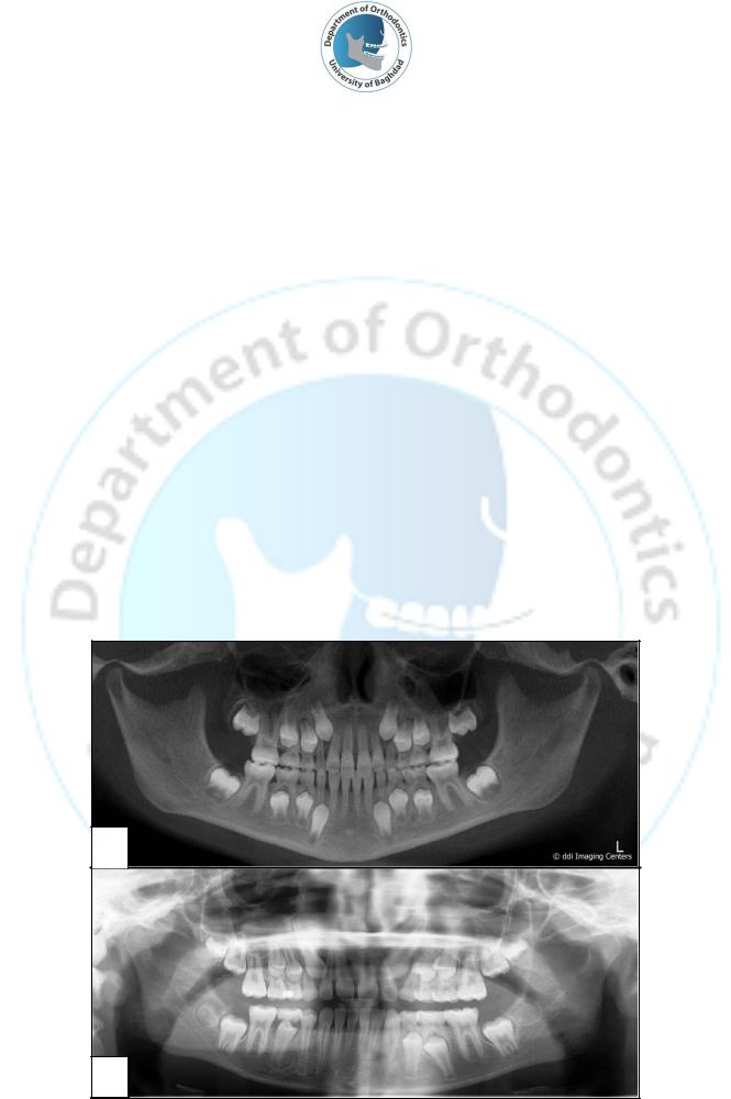
Figure 1: Orthopantomogram (OPG).
Figure 2: Precise information on periodontal membrane can be obtained from periapical radiograph.
Figure 3: Lower incisors region is better visualized from periapical radiograph.
Page 2 of 6
University of Baghdad |
Department of Orthodontics |
|
College of Dentistry |
Fifth year |
|
|
|
|
Lecture 6 |
|
Prof. Dr. Yassir Abdulkadhim Yassir |
|
|
|
Uses of an Orthopantomogram
1.Assessment of dental age, pathological conditions, growth and development: Stage of teeth formation, delayed tooth eruption, abnormality in eruption path, impaction, ankylosis, prolong retention, supernumerary teeth, congenitally missing teeth, abnormal resorption, cysts, tumors, density of the bone, etc..
2.The tempromandibular joint: The OPG provides a sharp and accurate profile view of the condyle and the articular eminence of the articular fossa itself.
3.Sinuses evaluation: the importance of maxillary sinuses is very recognized by orthodontist since a reduced sinuses size related to mouth breather and collapse of maxillary segments.
4.Mandibular morphology: The OPG gives a clear picture about the bony mass of the mandible, the extent of the alveolar bone, height and width of the ramus, in addition to the presence of teeth or any pathology.
5.Adjunct to serial extraction: The OPG is of great benefit during the serial extraction procedure which requires the removal of some deciduous teeth followed by some permanent teeth and this require knowledge about the stages of root formation of the teeth.
6.Investigation of facial asymmetries and swelling.
7.Investigation of suspected fractures of mandible and maxilla.
A
B
Figure 4: Dental age can be estimated from OPG. A: early mixed dentition 8-10 years, B: late mixed dentition 10-12 years.
Page 3 of 6

University of Baghdad |
Department of Orthodontics |
|
College of Dentistry |
Fifth year |
|
|
|
|
Lecture 6 |
|
Prof. Dr. Yassir Abdulkadhim Yassir |
|
|
|
Figure 5: Congenital missing lower second premolars.
Figure 6: Pathological lesions of the mandible and maxilla.
Figure 7: Fracture of the mandible.
Page 4 of 6

University of Baghdad |
Department of Orthodontics |
|
College of Dentistry |
Fifth year |
|
|
|
|
Lecture 6 |
|
Prof. Dr. Yassir Abdulkadhim Yassir |
|
|
|
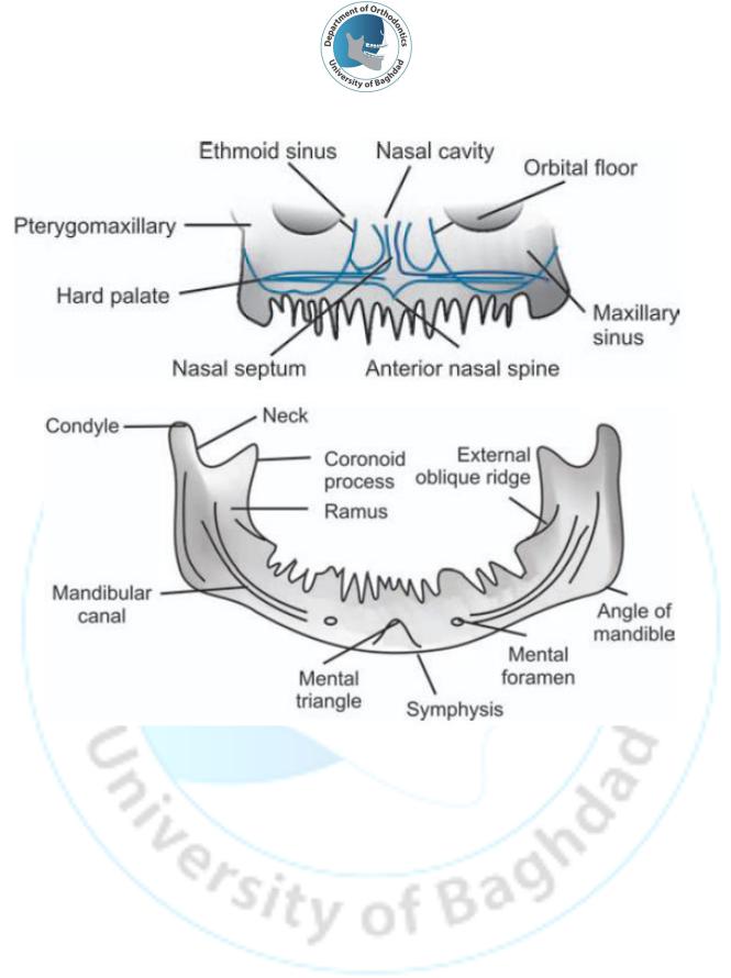
Steps of Reading/Interpreting Orthopantomogram
It is essential to be able to correctly read and interpret an orthopantomogram without missing out any important diagnostic detail. The followings should be examined:
1.Examine the mandibular bony outline from the right condylar head to the left condylar head.
2.Note the thickness and density of the mandibular cortex and the other structures including the mandibular canals, mental foramina, and the coronoid process
3.Examine the cortical outline of the maxilla starting on the right side. Check the hard palate with the anterior nasal spine. Examine the nasal cavities and the nasal septum followed by the maxillary sinuses. It is advisable to compare the right and left sides.
4.Compare the structure outline for any discontinuity, radiopacity or radiolucency, and most importantly assess symmetry from an orthodontic perspective.
5.Evaluate the teeth for; presence, stage of development, state of eruption unerupted or impacted teeth, placement, root morphology and position, cavities, fractures, contacts, any pathology. These findings have to be clinically correlated and/or with IOPAs or bitewing radiographs .
6.Note the third molar development and position.
7.Check any apparent soft tissue.
Note: Radiopaque shadows, which superimpose on normal anatomic structures are called
“ghosts” and are actually artifacts. These can sometimes pose a problem in radiographic interpretation. These are created when the X-ray beam projects through a dense object, e.g. the spinal cord and the opaque shadow of the object projects onto the opposite side of the radiograph.
Page 5 of 6
|
University of Baghdad |
Department of Orthodontics |
||
|
College of Dentistry |
Fifth year |
||
|
|
|
|
|
Lecture 6 |
|
Prof. Dr. Yassir Abdulkadhim Yassir |
||
|
|
|
|
|
|
|
|
|
|
|
|
|
|
|
Figure 8: Bony outline of the structures seen in the OPG.
Page 6 of 6