

Pathophysiology_FULL
.pdf1.Ischemic cell injury. The causes and pathogenesis. Reversible and irreversible injury.
Ишемия – уменьшение кровенаполнения органа или ткани вследствие недостаточного притока. Возникает при непроходимости приводящей артерии и отсутствии (или недостаточности) коллатерального притока крови в данную сосудистую территорию.
1)thrombi, emboli;
2)angiospasms;
3)sclerosis or inflammation of vessel walls
4)pressure from the outside from the artery.
Special: collateral ischemia with fast blood redistribution (for example in brain vessels in case of collapse)
Pathogenesis: Blood velocity and blood pressure drop -> erythrocyte redistribution -> blood poor with erythrocytes -> no division into axial and plasmatic bloodstream -> functional capillaries turn into plasmatic.
Low pressure -> capillaries shut down -> functional numbers DECREASE -> NO OXEGENATION AND ENERGY RESOURSES, WASTE ACCUMULATION -> HYPOXIA, HYPERCAPNIA, METABOLIC ACIDOSIS
Low pressure -> low filtration -> low tissue fluid levels -> decrease of lymphodrainage
Consequenses: short-term ischemia – decrease of function, long-term – hypoxic necrobiosis. Local ischemic necrosis = infarction, in some time infarction turns into post necrotic sclerosis 2. Role of ionized intracellular calcium and lipid peroxidation in aggravation of this pathological condition.
Increased permeability for Na, K and Ca ions -> increased intracellular Ca (in cytoplasm and mitochondria!!).
Deficit of energy -> activation of lipid peroxidation (+ increase of its substrate) and catecholamine-ergic system, inhibition of Ca2+-ATPase, Na/K-ATPase, increase intracellular Na - > increase intracellular Ca -> tetanus (muscle contraction) -> decrease of ATP
Increased Ca in cells -> activation of Ca-dependent proteases and phospholipases in mitochondrial membranes -> decreased transmembrane potential -> decreased Ca/H+ exchange in Mch membrane
Too much Ca in Mch -> formation of Ca salts -> malfunction of Mch
Too much Ca in Mch -> Increased osmotic pressure -> swelling -> disconnection of processes of cell respiration and phosphorylation, decrease in ATP synthesis.
«Нарушение барьерной функции клеточных мембран и повышение их пассивной проницаемости для ионов Na+, К+ и Са2+ сопровождается резким увеличением содержания Са2+ в цитоплазме и в матриксе МХ, что является важнейшим фактором повреждения клетки. При дефиците энергии активируется ПОЛ и катехоламинергическая система, ингибируются Са2+-АТФазы, Na+/K+-АТФазы, повышается внутриклеточный уровень Na+, что обусловливает повышение концентрации Ca2+ в цитоплазме, развитие мышечной контрактуры и дальнейшее снижение запасов АТФ в клетке. Активируются Ca2+-зависимые протеазы и фосфолипазы мембран МХ, повышается их проницаемость для Ca2+. Снижение трансмембранного потенциала внутренней мембраны МХ нарушает обмен Са2+ матрикса МХ на Н+. При перенасыщении митохондрий Ca2+ образуются фосфорные соли кальция, выпадающие в осадок, что необратимо нарушает функцию МХ. Увеличение осмотического давления в матриксе МХ сопровождается поступлением в них воды и набуханием органелл, разобщением процессов дыхания и фосфорилирования, дальнейшим снижением синтеза АТФ.

В определенных концентрациях Ca2+ стимулирует ПОЛ. Активируя фосфолипазы в очаге повреждения, Ca2+ повышает содержание полиненасыщенных жирных кислот, являющихся субстратом ПОЛ, а также снижает активность антиоксидантной системы. Повышение осмотического давления в клетке при избыточной кальциевой нагрузке может привести к осмотической гибели клетки.»
3. The mediators of inflammation which are the metabolites of arachidonic acid. Their role in inflammation.
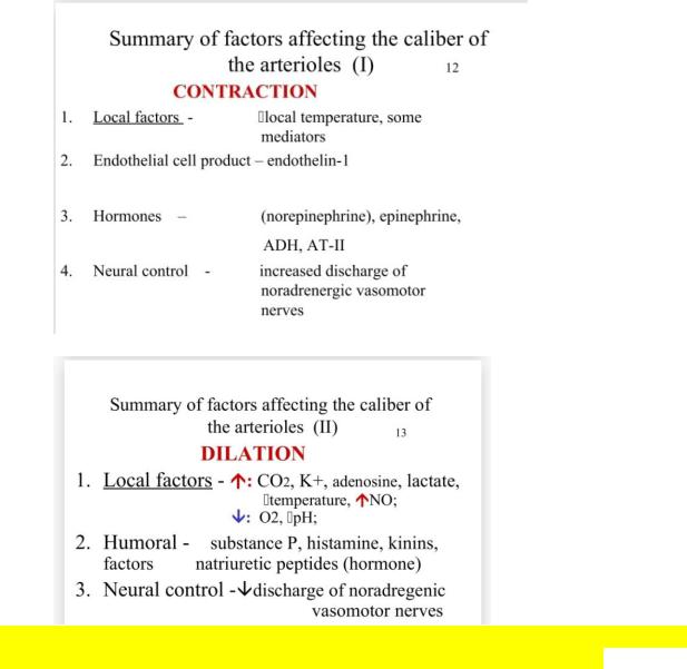
Dilation and increased permeability of microcirculatory vessels
Bronchodilation
Lymphocyte and polymorph-nucl leukocyte function suppression
Nociceptive sencitization
Bronchoconstriction
Dilation and increased permeability of microcirculatory vessels, Leukocyte function suppression
PLT adhesion+agregation inhibition, bronchodilatation
Bronchoconstriction, mictocirculatory vasoconstriction
Bronchoconstriction, mictocirculatory vasoconstriction, PLT adhesion+agregation stimulation, leukocyte/endothelium adhesion stimulation
Leukotrienes – bronchospasm, increased mucosa secretion, vasospasm of pulmonary vessels, decreased activity of ciliated epithelium. They play an important role in pathogenesis of asthma and other pulmonary diseases

Platelet Activation Factor -
4. Acute inflammation. Definition. Clinical signs of inflammation and mechanisms of their appearance.
Воспаление – развивающийся в месте повреждения реактивный патологический процесс (local reaction, typical pathological process).
Pain |
|
Vessel reaction |
Heat |
|
Exudation |
Redness |
OR |
Cell reactions (leukocyte |
Swelling |
|
emigration and phagocytosis) |
Loss of function |
|
|
Heat/redness – hyperemia, swelling – increased permeability of vessels, pain – nociceptors stimylation
There are three stages are known in high vessels permeability:
1. First, early stage is associated with release of histamine in course of mast cells degranulation provoked either by primary injuring factor or secondary. Secondary mast cell degranulation with histamine release associated with such biological active factors as anaphilatoxins (C3a and C5a activated complement fractions), different proteolytic enzymes, which appear in injured tissue later. It’s short-termed period that usually lasts less than 30-40 minutes. Histamine initiates an appearance of the gaps between endothelial cells which in normal are cemented in vast majority of the capillaries. Acting through the special histamine H1 receptors, it transforms the oval shapes of the endothelial cells into the round ones, that ultimately leads to appearance of the small spaces (gaps) between the endothelial cells. The matter of the fact, that stimulation of H1-receptors provokes accumulation of intracellular Ca ++ and then interaction of contractile structures inside the endothelium. As was said, this phenomenon is very short in time and permits only liquor and small proteins reversibly to pass through the small vessels wall. It is a start of inflammatory exudation. If we inject histamine in a volunteer intracutaneously we can observe so called Lewis triad of skin inflammatory symptoms: redness, swelling, and pain in form of itching. These symptoms last lesser than 30-40 minutes and imitate classical inflammatory signs. Histamine is the first vasoactive substance which dilates (via NO action) the microcirculatory vessels and increases their permeability. The matter of the

fact, that histamine triggers the synthesizing of NO in endothelium and the last, in turn, acting to the nearby situated smooth muscle cells, provokes their relaxation and arterioles dilation
2.Second stage is known as the late and prolong because, not at once, but for a long time (hours and days) can support high vessels permeability and their dilation. It is mostly associated with bradykinin formation from the kininogens of plasma. In addition must be said that bradykinin is responsible for nociceptive receptors stimulation, and such way it provokes severe pain in the site of inflammation. It’s a very strong algesiogenic substance.
3.Third stage is called postponed component of late response. It seems to be supported by PgE-2
5. Vascular reactions in the site of an acute inflammation. The role of biological active substances in these processes.
Short-term angiospasm
arterial hyperemia
o myoparalytic (smooth muscle damage, mediator action)
o neuroparalytic (norepinephrine exhaustion, damage to vasopressor endings) o neurotonic (axon reflex)
venous hyperemia
o cause: outflow imbalance

ointernal factors: decreased blood viscosity, RBC aggregation, endothelium swelling, fibrin, marginal standing of white blood cells
oexternal factors: serotonin (vasoconstriction), exudation, venous structure damage
stasis
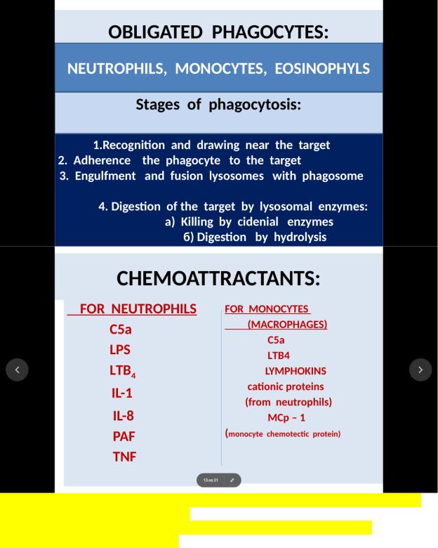
6. Phagocytosis. Stages and the events characteristic of each stage. The most important chemo attractants and their origin. The role of the phagocytosis in course of inflammation.
The steps of phagocytosis
1st step. Recognition and attachment of the foreign particles to be ingested by the leukocyte
2nd Its engulfment with subsequent formation of the phagocytic vacuole
3rd Killing and degradation of the ingested material
Recognition
The moving of the leukocyte toward an injured tissue named chemotaxis. Its locomotion activity may be explained by positive chemical gradient of biological active substances named the chemoattractants. Both exogenous and endogenous substances can act as chemoattractants. Mostly, exogenous agents are the bacterial products; lipopolysaccharides and proteins. Endogenous chemical mediators include:
Activated complement system components, especially C5a and C3a - anaphylatoxins
Products of lipoxygenase pathway metabolism, mainly LTB-4
PAF
Interleukin 1 and TNF-alpha as the late chemoattractants
All listed above substances are polyvalent chemoattractants because they attract all types of obligatory phagocytes, including the neutrophils, eosinophils, monocytes and macrophages.

Neutrophils – IL-8 and neutrophil chemotactic factors (mast cells)
Eosinophils – histamine, eosinophil chemotactic factor (mast cells) and eotaxin (endothelium)
Monocytes – monocytic chemotactic factor
lymphocytes – lymphotaxin
Attachment of the particles to be ingested by the leukocyte
It’s better if the opsonins are present. The role of opsonins fulfil such substances as activated C3b component of complement system, IgGs, IgMs, and C-reactive plasma protein. Via specific receptors on the phagocytes these substances opsonize them acting like a clay, gathering the foreign particles on the surface of phagocyte. For this reason, in the immunized organisms which reach of the immunoglobulins, phagocytosis is more active and, moreover, the treatment with specific serum immunoglobulins is strongly recommended in case of a pyogenic infection, especially in children.
Engulfment
A pseudopod flows around (hugs) the object to be engulfed and enclosed completely a particle within a phagosome created by a phagocyte membrane. Then the lysosomes become fused with limited wall of phagosome, resulting in release of granules content with following formation of phagolysosome. Degranulation of activated inflammatory neutrophils, monocytes and other obliged phagocytes leads to digestion of foreign particles, and very often to death of the phagocytes. Many of them are found in the pus (special purulent exudate consisted of died leukocytes and tissue debris).
Killing and degradation
Killing may be realized by two pathways: oxygen dependent and oxygen non-dependent. Phagocytes are not the “cannibals”, and before digestion of alive particles the lasts must be killed.
The steps of oxygen dependent killing proceeding in obligate phagocytes
1.Chemoattractants initiate the respiratory burst of a leukocyte, when it uptakes greedy an oxygen. It must be noted that in normal, not being activated, any leukocyte supports its energetic needs by the glycogenolysis with participation of hexosomonophosphate shunt.
2.After phagosome is arranged, the external membrane of the leukocyte contacts with cell cytoplasm due to cytoplasm invagination. That time, two components of NADPH-system (cytoplasm and membrane) meet each other, and it results in activation of NADPH-ase.
3.Activated NADPH-ase, in turn, transfers a single electron on an oxygen molecule with following superoxide radical formation.
4.Then part of the superoxide dismutase with such end product as hydrogen peroxide formation
5.Eventually, in Fenton’s reaction when transient metals iron and cooper are involved, hydroxyl radical OH’ is formed.
6.Reduction of NADP up to NADPH is realized through hexose-mono-phosphate shunt activity Described above reactions, result in the three primary radicals or active oxygen species formation. They possess by very strong bactericidal properties. These reactions are going in any type of the obliged phagocytes, but the granulocytes possess by stronger weapon in their fighting against a pathogen. It is myeloperoxidase system.
The granulocytes: neutrophils and eosinophils, except the primary oxidants, produce the secondary oxidants, because they possess by the enzyme myeloperoxidase in their azurophilic granules. It’s their marker, and yet in the bone marrow, they get it during maturation. As was said, granulocytes possess by myeloperoxidase system. It consists of enzyme myeloperoxidase, hydrogen peroxide and halids (chlorides and bromines); the system doesn’t possess by specify, acting mortally to any pathogen. Assembly of hydrogen peroxide, myeloperoxidase and chlorides results in hypochloric anion (OCL-) and hypochloric acid (HOCL-) formation; those in further interact with the aminogroups of proteins and form chloramines, very strong oxidants. Hydroperoxide-myeloperoxidase-halide system is not specific of organism, but very effective in the struggle against such pathogens as worms, protozoa, fungi, and cells infected by the viruses.
Oxygen non-dependent mechanisms of killing
It includes both, enzymatic and non-enzymatic factors containing in neutrophil granules: cationic protein, granzymes, BIP-factor (bacterial permeability increasing protein), besides, the lysozomal content and lactic acid in their vacuoles.
Major basic protein in eosinophil granules is the best bactericidal weapon in the fight against the worms and protozoa. Some substances, releasing from neutrophil granules, possess by bacteriostatic properties. That is nonless important as the protective mechanisms due to retarding the growth of animate pathogens. So, lactoferrin, binding iron, inhibits bacterial respiration, but protein, binding vitamin B12, interferes with proper bacterial growth. And, eventually, the leukocyte hydrolyses complete a process of foreign bodies destruction by lysing of dead particles; the lasts are presented by the tissue debris and dead phagocytes.
As for phagocytosis, it may be non-completed when bacteria or other particles remained inside the phagocytic vacuole as the residents, for example, meningococcal or gonococci. It’s a very danger for an organism, because one day the weakness of its immunity may result in dissemination of infection all over the organism. The other variant is interrupted phagocytosis when the object is too large to be ingested by phagocyte. In this case the phagocyte acts as a “valiant”, attacking the enemy by releasing of the oxygen species and enzymes via bombarding an object. This fighting may injury not only a pathogen but normal surrounding tissue too. Such kind of injury may be observed in chronic bacterial or immune inflammation: allergic dermatitis, psoriasis, and skin lesions in lupus erythematosus. For this reason, the drugs, including the antioxidants, may become very useful in case of these diseases treatment.
7.Leukocytes emigration in acute inflammation. The stages and characteristic of each stage. Biological significance of the phenomena.
8.Cellular events in acute inflammation. Emigration of the leukocytes: stages and corresponding events in their dynamic.
The chain of the events touching on the leukocyte behavior at that moment may be presented in such consequence: margination—rolling—activation—adhesion—transmigration
Mechanisms of leukocyte adhesion
In leukocyte adhesion both leukocytes and vessels wall are involved. Such substances as microbial polysaccharides and some mediators of inflammation, but later the mediators of an acute phase response are capable to express the adhesion molecules both on the leukocytes and endothelium membranes. As for inflammatory mediators, the following must be called:
LTB-4, PAF, C3a and C5a anaphylatoxins. At the same time, IL-1 and TNF-alpha also become
responsible for these phenomena but some later, when the macrophages appear on the
“field of the battle”.
There are three classes of adhesion molecules are known: selectins, integrins and superfamily of the globulins.
P-selectins |
On endothelium, preformed in |
|
|
|
vesicles, appear if stimulated by |
Only for rolling of |
|
|
histamine or thrombin |
||
|
leukocytes |
||
E selectins |
On endothelium |
||
|
|||
L selectins |
On leukocytes |
|
More firm adhesion needs elaboration of beta-integrins by the leukocytes and ICAM1 and ICAM-2 molecules by the endothelial cells with their expression on the corresponding cell membrane. Needless to say, that they are producing later, and their appearance precedes a leukocyte transmigration. Both are synthesized “de novo”.
The common features of leukocyte emigration
The law of Mechnikov sounds so:
1.Firstly, polynuclear phagocytes emigrate, mostly neutrophils and then mononuclear formsmonocytes.
2.There are three steps in leukocyte emigration: margination and adhesion, transmigration, and residence of the leukocytes in tissue.
The fact, that adhesion molecules are very important for the first step of emigration must be proved by the inhibition of leukocyte emigration when get used the antibodies to adhesion molecules. These monoclonal antibodies against the adhesion molecules retard an acute inflammation and, such way, can predispose an organism to chronic inflammation. There are some hereditary deficiencies in their synthesizing.
adhesion molecules deficiencies named DAL-1 and DAL-2 (defect adhesion of leukocytes).
DAL-1- corresponds to deficiency of b-chains of integrins
DAL-2- results to the deficiency in the receptors to L-selectins on the leukocytes.
Through the vessels wall neutrophil moves by extending its pseudopods that pull the remainder of the cell in the direction of extension likes automobile with its wheels. Locomotion is a genetic property of the leukocyte. It was estimated that neutrophil can’t be in the rest even in normal and persists in form of “chaotic dance” I when it throws out the pseudopods. But emigrated leukocyte has difficulties in navigation problem to arrive to correct extravascular location, and both chemoattractants and their receptors on the leukocyte membrane are engaged in this moving. Chemokines or chemoattractants in step by step manner call the leukocytes in the site of their high concentration via stimulation leukocyte locomotion. At first, the neutrophil enters the gap between the endothelial cells and then, being activated before by the chemoattractants, releases some hydrolyzes on the basal membranes. These are the elastase, collagenase, depolarizing the mucopolysaccharides of the vessels walls. The proteolytic enzymes make vessels wall more permeable, and such way, facilitate a leakage of the leukocytes through the small vessels. If you try to observe the vessels wall in electron microscope before and after leukocyte passage, you hardly ever find any defect in the vessels wall. Monocytes can emigrate by the same way or by pinocytosis using their glycolytic reserve. Obviously, the antagonists of glycolysis can arrest a pinocytosis due to creating the lack of energy for monocyte active transport. In contrast to the neutrophils and monocytes, the lymphocytes emigrate only in small venules with special tall type of endothelium. Such endothelial cells possess by so called “homming” receptors that are necessary for lymphocyte transmigration. It must be added, that in opposite to the phagocytes, which have got “one way