

- •Questions for examination and intersessional control of knowledge in pathological anatomy for 3rd year students of the medical and preventive faculty.
- •History of the Department of Pathological Anatomy of the Rostov State Medical University.
- •Biopsy: definition of the concept, types, goals and objectives. The role of the pathologist in the life-time diagnosis of various diseases.
- •According to the method of obtaining the material
- •Sampling of material for histological examination
- •Collection of material for cytological examination
- •By type of accuracy control:
- •Autopsy: definition of the concept, methodology, goals and objectives.
- •Pathology of the cell nucleus, mitosis, chromosomal apparatus: classification, structural changes, examples of diseases.
- •Reversible cell damage: definition, classification, causes, mechanisms of development.
- •Parenchymal proteinaceous dystrophy: definition, causes, mechanisms of development, macro- and microscopic signs. Disease examples.
- •Parenchymal fatty degeneration: definition, causes, mechanisms of development, macro-microscopic signs. Disease examples.
- •Fatty degeneration of the liver: terminology, causes, mechanisms of development, macro- and microscopic changes, clinical manifestations, outcomes, complications.
- •Myocardial fatty degeneration: terminology, causes, mechanisms of development, macro- and microscopic changes, clinical manifestations, outcomes, complications.
- •Mesenchymal dystrophies: definition, classification, causes, mechanisms of development. Disease examples.
- •Mesenchymal protein dystrophies: stages of connective tissue disorganization, causes, mechanisms. Macro- and microscopic changes in the connective tissue. Disease examples.
- •Hyalinosis: definition, classification, causes, mechanisms of development. Macro- and microscopic changes in the connective tissue. Disease examples.
- •Amyloidosis: definition, classification, mechanisms of development, structure of amyloid. Methods for detecting amyloid in tissues.
- •Amyloid consists of two components with antigenic properties:
- •4) Classification of amyloidosis:
- •By reason (origin):
- •Primary amyloidosis: causes, chemical structure of amyloid, mechanisms of development, examples of diseases. Macro- and microscopic changes, outcomes, clinical significance.
- •Secondary amyloidosis: causes, chemical structure of amyloid, mechanisms of development, examples of diseases. Macro- and microscopic changes, outcomes, clinical significance.
- •General obesity: definition, classification, causes. Macro- and microscopic changes in organs, related diseases and complications.
- •Mixed dystrophies: definition, classification. Types of hemoglobinogenic pigments, their significance for the body.
- •Local hemosiderosis: causes, mechanisms of development. Macro- and microscopic changes in organs, detection methods. Examples of pathological processes.
- •Pathology of hematins: types, structural features, examples of diseases. Macro- and microscopic changes in organs with the accumulation of malarial pigment.
- •Jaundice: definition, classification, normal bilirubin metabolism. Prehepatic jaundice: causes, macro-, microscopic changes in organs, clinical signs, complications, outcomes.
- •Hepatic jaundice: definition, causes, bilirubin metabolism, macro- and microscopic changes in the liver, clinical signs, complications, outcomes.
- •Subhepatic jaundice: definition, causes, bilirubin metabolism, macro- and microscopic changes in the liver, clinical signs, complications, outcomes.
- •Pathology of lipidogenic pigments: types, causes of formation, examples of diseases. Macro- and microscopic changes in organs with lipofuscinosis, outcomes.
- •Pathology of tyrosinogenic pigments: types, role in normal and pathological conditions. Violation of melanin metabolism: melanin metabolism is normal, classification.
- •Types and origin of proteinogenic (tyrosinogenic) pigments:
- •Causes of common acquired melanosis (melanodermia):
- •General and local hypermelanosis: causes, mechanisms of development, macro- and microscopic signs, clinical significance.
- •General and local hypomelanosis: causes, mechanisms of development, macro- and microscopic signs, clinical significance.
- •Calcifications: definition of the concept, types. Metabolism and regulation of calcium is normal.
- •Calcium metabolism:
- •Dystrophic calcification: definition, causes, mechanisms of development. Macro- and microscopic changes in organs, clinical significance. Disease examples.
- •Metastatic calcification: definition, causes, mechanisms of development. Macro- and microscopic changes in organs, clinical significance. Disease examples.
- •Formation of stones: definition of the concept, causes, mechanisms of development. Forms and chemical composition of urinary and biliary tract stones, complications, clinical significance.
- •Violation of the metabolism of nucleoproteins - gout: definition of the concept, types. Macro- and microscopic changes in organs, clinical significance, complications, outcomes.
- •Necrosis : definition of the concept, etiology and pathogenesis, classification by etiology and pathogenesis.
- •Necrosis: stages of morphogenesis, clinical and morphological criteria of cell death, pathoanatomical types. Macro-, microscopic signs of necrosis, outcomes.
- •Clinical and morphological forms of necrosis: macro-, microscopic characteristics, examples of diseases.
- •Gangrene: definition, causes, types, macro-microscopic signs, outcomes, clinical significance.
- •Heart attack: definition, causes, classification, conditions of development. Macro- and microscopic signs, outcomes, clinical significance.
- •Ischemic infarction: definition, causes, localization. Macro- and microscopic changes in organs, outcomes. Clinical significance.
- •Hemorrhagic infarction: definition, causes, localization. Macro- and microscopic changes in organs, outcomes. Clinical significance.
- •Apoptosis: definition, causes, pathogenesis - biochemical and microscopic features.
- •Apoptosis: definition, activation pathways, activator genes, receptors, role of caspases. Variants of apoptosis regulation disorders, role in pathology, examples of pathological processes.
- •Morphology of apoptosis: ultrastructural features. Comparative characteristics of necrosis and apoptosis.
- •Dysregulation of apoptosis in pathology, types, clinical significance. Examples of pathological processes.
- •Gangrene: types, causes, pathological characteristics, significance for the body.
- •Violations of the content of tissue fluid: definition, types, pathogenetic factors. Types of edema depending on the cause of the disease. Clinical significance.
- •Arterial hyperemia: definition, types. Types of pathological arterial hyperemia, significance for the body.
- •Venous hyperemia: definition, classification. Pathological and anatomical characteristics of general venous plethora, causes, mechanisms of development.
- •Acute general venous plethora: definition, causes, pathogenesis. Pathological changes in organs, outcomes, clinical significance.
- •Chronic general venous plethora: definition, causes, pathogenesis. Pathological changes in organs, outcomes, clinical significance.
- •Brown induration of the lungs: definition of the concept, causes, pathogenesis. Macro- and microscopic changes in the lungs, outcomes, clinical significance.
- •Nutmeg liver: definition of the concept, causes, pathogenesis. Macro- and microscopic changes in the liver, outcomes, clinical significance.
- •Bleeding, hemorrhage: definition of concepts, types, mechanisms. Examples of diseases depending on the mechanism of development. Outcomes, clinical significance.
- •Thrombosis: definition of the concept, general and local factors of thrombus formation, stages of thrombus development. Types and structure of blood clots, their outcomes.
- •Shock: definition of concept, types, stages. Macro- and microscopic changes in organs during shock.
- •Embolism: definition, classification, complications of embolism..
- •Thromboembolism of the arteries of the pulmonary circulation: types, causes, significance for the body.
- •Thromboembolism of the arteries of the systemic circulation: causes, significance for the body
- •Tissue and bacterial embolism: causes, significance for the body.
- •Hypovolemic shock: definition, etiology, pathogenesis, pathological anatomy.
- •Cardiogenic shock: definition, etiology, pathogenesis, pathological anatomy.
- •Vascular shock: definition, etiology, pathogenesis, pathological anatomy.
- •Infectious-toxic shock: etiology, pathogenesis, pathological anatomy.
- •Inflammation: definition of the concept, etiology, classification, pathoanatomical characteristics of the phases of inflammation, outcomes, clinical significance.
- •Exudative inflammation: definition, causes, types. Pathological anatomical characteristics of serous inflammation, causes, localization, outcomes, clinical significance.
- •Fibrinous inflammation: definition, causes, mechanisms, types. Pathological anatomical characteristics, localization, complications, outcomes, clinical significance.
- •Purulent inflammation: definition, causes, mechanisms, types. Pathological anatomical characteristics, complications, outcomes, clinical significance.
- •Catarrhal inflammation: definition, causes, mechanisms. Pathological anatomical characteristics, complications, outcomes, clinical significance.
- •Hemorrhagic inflammation: definition, causes, mechanisms. Pathological anatomical characteristics, complications, outcomes, clinical significance.
- •Proliferative inflammation: definition, types, causes, mechanisms of development. Pathological anatomical characteristics, complications, outcomes, clinical significance.
- •Acute inflammation: definition, causes, types. Pathological anatomy of acute productive inflammation, outcomes, clinical significance, examples of diseases.
- •Chronic inflammation: definition, causes, types. Pathological anatomy, outcomes, clinical significance, examples of diseases.
- •Granulomatous inflammation: definition, causes, types, conditions of formation, mechanisms. Pathological anatomy, outcomes, clinical significance, examples of diseases.
- •Specific granulomas: definition of the concept, conditions of formation, causes. Macro- and microscopic structure, outcomes, complications, clinical significance.
- •Nonspecific granulomas: definition of the concept, conditions of formation, causes. Macro- and microscopic structure, outcomes, complications, clinical significance.
- •Granulomatous inflammation in tuberculosis: etiology, pathogenesis, conditions of development. Morphological characteristics, outcomes, complications, clinical significance.
- •Granulomatous inflammation in syphilis: etiology, pathogenesis, conditions of development. Morphological characteristics, outcomes, complications, clinical significance.
- •Granulomatous inflammation in leprosy: etiology, pathogenesis, conditions of development. Morphological characteristics, outcomes, complications, clinical significance.
- •Stimulation of the humoral link of immunity: participants in immunity, causes. Pathological anatomy of changes in the organs of the immune system (in the lymph nodes, spleen, bone marrow, thymus).
- •Variants:
- •Stimulation of the cellular link of immunity: participants in immunity, causes. Pathological anatomy of changes in the organs of the immune system (in the lymph nodes, spleen, bone marrow, thymus).
- •II hypersensitivity reaction , mechanism and scheme of the reaction, examples of diseases.
- •III hypersensitivity reaction , mechanism and scheme of the reaction, examples of diseases.
- •IV hypersensitivity reaction , mechanism and scheme of the reaction, examples of diseases.
- •Primary immunodeficiency syndromes: definition, classification, causes, changes in the organs of the immune system, complications.
- •Primary immunodeficiency may be associated with insufficiency:
- •2) Cellular immunity deficiency syndrome
- •3) The syndrome of insufficient humoral immunity.
- •Secondary immunodeficiency syndromes (acquired)-in connection with the disease or the type of treatment
- •Acquired immunodeficiency syndrome: etiology, pathogenesis, pathological anatomy of the organs of immunogenesis.
- •Adaptation, compensation: definition of concepts, classification, stages of development of compensatory processes.
- •Atrophy: definition of the concept, types, macro- and microscopic changes in organs, examples of diseases.
- •3) Morphology of general atrophy (cachexia, exhaustion):
- •4) Types of local atrophy:
- •The significance and outcomes of atrophy:
- •Hypertrophy: definition of the concept, types, macro- and microscopic changes in organs, examples of diseases.
- •1) Hypertrophy is an increase in the volume and mass of an organ.
- •2) Morphology of various types of hypertrophy:
- •Hyperplasia, metaplasia, dysplasia: definition of concepts, types. Macro- and microscopic changes in organs, examples of diseases.
- •Regeneration, reparation: definition of concepts, types, biological significance, morphological characteristics.
- •Morphogenesis of the regenerative process:
- •Physiological regeneration - occurs throughout life, includes:
- •Granulation tissue: causes, macro- and microscopic features, biological properties.
- •Wound healing by primary and secondary intention: definition of concepts, causes, pathogenesis, morphogenesis, outcomes, complications.
- •In order for the wound to heal by primary tension, the following conditions must be met:
- •Hypertrophy and hyperplasia: definition of the concept, types, significance for the body.
- •1.Physiological
- •2. Pathological(
- •Hypertrophy of the heart: definition of the concept, classification, causes, stages. Macro- and microscopic changes in the heart during hypertrophy, outcomes, complications, clinical significance.
- •Hypertrophy of the lv wall.
- •Hypertrophy of the pancreatic wall (pulmonary heart).
- •Intracardial causes:
- •III. “Bull's heart is an enlargement of the whole heart.
- •Local atrophy: definition of the concept, causes, types. Macro- and microscopic changes in organs with local atrophy, outcomes, clinical significance.
- •General atrophy: definition of the concept, causes. Macro- and microscopic changes in organs with local atrophy, outcomes, clinical significance.
- •Metaplasia, definition of the concept, causes, role in the development of tumor growth, examples.
- •Dysplasia (intraepithelial neoplasia) of the epithelium as a precancerous process: definition of the concept, types, causes, significance for the body, examples.
- •Definition of the concept and basic properties of the tumor. The difference between tumor growth and tissue growth during regeneration, hyperplasia, chronic inflammation.
- •Molecular genetic bases of carcinogenesis. Protooncogenes, suppressor genes, apoptosis regulator genes, their role in tumor development and progression.
- •Appearance and features of growth of tumors, The concept of the progression of tumors. Stages, types and ways of metastasis.
- •5) The growth of metastasis.
- •Local and general influence of the tumor on the body, examples.
- •Principles of classification of tumors. The role of the pathologist in the diagnosis of tumors.
- •Benign epithelial tumors: terminology, localization. Macro- and microscopic features of the structure, the nature of growth, outcomes, complications, clinical significance.
- •Cancer: definition, localization, basic principles of classification. Macro- and microscopic structural features, growth patterns, metastasis, outcomes, complications, clinical significance.
- •Sarcoma: definition, localization, basic principles of classification. Macro- and microscopic structural features, growth patterns, metastasis, outcomes, complications, clinical significance.
- •Precancerous processes. Obligate and facultative precancer. Stages of occurrence of cancer. Methods of pathoanatomical diagnostics of precancerous processes.
- •Tumors of the anterior pituitary gland: origin, terminology, types. Macro-microscopic structure, complications, clinical significance.
- •Tumors of the thyroid gland: origin, terminology, types. Macro-microscopic structure, complications, clinical significance.
- •Malignant epithelial and mesenchymal skin tumors: origin, terminology, types. Origin, terminology, types. Macro-microscopic structure, complications, clinical significance.
- •Benign and malignant tumors of connective tissue origin: origin, terminology, types. Macro-microscopic structure, complications, clinical significance.
- •Tumors of vascular origin: origin, terminology, types. Macro-microscopic structure, complications, clinical significance.
- •Tumors of osteoarticular origin: origin, terminology, types. Macro-microscopic structure, complications, clinical significance.
- •Tumors of muscular origin: origin, terminology, types. Macro-microscopic structure, complications, clinical significance.
- •Tumors of melanin-forming tissue: origin, terminology, types. Macro-microscopic structure, complications, clinical significance.
- •Teratomas: origin, terminology, types. Macro-microscopic structure, complications, clinical significance.
- •Anemia: definition, classification, types, causes, pathological anatomy, outcomes, complications.
- •Posthemorrhagic anemia: definition, causes, pathological anatomy, outcomes, causes of death.
- •Clinical and anatomical classification of leukemias. Pathological differences between acute and chronic leukemias.
- •Pathological anatomy of acute leukemia: definition, classification, changes in the hematopoietic organs. Outcomes, complications, clinical significance.
- •Pathological anatomy of chronic leukemia: definition, classification, changes in the hematopoietic organs. Outcomes, complications, clinical significance.
- •Lymphomas: definition, classification, pathological anatomy, immunohistochemical diagnosis, complications, causes of death.
- •Rheumatic diseases: definition of the concept, classification, general characteristics. Stages of disorganization of connective tissue.
- •Rheumatism: definition, clinical and morphological forms, pathoanatomical changes in the heart, joints, skin, nervous system.
- •Rheumatic endocarditis: definition, types, pathological anatomy, outcomes, complications, causes of death in patients.
- •Changes in the heart, large and small circles of blood circulation with mitral defects.
- •Changes in the heart, systemic and pulmonary circulation in aortic malformations.
- •Myocarditis: definition, classification, etiology, pathogenesis, pathological anatomy, complications, outcomes.
- •Cardiomyopathy: definition, causes, classification, pathological anatomy, complications, outcomes.
- •Systemic vasculitis: definition, etiology, classification, pathological anatomy, examples of diseases.
- •Atherosclerosis: definition, etiology, pathogenesis. Macro- and microscopic changes in arteries, complications, outcomes, clinical significance .
- •Atherosclerosis: definition of the concept, risk factors, developmental theories. Macro- and microscopic stages of atherosclerosis.
- •Atherosclerosis: definition, structure of stable and unstable atherosclerotic plaques. Complications and causes of death of patients.
- •Clinical and anatomical forms of atherosclerosis and related complications.
- •Symptomatic arterial hypertension: causes, mechanisms of development, complications, causes of death in patients.
- •Changes in the brain in arterial hypertension and related complications.
- •Hypertension: etiology, pathogenesis, pathological anatomy, causes of death.
- •Clinical and morphological forms of hypertension, pathological anatomy, causes of death.
- •Pathological anatomy of benign hypertension, causes of death.
- •Pathological anatomy of malignant hypertension, causes of death.
- •Ischemic heart disease (chd): definition, causes, forms. Risk factors, pathogenesis. The role of unstable atherosclerotic plaque in the morphogenesis of ihd.
- •Myocardial infarction: definition, causes, classification, pathogenesis. Stages of development and outcome.
- •. Complications of myocardial infarction: early and late, pathological anatomy, causes of death of patients.
- •Chronic ischemic heart disease (hihd): definition, causes, forms. Pathological anatomy of cihd, complications, outcomes, clinical significance.
- •Cerebrovascular diseases: definition, etiology, types, morphological characteristics. Changes in the brain in hypertension and related complications.
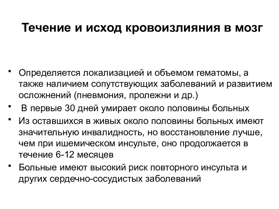
- •Cerebral hemorrhage: classification, causes, pathological anatomy, complications, outcomes, clinical significance.
- •Ischemic cerebral infarction: causes, pathological anatomy, complications, outcomes, clinical significance.
- •Pneumococcal pneumonia: pathological anatomy, complications and pathomorphosis.
- •Bronchopneumonia: etiology, pathoanatomical characteristics of pneumococcal, staphylococcal, streptococcal, fungal, viral pneumonia. Features of pneumonia in children.
- •Bronchiectasis: definition, classification, pathogenesis, morphogenesis, pathological anatomy, complications, clinical significance.
- •Pulmonary emphysema: definition, types, mechanisms of development, pathological anatomy, outcomes, complications, clinical significance.
- •Bronchial asthma: definition, etiology, mechanism of development, pathological anatomy, outcomes, complications.
- •Interstitial lung diseases: definition, etiology, pathogenesis, morphogenesis, classification, pathological anatomy, complications, clinical significance.
- •Sarcoidosis of the lungs: definition, etiology, pathogenesis, macro- and microscopic changes in the lungs, complications, outcomes.
- •Lung cancer: classification, localization, morphological characteristics, features of metastasis, complications, causes of death in patients.
- •Precancer and cancer of the esophagus: pathological anatomy. Forms of growth, features of esophageal cancer metastasis, complications, outcomes, clinical significance.
- •Chronic gastritis: definition, classification. Role of Helicobacter pylori in the morphogenesis of chronic gastritis. Complications, clinical significance.
- •Peptic ulcer of the stomach and duodenum: definition, etiology, pathogenesis, localization, macro- and microscopic characteristics of the ulcer, complications.
- •Gastric cancer: localization, classification. Features of metastasis, complications and causes of death in patients with gastric cancer.
- •Acute appendicitis: definition, etiology, macro- and microscopic signs, complications, outcomes.
- •Precancer and colon cancer: predisposing factors, pathological anatomy. Forms of growth, features of colon cancer metastasis, complications, outcomes, clinical significance.
- •Massive liver necrosis: causes, macro- and microscopic characteristics, complications, outcomes.
- •Alcoholic liver damage: types, macro- and microscopic signs, complications, outcomes.
- •Hepatitis: principles of classification, morphological features depending on the etiology, complications, outcomes.
- •Viral hepatitis b: etiology, pathogenesis, ways of infection, forms, pathological anatomy, outcomes.
- •Cirrhosis of the liver: classification, pathological anatomy, complications.
- •Acute and chronic cholecystitis: definition, etiology, classification, patho- and mophogenesis, complications.
- •Glomerulonephritis: principles of classification, morphological characteristics, leading clinical symptoms, complications.
- •Urolithiasis: etiology, chemical composition of stones, mechanism of stone formation. Macro- and microscopic changes in the kidneys, complications, outcomes.
- •Uremia: etiology, pathogenesis, macro- and microscopic changes in the kidneys. Complications, causes of death, clinical significance.
- •Inflammatory diseases of the female and male genital organs: causes, types, pathological anatomy, complications, outcomes, clinical significance.
- •Cervical cancer, the role of viral infections in its development. Pathological anatomical characteristics (macro- and microscopic signs), features of metastasis, complications, causes of death.
- •Diseases of the thyroid gland (goiter, thyrotoxicosis, thyroiditis, tumors): macro- and microscopic signs, complications, clinical significance.
- •Diabetes mellitus type I and II : definition, etiology, macro- and microscopic changes in the pancreas, blood vessels, kidneys, liver.
- •Covid -19: etiology, pathogenesis, pathological anatomy, complications, causes of death.
- •Influenza: etiology, pathogenesis, pathological anatomy, complications, causes of death.
- •Measles: etiology, pathogenesis, morphological characteristics, complications, causes of death.
- •Typhoid fever: etiology, pathogenesis, characteristics of intestinal changes and their outcomes.
- •Dysentery: etiology. Pathogenesis, morphological characteristics, intestinal complications, and their outcomes.
- •Cholera: etiology, pathogenesis, morphological characteristics, outcomes.
- •Acute enteritis (salmonellosis, staphylococcal, caused by Escherichia coli).
- •Diphtheria: etiology, pathogenesis, pathological anatomy. Complications, causes of death.
- •Scarlet fever: etiology, pathogenesis, pathological anatomy of the first and second periods, complications, causes of death.
- •Pathological anatomy of infection caused by hiv.
- •Primary tuberculosis complex in the lung and its complications.
- •Forms of hematogenous generalized tuberculosis and their morphological characteristics.
- •Secondary tuberculosis: its forms, clinical and morphological characteristics, complications.
- •Clinical and morphological characteristics of primary, secondary, tertiary and congenital syphilis. Complications, causes of death.
- •Pathological anatomy of septicemia and septicopyemia.
- •Acute infective endocarditis: pathomorphological characteristics, complications, causes of death in patients.
- •Protracted infective endocarditis: pathological anatomy and pathogenesis, complications.
Cerebral hemorrhage: classification, causes, pathological anatomy, complications, outcomes, clinical significance.
Cerebral hemorrhage is damage to the parenchyma of the brain caused by bleeding due to rupture of the intracerebral artery, leading to compression and displacement of supra- and subtentorial structures.
Etiology:
-Arterial hypertension
-Malformations of the arterial system of the brain — aneurysms, angiomas,
- Coagulopathy
-Hemorrhage into the tumor
-Hemorrhagic transformation of a heart attack
- Injuries
- Vasculitis
Types: Epidural hematoma - blood accumulates between the bones of the cranial vault and the meninges, i.e. in the epidural space. The source is the middle shell a or its branches Subdural hematoma - blood is poured out between the inner meninges outside and the arachnoid inside Subarachnoid hematoma - between the arachnoid and soft meninges Intracerebral - bleeding from the vessels of the brain tissue Intraventricular hematoma - accumulation of blood in the ventricles GM
They are also classified into: petechiae, hematomas, hemorrhagic infiltrations.
Complications:
Loss or speech disorders.
Changes in behavior, emotional component.
Problems in the work of the senses.
Cognitive deviations. Thinking, memory, perception, and concentration suffer.
Disability.
Deep vegetative state.
Death.
Ischemic cerebral infarction: causes, pathological anatomy, complications, outcomes, clinical significance.
Ischemic stroke is the formation of foci of necrosis in the GM as a result of metabolic disorders caused by insufficient blood supply; acute cerebrovascular accident with damage to brain tissue, damage to its functions or cessation of blood flow. It is accompanied by softening of areas of brain tissue - a cerebral infarction
Classification
1. By type
- White (ischemic) - occurs in conditions of insufficient collateral circulation
- Red (hemorrhagic) - occurs in conditions of a double type of blood supply to the tissue
- Mixed
2. By size
- Extensive, Large, Medium, Small Deep
3. By localization
- Superficial, Deep
Etiology
- CHD and blood vessels
- High blood viscosity
- The act of rheumatic endocarditis
- IBS
- Myocardial infarction
- Age over 60
- Alcoholism, smoking
- Coagulopathy
- SD
The form of AI development
1- Acute - symptoms increase within a few minutes
2- Subacute - symptoms increase within a few hours /days
3- Chronic - the course is gradual - the symptoms increase for 1 or more months
4 - Intermittent - symptoms increase gradually over 2-3 weeks, with short intervals between each deterioration
Morphological changes in transient ischemic attack of the brain are manifested by vascular disorders (spasm of arterioles, plasma leakage of their walls, perivascular edema and single small hemorrhages), as well as the appearance of foci of altered brain tissue (edema, dystrophic changes in cell groups). These changes are reversible.
Ischemic heart attack is the most common manifestation (75% of cases) of ischemic stroke. It looks like a focus of gray softening of brain tissue. During microscopic examination, dead neurons are found among the necrotized masses.
Hemorrhagic cerebral infarction looks similar to the focus of hemorrhagic stroke, but has a different mechanism of occurrence. Ischemia of the brain occurs primarily; hemorrhage into ischemic tissue occurs secondarily. Hemorrhagic infarction is more common in the cerebral cortex, less often in the subcortical nodes.
With a mixed infarction, which always occurs in the gray matter of the brain, it is possible to find areas of both ischemic and hemorrhagic infarction.