

5 курс / Пульмонология и фтизиатрия / Clinical_Manifestations_and_Assessment_of_Respiratory
.pdfFIGURE 2.5 Topographic location of lung fissures projected on the anterior chest (A) and posterior chest (B).
The right lung is separated into the upper, middle, and lower lobes by the horizontal fissure and the oblique fissure. The horizontal fissure runs anteriorly from the fourth rib at the sternal border to the fifth rib at the midaxillary line. The horizontal fissure separates the right anterior upper lobe from the middle lobe. The oblique fissure runs laterally from the sixth or seventh rib and the midclavicular line to the fifth rib at the midaxillary line. From this point, the oblique fissure continues to run around the chest posteriorly and upward to about the level of T-3. Anteriorly, the oblique fissure divides the lower lobe from the lower border of the middle lobe. Posteriorly, the oblique fissure separates the upper lobe from the lower lobe.
The left lung is separated into the upper and lower lobes by the oblique fissure. Anteriorly, the oblique fissure runs laterally from the sixth or seventh rib and the midclavicular line to the fifth rib at the midaxillary line. The fissure continues to run around the chest posteriorly and upward to about the level of T-3.
Inspection
The inspection of the patient is an ongoing observational process that begins with the history and continues throughout the patient interview, taking of vital signs, and physical examination. The inspection consists of a series of observations to gather clinical manifestations—signs and symptoms—that are directly or indirectly related to the patient's respiratory status. For example, during the patient interview and physical examination, the respiratory therapist might observe the patient demonstrating an abnormal ventilatory pattern, the use of accessory muscles, pursed-lip breathing, nasal flaring, splinting of the chest, a productive cough, or having clubbed fingers or lip or digital cyanosis.
A more in-depth discussion of the pathophysiologic basis for commonly seen respiratory disease clinical manifestations can be found in Chapter 3, The Pathophysiologic Basis for Common Clinical Manifestations.
Palpation
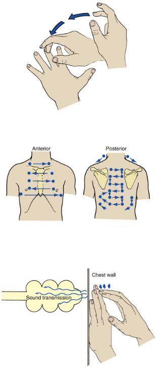
Palpation is the process of touching the patient's chest to evaluate the symmetry of chest expansion, the position of the trachea, skin temperature, muscle tone, areas of tenderness, lumps, depressions, and tactile fremitus and vocal fremitus. When palpating the chest, the clinician may use the heel or ulnar side of the hand, the palms, or the fingertips. As shown in Fig. 2.6, both the anterior and posterior chest should be palpated from side to side in an orderly fashion, from the apices of the chest down.
Данная книга находится в списке для перевода на русский язык сайта https://meduniver.com/

FIGURE 2.6 Path of palpation for vocal or tactile fremitus.
To evaluate the position of the trachea, the examiner places an index finger over the sternal notch and gently moves it from side to side. The trachea should be in the midline directly above the sternal notch. A number of abnormal pulmonary conditions can cause the trachea to deviate from its normal position. For example, a tension pneumothorax, pleural effusion, or tumor mass may push the trachea to the unaffected side, whereas atelectasis and pulmonary fibrosis pull the trachea to the affected side.
Chest Excursion
The symmetry of chest expansion is evaluated by lightly placing each hand over the patient's posterolateral chest so that the thumbs meet at the midline at about the T-8 to T-10 level. The patient is instructed to exhale slowly and completely and then inhale deeply. As the patient is inhaling, the examiner evaluates the distance that each thumb moves from the midline. Normally, each thumb tip moves equally about 3 to 5 cm from the midline (Fig. 2.7).
FIGURE 2.7 Assessment of chest excursion. (A) Anterior. (B) Posterior. Note that the thumbs move apart on inspiration as the volume of the thorax increases.
The examiner next faces the patient and lightly places each hand on the patient's anterolateral chest so that the thumbs meet at the midline along the costal margins near the xiphoid process. The patient is again instructed to exhale slowly and completely and then to inhale deeply. As the patient is inhaling, the examiner observes the distance each thumb moves from the midline. Again, an excursion of 3 to 5 cm from the midline (xiphoid process) is normally seen.
A number of pulmonary disorders can alter the patient's chest excursion. For example, bilaterally decreased chest expansion (excursion) may be caused by both obstructive and restrictive lung disorders. An unequal chest expansion may occur when one or more of the following develop in or around one lung only: alveolar consolidation (e.g., pneumonia), lobar atelectasis, pneumothorax, large pleural effusions, or chest trauma (e.g., fractured ribs).
Tactile and Vocal Fremitus
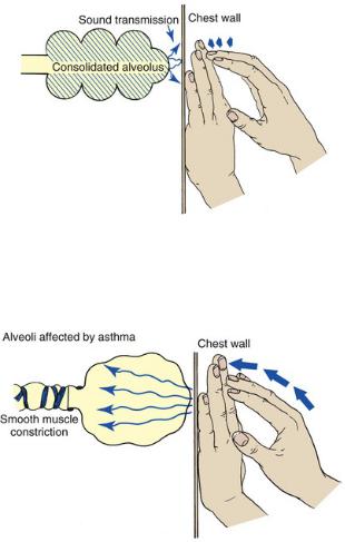
Vibration that can be perceived by palpation over the chest is called tactile fremitus (also known as rhonchial fremitus). This condition is commonly caused by gas flowing through thick secretions that are partially obstructing the large airways. Tactile fremitus is often noted during inhalation and exhalation and may clear after a strong cough. It is often associated with coarse, low-pitched crackles that are audible without a stethoscope. Vibration that can be perceived by palpation or auscultation over the chest during phonation is called vocal fremitus. Sounds produced by the vocal cords are transmitted down the tracheobronchial tree and through the lung parenchyma to the chest wall, where the examiner can feel the vibration. Vocal fremitus can often be elicited by having the patient repeat the phrase “ninety-nine” or “blue moon.” These are resonant phrases that produce strong vibrations. Normally, fremitus is most prominent between the scapulae and around the sternum, sites where the large bronchi are closest to the chest wall.
Tactile and vocal fremitus decreases when anything obstructs the transmission of vibration. Such conditions include chronic obstructive pulmonary disease, tumors or thickening of the pleural cavity, pleural effusion, pneumothorax, and a muscular or obese chest wall. Tactile and vocal fremitus increases in patients with alveolar consolidation, atelectasis, pulmonary edema, lung tumors, pulmonary fibrosis, and thin chest walls.
Crepitus (also called subcutaneous emphysema) is a coarse, crackling sensation that may be palpable over the skin
surface. It occurs when air escapes from the thorax and enters the subcutaneous tissue. It may occur after a tracheostomy and mechanical ventilation, open thoracic injury, or thoracic surgery. In severe cases, crepitus also may be felt over the abdomen, genitalia, extremities, and face.
Percussion
Percussion over the chest wall is performed to determine the size, borders, and consistency of air, liquid, or solid material in the underlying lung. When percussing the chest, the examiner firmly places the distal portion of the middle finger of the nondominant hand between the ribs over the surface of the chest area to be examined. No other portion of the hand should touch the patient's chest. With the end of the middle finger of the dominant hand, the examiner quickly strikes the distal joint of the finger positioned on the chest wall and then quickly withdraws the tapping finger (Fig. 2.8). The examiner should perform the chest percussion in an orderly fashion from top to bottom, comparing the sounds generated on both sides of the chest, both anteriorly and posteriorly (Fig. 2.9).
FIGURE 2.8 Chest percussion technique.
FIGURE 2.9 Path of systematic percussion to include all important areas.
In the normal lung the sound created by percussion is transmitted throughout the air-filled lung and is typically described as loud, low in pitch, and long in duration. The sounds elicited by the examiner vibrate freely throughout the large surface area of the lungs and create a sound similar to that elicited by knocking on a watermelon (Fig. 2.10).
FIGURE 2.10 Chest percussion of a normal lung.
Resonance may be muffled somewhat in the individual with a heavily muscular chest wall and in the obese person. When percussing the anterior chest, the examiner should take care not to confuse the normal borders of cardiac dullness with pulmonary pathologic conditions. In addition, the upper border of liver dullness is normally located in the right fifth intercostal space and midclavicular line. Over the left side of the chest, tympany (hyperresonance) is produced over the gastric space. When percussing the posterior chest, the examiner should avoid the damping effect of the scapulae.
Данная книга находится в списке для перевода на русский язык сайта https://meduniver.com/
Abnormal Percussion Notes
A dull percussion note is heard when the chest is percussed over areas of pleural thickening, pleural effusion, atelectasis, and consolidation. When these conditions exist, the sounds produced by the examiner do not freely vibrate throughout the lungs. A dull percussion note is described as flat or soft, high in pitch, and short in duration, similar to the sound produced by knocking on a full barrel (Fig. 2.11).
FIGURE 2.11 A short, dull, or flat percussion note is typically produced over areas of alveolar consolidation.
When the chest is percussed over areas of trapped gas, a hyperresonant note is heard. These sounds are described as very loud, low in pitch, and long in duration, similar to the sound produced by knocking on an empty barrel (Fig. 2.12). A hyperresonant note is commonly elicited from air trapping in the patient with chronic obstructive pulmonary disease or pneumothorax.
FIGURE 2.12 Percussion becomes more hyperresonant with alveolar hyperinflation.
Diaphragmatic Excursion
The relative position and range of motion of the hemidiaphragms also can be determined by percussion. Clinically, this evaluation is called the determination of diaphragmatic excursion. To assess the patient's diaphragmatic excursion, the examiner first maps out the lower lung borders by percussing the posterior chest from the apex down and identifying the point at which the percussion note definitely changes from a resonant to flat sound. This procedure is then performed at maximal inspiration and again at maximal expiration. Under normal conditions the diaphragmatic excursion should be equal bilaterally and should measure approximately 4 to 8 cm in the adult.
When severe alveolar hyperinflation is present (e.g., severe emphysema, asthma), the diaphragm is low and flat in position and has minimal excursion. Lobar collapse of one lung may pull the diaphragm up on the affected side and reduce excursion. The diaphragm also may be elevated and immobile in neuromuscular diseases that affect it.
Auscultation
Auscultation of the chest provides information about the heart, blood vessels, and air flowing in and out of the tracheobronchial tree and alveoli. A stethoscope is used to evaluate the frequency, intensity, duration, and quality of the sounds. During auscultation the patient should ideally be in the upright position and instructed to breathe slowly and deeply through the mouth. The anterior and posterior chest should be auscultated in an orderly fashion from the apex to base while the right side of the chest is compared with the left (Fig. 2.13). When examining the posterior chest, the examiner should ask the patient to rotate the shoulders forward so that a greater surface area of the lungs can be auscultated. Lung sounds are classified as either normal breath sounds or abnormal lung sounds (also called adventitious lung sounds).

FIGURE 2.13 Path of systematic auscultation to include all important areas. Note the exact similarity of this pathway to that in Fig. 2.6.
Normal Breath Sounds
Three different normal breath sounds can be auscultated over the normal chest. They are called bronchial, bronchovesicular, and vesicular breath sounds. Important characteristics of breath sounds include the pitch (vibration frequency), amplitude or intensity (loudness), and the duration of inspiratory sounds compared with expiration. Fig. 2.14 presents a sound diagram that illustrates the audio characteristics of the normal vesicular breath sound. Table 2.8 provides an overview of the normal breath sounds in regard to their location, pitch, intensity, and sound diagram.
FIGURE 2.14 The normal vesicular breath sound. The blue upward arrow represents inhalation. The red downward arrow symbolizes exhalation. The length of the arrow signifies duration. The thickness of the arrow denotes intensity or loudness. The angle between the blue inhalation arrow and the horizontal line symbolizes pitch (i.e., fast or slow vibration frequency).
TABLE 2.8
Normal Breath Sounds
Breath Sound |
Location |
Pitch |
Intensity Sound Diagram* |
|
Bronchial |
Over trachea |
High |
Loud |
|
|
|
|
|
|
Bronchovesicular |
Upper portion of anterior sternum, between |
Moderate |
Moderate |
|
|
scapulae |
|
|
|
|
|
|
|
|
Vesicular |
Peripheral lung regions |
High |
Soft |
|
|
|
|
|
|
*For the sound diagrams above, the blue upward arrow represents inhalation. The red downward arrow symbolizes exhalation. The length of the arrow signifies duration. The thickness of the arrow denotes intensity or loudness. The angle between the blue inhalation arrow and the horizontal line symbolizes pitch (i.e., fast or slow vibration frequency).
Bronchial Breath Sounds.
Bronchial breath sounds are normally auscultated directly over the trachea and are caused by the turbulent flow of gas through the upper airway. Bronchial breath sounds have a harsh, hollow, or tubular quality. They are loud, high in pitch, and about equal in duration in length of inspiration and expiration. A slight pause occurs between these two components. These sounds are also called tracheal, tracheobronchial, and tubular breath sounds.
Bronchovesicular Breath Sounds.
Bronchovesicular breath sounds are auscultated directly over the mainstem bronchi. They are softer and lower in pitch and intensity than bronchial breath sounds and do not have a pause between the inspiratory and expiratory phase. These sounds are reduced in pitch and intensity as a result of the filtering of sound that occurs as gas moves between the large airways and alveoli. Anteriorly, bronchovesicular breath sounds can be heard directly over the mainstem bronchi between the first and second ribs. Posteriorly, they are heard between the scapulae near the spinal column between the first and sixth ribs, especially on the right side (Fig. 2.15A).
Данная книга находится в списке для перевода на русский язык сайта https://meduniver.com/
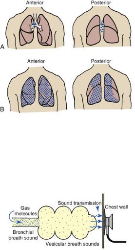
FIGURE 2.15 The location at which bronchovesicular breath sounds (A) and vesicular breath sounds (B) are normally auscultated.
Vesicular Breath Sounds.
Vesicular breath sounds are the normal sounds of gas rustling or swishing through the small bronchioles and the alveoli. Under normal conditions, vesicular breath sounds are auscultated over most lung fields, both anteriorly and posteriorly (see Fig. 2.15B). They are primarily heard during inspiration. As the gas molecules enter the alveoli, they are able to spread out over a large surface area and, as a result of this action, create less gas turbulence. Vesicular breath sounds also are heard during the initial third of exhalation as gas leaves the alveoli and bronchioles and moves into the large airways (Fig. 2.16).
FIGURE 2.16 Auscultation of vesicular breath sounds over a normal lung unit.
Abnormal Lung Sounds.
Abnormal lung sounds (ALS) are atypical, or uncharacteristic, lung sounds that are not normally heard over a specific area of the thorax. To describe the pitch of an ALS, the experts recommend the use of such words as high, moderate, or low—for example, “high-pitched wheezes were auscultated.” For the intensity or loudness of the ALS, words such as faint, soft, mild, moderate, or loud should be used—for example, “loud bronchial breath sounds were auscultated.” The part of the respiratory cycle in which the ALS occurs should be recorded—for example, “crackles were heard both during inspiration and expiration.” In addition, include mention of when the ALS occurs during inspiration—for example, “late-inspiratory crackles.” Also, document the magnitude of the ALS—for example, “small, scant, or profuse crackles.” Always record the precise location over the chest the ALS is auscultated—for example, “expiratory wheezes were noted over the anterior right lower lobe.”
Although the experts (American Thoracic Society [ATS] and the American College of Chest Physicians [ACCP] Joint Committee on Pulmonary Nomenclature) have debated for many years the value of some of the terms and adjectives used to describe ALS, they have agreed—for the most part—that several terms or phrases are either ambiguous or very subjective and therefore should not be used for clinical reports, charting, and/or electronic documentation. Terms, adjectives, and phrases not recommended at present are wet, dry, rales, rhonchi, crepitations, sonorous rales, musical rales, and sibilant rales.
Currently, the recommended terms, adjectives, and phrases for ALS are the following: fine crackles, medium crackles, or coarse crackles, wheezes, bronchial breath sounds, stridor, pleural friction rub, diminished breath sound, and whispering pectoriloquy. Fig. 2.17 illustrates the general location and cause for these ALS. Table 2.9 provides an overview and description of the common ALS.
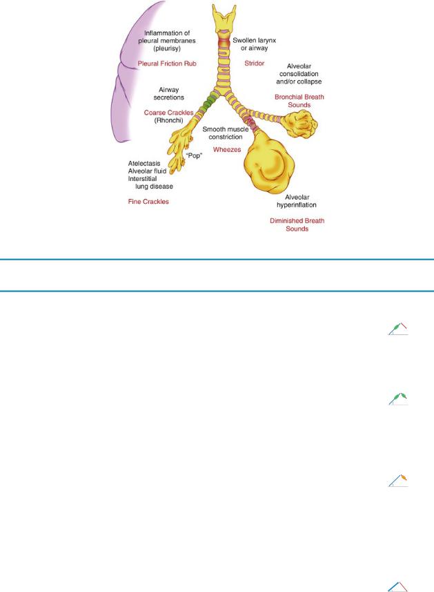
FIGURE 2.17 General location and origin of abnormal lung sounds.
TABLE 2.9
Abnormal Lung Sounds
Abnormal Lung Sound |
Sound Diagrams* |
|
Crackles |
|
|
Crackles (previously called rales) can be categorized as fine, medium, and coarse crackles. |
|
|
Fine crackles are discontinuous, high-pitched, crackling, and popping sounds similar to |
|
|
popping of bubble wrap or the sound created by rolling hair between fingers near one's ear |
|
|
and is heard near the end of inspiration (see Sound Diagram A). Fine crackles are produced |
A. Fine crackles. |
|
by the rapid equalization of gas pressure when collapsed alveoli or terminal bronchioles |
||
suddenly snap open (see Fig. 2.17). Fine crackles usually do not clear after a strong cough. |
The green oval |
|
Fine crackles are associated with alveolar collapse (atelectasis), interstitial fibrosis, early |
shape represents |
|
pulmonary edema, and pneumonia. |
crackles. |
|
Medium crackles are the same as fine crackles but are medium in pitch and have a moist |
|
|
quality as the disease process worsens. |
|
|
Coarse crackles (previously called rhonchi†) are discontinuous, low-pitched, rumbling, |
|
|
bubbling, or gurgling sounds that start early during inspiration and extend into exhalation. |
|
|
These sounds are caused by air moving through excessive airway secretions in the larger |
B. Coarse |
|
airways (see Sound Diagram B). Coarse crackles are commonly associated with severe |
||
crackles. The |
||
chronic obstructive pulmonary disease, cystic fibrosis, bronchiectasis, and congestive heart |
||
green oval shape |
||
failure (pulmonary edema). Coarse crackles may or may not change in nature after a strong, |
||
represents |
||
vigorous cough. |
||
crackles. |
||
|
||
Wheezing |
|
|
Wheezing is the characteristic sound produced by airway obstruction. Found in all |
|
|
bronchospasm disorders, it is one of the cardinal findings in asthma (see Fig. 2.17). |
|
|
Wheezes are continuous, high-pitched, musical whistles generally heard on expiration (see |
|
|
Sound Diagram). In severe cases, they may be heard during inspiration. In addition to |
Wheezing |
|
bronchospasm, other common causes of partial or total airway obstruction include mucosal |
||
edema, inflammation, tumors, and foreign bodies. |
The orange oval |
|
Partial airway obstruction is often made greater by this mechanism: When the bronchial |
shape represents |
|
airway is narrowed, the velocity of air flow through the constricted airway increases, |
wheezing. |
|
which, in turn, causes the lateral airway wall pressure to decrease. This condition causes |
|
|
the airways to narrow even further and/or collapse. As the airways narrow, they vibrate |
|
|
similar to a reed on a woodwind instrument, thereby producing a wheezing sound (Fig. |
|
|
2.18). |
|
|
Bronchial Breath Sounds |
|
|
Bronchial breath sounds have a harsh, hollow, or tubular quality. They are loud, high in pitch, |
|
|
and about equal in duration during inspiration and expiration. The inspiratory phase is |
|
|
louder (see Fig. 2.17 and Sound Diagram). Bronchial breath sounds are associated with |
|
|
alveolar consolidation and atelectasis (Fig. 2.19). |
Bronchial breath |
|
|
sound |
|
|
Note the thickness |
|
|
of the |
|
|
inspiratory and |
|
|
expiratory |
|
|
arrows, which |
|
|
denote |
|
|
loudness. |
|
Stridor |
|
|
|
|
Данная книга находится в списке для перевода на русский язык сайта https://meduniver.com/
Stridor is a continuous, loud, high-pitched sound caused by an upper obstruction in the |
|
|
trachea or larynx (see Fig. 2.17 and Sound Diagram). It is generally heard during |
|
|
inspiration. Stridor is usually loud enough to hear without a stethoscope, as in infantile |
|
|
croup. Stridor indicates a neoplastic or inflammatory condition, including glottic edema, |
Stridor |
|
diphtheria, laryngospasm croup, and papilloma (a benign epithelial neoplasm of the |
The orange shapes |
|
larynx). |
represent |
|
|
stridor sounds. |
|
|
|
|
Pleural Friction Rub |
|
|
If pleurisy accompanies a respiratory disorder, the inflamed pleural membranes resist |
|
|
movement during breathing and create a peculiar and characteristic sound known as a |
|
|
pleural friction rub (see Fig. 2.17 and Sound Diagram). It is a continuous, low-pitched, |
|
|
coarse creaking or grating-type sound reminiscent of that made by a creaking shoe. It is |
Pleural friction |
|
usually heard throughout inspiration and expiration over the area where the patient |
||
complains of pain. The intensity of a pleural rub often increases with deep breathing. A |
rub |
|
pleural friction rub does not clear with cough. A pleural friction rub is associated with |
The orange shapes |
|
pleurisy, tuberculosis, pneumonia, pulmonary fibrosis, pulmonary infarction, or after |
represent |
|
thoracic surgery. |
pleural friction |
|
|
rub sounds. |
|
Diminished Breath Sounds |
|
|
Breath sounds are diminished or distant in any respiratory disorder that reduces the sound |
|
|
intensity of air flow as in obesity. In another example, chronic obstructive pulmonary |
|
|
disease leads to air trapping, an increased function residual capacity, and hypoventilation, |
|
|
which, in turn, result in diminished breath sounds (Fig. 2.20). Heart sounds also may be |
Diminished |
|
diminished in patients with air trapping. |
breath sound |
|
The intensity of breath sounds is also reduced in any condition that causes shallow or slow |
Note the |
|
breathing patterns—for example, drug overdose, major sedation, or neuromuscular |
decreased |
|
diseases such as Guillain-Barré syndrome or myasthenia gravis. Diminished breath sounds |
angle on the |
|
are also found in respiratory disorders that cause hypoventilation by compressing the lung, |
inspiratory and |
|
such as flail chest, pleural effusion, and pneumothorax. |
expiratory |
|
|
arrows, which |
|
|
represent |
|
|
decreased |
|
|
intensity or |
|
|
loudness. |
|
Whispering Pectoriloquy |
|
|
Whispering pectoriloquy is the term used to describe the unusually clear transmission of |
|
|
the whispered voice of a patient as heard through the stethoscope. When the patient |
|
|
whispers “one, two, three,” the sounds produced by the vocal cords are transmitted not |
|
|
only toward the mouth and nose but also throughout the lungs. As the whispered sounds |
Normal vesicular |
|
travel down the tracheobronchial tree, they remain relatively unchanged, but as the sound |
||
breath sounds |
||
disperses throughout the large surface area of the alveoli, it diminishes sharply. |
||
—unclear, |
||
Consequently, when the examiner listens with a stethoscope over a normal lung while the |
||
muffled words |
||
patient whispers “one, two, three,” the sounds are diminished, distant, muffled, and |
||
(“one, two, |
||
unintelligible (Fig. 2.21). |
||
three”) |
||
When a patient who has atelectasis or consolidated lung areas whispers “one, two, three,” the |
||
auscultated. |
||
sounds produced are prevented from spreading out over a large alveolar surface area. |
||
Even though the consolidated area may act as a sound barrier and diminish the sounds |
|
|
somewhat, the reduction in sound is not as great as it would be if the sounds were allowed |
|
|
to dissipate throughout a normal lung. Consequently, the whispered sounds are much |
|
|
louder and more intelligible over the affected lung areas (Fig. 2.22). |
Consolidation |
|
|
||
|
and/or |
|
|
atelectasis— |
|
|
clear words |
|
|
(“one, two, |
|
|
three”) |
|
|
auscultated. |
*For the sound diagrams, the blue upward arrow represents inhalation. The red downward arrow symbolizes exhalation. The length of the arrow signifies duration. The thickness of the arrow denotes intensity or loudness. The angle between the blue inhalation arrow and the horizontal line symbolizes pitch (i.e., fast or slow vibration frequency).
†Historically, coarse crackles have also been called rhonchi, a term not currently recommended. Coarse crackles often produce palpable vibrations called tactile fremitus, also known as rhonchial fremitus. Coarse crackles are sometimes referred to as a “death rattle.” It is this abnormal lung sound that every practicing respiratory therapist knows all too well—that is, the patient, whose loud, rumbling, gurgling secretions can be heard across the patient's room, which clearly signals the immediate need for tracheal suctioning or chest physical therapy.
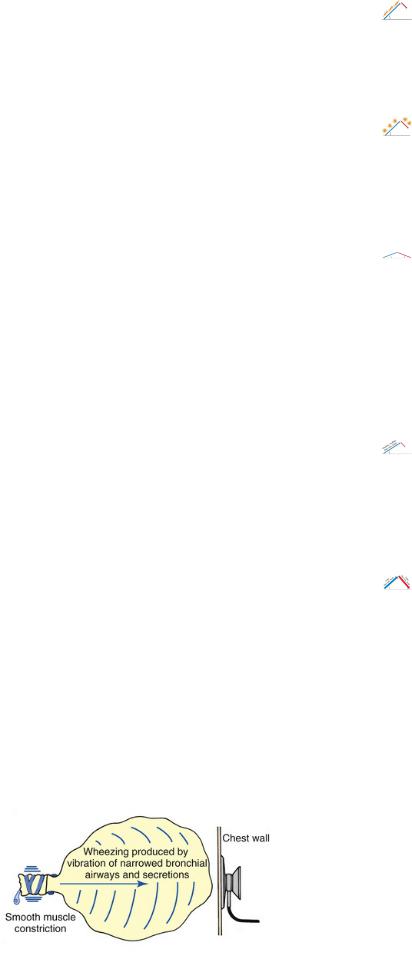
FIGURE 2.18 Wheezing and coarse crackles often develop during an asthmatic episode because of smooth muscle constriction, wall edema, and mucous accumulation.
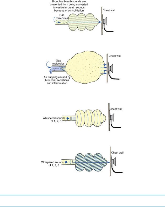
FIGURE 2.19 Auscultation of bronchial breath sounds over a consolidated lung unit.
FIGURE 2.20 As air trapping and alveolar hyperinflation develop in obstructive lung diseases, breath sounds progressively diminish.
FIGURE 2.21 Whispered voice sounds auscultated over a normal lung are usually faint and unintelligible.
FIGURE 2.22 Whispering Pectoriloquy. Whispered voice sounds heard over a consolidated lung are often louder and more intelligible compared with those of a normal lung.
Table 2.10 summarizes the common assessment abnormalities found during inspection, palpation, percussion, and auscultation.
TABLE 2.10
Common Assessment Abnormalities
Finding |
Description |
Possible Cause and Significance |
Inspection |
|
|
Pursed-lip |
Exhalation through mouth with lips pursed together to slow |
COPD, asthma. Suggests ↑ |
breathing |
exhalation. |
breathlessness. Strategy taught |
|
|
to slow expiration, ↓ dyspnea. |
Tripod |
Leaning forward with arms and elbows supported on overbed |
COPD, asthma in exacerbation, |
position; |
table. |
pulmonary edema. Indicates |
inability to lie |
|
moderate to severe respiratory |
flat |
|
distress. |
Accessory |
Neck and shoulder muscles used to assist breathing. Muscles |
COPD, asthma in exacerbation, |
muscle use; |
between ribs pull in during inspiration. |
secretion retention. Indicates |
intercostal |
|
severe respiratory distress, |
retractions |
|
hypoxemia. |
|
|
|
Splinting |
Voluntary ↓ in tidal volume to ↓ pain on chest expansion. |
Thoracic or abdominal incision |
|
|
pain. Chest trauma, pleurisy. |
|
|
|
Данная книга находится в списке для перевода на русский язык сайта https://meduniver.com/

↑ AP diameter |
AP chest diameter equal to lateral. Slope of ribs more horizontal |
COPD, asthma, cystic fibrosis. |
|
(90 degrees) to spine. |
Lung hyperinflation. Advanced |
|
|
age. |
Tachypnea |
Rate >20 breaths/min; >25 breaths/min in elderly. |
Fever, anxiety, hypoxemia, |
|
|
restrictive lung disease. |
|
|
Magnitude of ↑ above normal |
|
|
rate reflects magnitude of |
|
|
increased work of breathing. |
Kussmaul's |
Regular, rapid, and deep respirations. |
Metabolic acidosis; ↑ in rate aids |
respiration |
|
body in ↑ CO2 excretion. |
Cyanosis |
Bluish color of skin best seen in earlobes, under the eyelids, or in |
↓ Oxygen transfer in lungs, ↓ |
|
nail beds. |
cardiac output. Nonspecific, |
|
|
unreliable indicator. |
Clubbing of |
↑ Depth, bulk, sponginess of distal digits of fingers. |
Chronic hypoxemia. Cystic fibrosis, |
fingers |
|
lung cancer, bronchiectasis. |
Peripheral |
Pitting edema. |
Congestive heart failure, cor |
edema |
|
pulmonale. |
Distended neck |
Jugular venous distention. |
Cor pulmonale, flail chest, |
veins |
|
pneumothorax. |
Cough |
Productive or nonproductive. |
Bronchial airway and alveolar |
|
|
disease. |
Sputum |
See Table 3.4. |
COPD, asthma, cystic fibrosis, |
|
|
pneumonia. |
Abdominal |
Inward (rather than normal outward) movement of abdomen |
Inefficient and ineffective |
paradox |
during inspiration. |
breathing pattern. Nonspecific |
|
|
indicator of severe respiratory |
|
|
distress. |
Palpation |
|
|
Tracheal |
Leftward or rightward movement of trachea from normal midline |
Nonspecific indicator of change in |
deviation |
position. |
position of mediastinal |
|
|
structures. Medical emergency |
|
|
if caused by tension |
|
|
pneumothorax. |
Altered tactile |
Increase or decrease in examiner-felt vibrations. |
↑ In pneumonia, atelectasis; |
fremitus |
|
pulmonary edema; ↓ in pleural |
|
|
effusion, lung hyperinflation; |
|
|
absent in pneumothorax. |
Altered chest |
Unequal or equal but diminished movement of two sides of chest |
Unequal movement caused by |
movement |
with inspiration. |
atelectasis, pneumothorax, |
|
|
pleural effusion, splinting; |
|
|
equal but diminished movement |
|
|
caused by barrel chest, |
|
|
restrictive disease, |
|
|
neuromuscular disease. |
|
|
|
Percussion |
|
|
Hyperresonance |
Loud, lower-pitched sound over areas that normally produce a |
Lung hyperinflation (COPD), lung |
|
resonant sound on chest percussion. |
collapse (pneumothorax), air |
|
|
trapping (asthma). |
Dullness/flatness |
Medium-pitched sound over areas that normally produce a |
↑ Density (pneumonia, large |
|
resonant sound on chest percussion. |
atelectasis), ↑ fluid pleural |
|
|
space (pleural effusion). |
Auscultation |
|
|
Fine crackles |
Series of discontinuous short, crackling, and popping sounds, |
Loss of lung volume (atelectasis), |
|
high-pitched sounds heard just before the end of inspiration; |
interstitial fibrosis (asbestosis), |
|
result of rapid equalization of gas pressure when collapsed |
interstitial edema (early |
|
alveoli or terminal bronchioles suddenly snap open; similar |
pulmonary edema), alveolar |
|
sound to that made by rolling hair between fingers just behind |
filling (pneumonia), early phase |
|
ear. |
of congestive heart failure. |
Coarse crackles |
Series of discontinuous short, low-pitched bubbling or gurgling |
COPD, cystic fibrosis, congestive |
|
sounds caused by air passing through airway intermittently |
heart failure, bronchiectasis, |
|
occluded by mucus, unstable bronchial wall, or fold of mucosa; |
pulmonary edema, pneumonia |
|
evident on inspiration and, in more severe cases, expiration; |
with severe congestion, COPD. |
|
similar sound to blowing through straw under water; increase |
|
|
in bubbling quality with more fluid. |
|
Wheezes |
Continuous high-pitched whistling sound caused by rapid |
Bronchospasm (caused by |
|
vibration of bronchial walls; first evident on expiration but also |
asthma), airway obstruction |
|
possibly evident on inspiration as obstruction of airway |
(caused by mucosal edema, |
|
increases; possibly audible even without a stethoscope. |
inflammation, foreign body, |
|
|
tumor), COPD. |
Bronchial breath |
A harsh, hollow, or tubular breath sound. They are loud, high in |
Alveolar consolidation and alveolar |
sound |
pitch, and about equal in duration during inspiration and |
collapse (atelectasis). |
|
expiration. Inspiration is louder. |
|
Stridor |
Continuous, loud, high-pitched sound caused by a partial |
Croup, epiglottitis, vocal cord |
|
obstruction of the larynx or trachea. Generally heard during |
edema after extubation, foreign |
|
inspiration. |
body. |
|
|
|